FIGURE 55-1. Pathophysiology of the diabetic foot leading to vascular complications.
(Modified from LoGerfo FW: Bypass grafts to the dorsalis pedis artery. In Whittemore AD, Bandyk DF, Cronenwett JL, Hertzer NR, White RA [eds]: Advances in Vascular Surgery, vol 10. Philadelphia, Mosby, 2002, p 174.)
NEUROPATHY
The neuropathy stemming from diabetes mellitus has multiple manifestations within the diabetic foot because it encompasses sensory, motor, and autonomic fibers. Sensory neuropathy affects small-diameter pain and temperature fibers first, and susceptibility to injury is increased because these patients are less sensitive to pressure-related trauma and other minor skin injuries. Motor neuropathy affects the longer fibers that innervate the foot, including intrinsic foot muscles and leg muscles. Atrophy, or muscle wasting, in the intrinsic foot muscles allows the strong flexor muscles to draw up the toes in a “clawed” position, and new pressure points emerge at the tips of the toes and the prominent metatarsal heads. Limited joint mobility from glycation of scleral proteins exacerbates the situation by further changing the normal weight distribution on the foot. Last, autonomic neuropathy causes the skin to become dry through loss of sweat and oil gland function. This dry skin has a markedly increased susceptibility to skin breakdown and fissures, thus creating a portal of entry for bacteria. Additionally, diabetic patients experience a blunted neuroinflammatory response and thus are missing a crucial component of the body’s natural first-line defense against pathogens.4
INFECTION
Diabetic patients typically have an altered response to infectious processes owing to defects in their host immune defense system.5 Wound healing is delayed in diabetic patients as a result of abnormal cellular and inflammatory pathways involving fibroblasts, neutrophils, and advanced glycation end products (AGEs). Glycation is a nonenzymatic chemical reaction whereby sulfhydryl protein linkages are replaced by glucose, causing impairment in normal cellular and tissue functions.6 AGEs increase the stiffness of precapillary vessel walls and contribute to the development of diabetic microangiopathy.7 Furthermore, neuropathic and ischemic deficiencies in diabetic patients predispose them to infection and then unfortunately compound the problem by potentiating infection once a pathogen has been introduced.
ISCHEMIA
Much progress has been made in identifying the cause of ischemia in the lower extremities of diabetic patients, and results have challenged long-standing misconceptions in the literature and in the medical community at large. It is imperative that practitioners continue to reject the “small-vessel disease” theory related to occlusions of the microcirculation, as espoused by a single histologic study in 1959,8 and instead embrace the notion that ischemia results from both atherosclerotic macrovascular disease and microcirculatory dysfunction.9 Diabetic patients typically suffer from tibial and peroneal arterial disease with sparing of the foot arteries, especially the dorsalis pedis and its branches (Fig. 55-2).
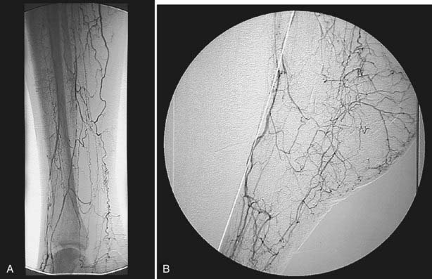
FIGURE 55-2. Intra-arterial digital subtraction arteriogram showing typical pattern of occlusive disease in a diabetic patient. A, Calf view: The posterior tibial, anterior tibial, and peroneal arteries are severely narrowed. Blood flow to the foot is entirely dependent on the small collateral vessels that are visible. B, Lateral foot view: The dorsalis pedis artery is widely patent with runoff into patent tarsal branches.
Research has shown that diabetes causes structural and functional changes within the arteriolar and capillary systems, notably thickening of the basement membrane. In spite of the thickened capillary basement membrane, no evidence reveals a decrease in capillary luminal diameter.10 A thickened membrane impairs the migration of leukocytes and hampers the normal hyperemic or vasodilatory response to injury, thus simultaneously increasing the susceptibility to injury while blunting the typical manifestations of such an injury.11 Overall, this dysfunction of the microcirculation in diabetic patients creates a functionally ischemic foot despite conditions that represent normal blood flow in healthy patients.
Besides causing specific structural changes in the microcirculation, diabetes also causes a compromise in the overall biology of the foot. When compared with nondiabetic individuals with normal biology, diabetes causes an undesirable shift in the natural balance that exists between stress/ulceration and resistance to stress/ulceration. The diabetic foot thus is more prone to ulcerate under the stress of daily life. Additionally, the presence of neuropathy usually mandates revascularization under conditions of perfusion that would not require revascularization in the absence of neuropathy. In other words, the pathophysiology underlying diabetes creates situations whereby the compromised foot requires even more perfusion than usual to resist ulceration or respond appropriately to injury (Fig. 55-3).
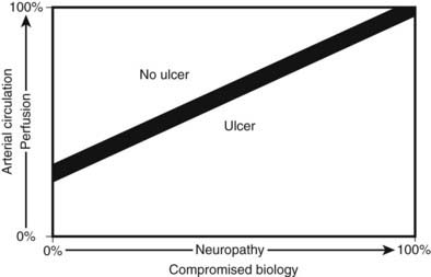
FIGURE 55-3. The relationship between ulceration, compromised biology, and arterial circulation. As perfusion decreases, even a foot with perfect biology will ulcerate. As neuropathy increases, even a well-perfused foot will ulcerate. With revascularization, the improvement in perfusion will allow healing of ulcers in diabetic patients with compromised biology.
Presentation and Diagnosis
All of the mechanisms described above place the diabetic patient at risk for foot complications, ranging from simple ulcers to severe life/limb-threatening infections or ischemic episodes. As with most diseases, patient education and access to health care are integral to the diagnosis and treatment of these complications. Patients who are aware of their foot care needs are more likely to seek help at the early stages of impending complications, and health care providers who regularly treat diabetic patients are more likely to catch a complication at its inception. To decrease the morbidity and mortality associated with diabetic foot complications, health care providers need to maintain heightened vigilance for these problems before they arise and similarly execute appropriate judgment when making the diagnoses in these patients.
ULCERS
The lifetime risk for acquiring foot ulcers has been estimated at 15% in diabetic patients, with an incidence of approximately 1.9% per year. Additionally, in more than 15% of diabetic patients, ulcers ultimately will lead to amputation.12 According to an American Diabetes Association consensus statement, the risk for foot ulcers is increased in diabetic patients who have had the disease for longer than 10 years, are male, have poor glycemic control, and already have other complications (cardiovascular, renal, or retinal).13 Many ulcers stem from the altered pressures created in the diabetic foot. These foot pressures are influenced by muscle atrophy, obesity, callous formation, other forms of local trauma (including improper footwear), and limited joint mobility. The presence of neuropathy, as was discussed previously, increases the risk for ulcer formation because diabetic patients might not be aware of the damage they are inflicting on their feet through their normal activities of daily living. Furthermore, impaired wound healing and blunted neuroinflammatory responses contribute to progression of the initial ulcer to a potentially more serious vascular complication.
Patients typically present with a wound that fails to heal or with pain at the site of a callus, pressure point, or other bony prominence. Important considerations for foot ulcers include the depth and extent of involvement, anatomic location, origin, presence of ischemia or infection, and clinical signs of systemic infection. Diagnosis of a foot ulcer stems from education and reliability on the part of both patient and physician. A patient must be instructed on the benefit of meticulous foot care and must seek treatment regularly from a health care provider who is familiar with the diabetic foot. Similarly, the health care provider must be learned in the field of diabetes management so as not to miss the sometimes confusing symptoms and signs of diabetic foot complications.
INFECTION/OSTEOMYELITIS
As was discussed earlier, diabetic patients have a blunted neuroinflammatory response and thus do not display the typical physiologic reactions to infection. In fact, the usual manifestations of infection (e.g., fever, tachycardia, elevated white blood cell count) are frequently absent in diabetic patients; therefore, these patients require extra vigilance so that providers do not overlook life-threatening conditions. Unexplained hyperglycemia should prompt an aggressive search for an infectious source in diabetic patients, because the elevated glucose might be the only sign of impending problems.14
Simple inspection of the diabetic foot or a patient’s history alone might not suffice to identify occult infection. Typically, patients will not recognize signs of infection until they smell a foul odor or notice drainage on a sock. At this point, the portal of entry for bacteria has already been well established, and a polymicrobial infection may have overcome the diabetic patient’s blunted host defense system. It is therefore imperative that all ulcers or calluses be carefully probed and inspected, followed by unroofing of superficial eschar to search for potential deep space abscesses. It is not uncommon for practitioners to find unexpected purulent material or necrotic tissue underneath an area of dry crust or dry gangrene. Again, it is important to emphasize that diabetic patients may lack the usual local signs of infection, such as erythema, rubor, cellulitis, or tenderness, and they similarly might not be capable of manifesting the usual systemic signs of infection.
Osteomyelitis occurs after the spread of superficial infection of the soft tissue to adjacent bone or marrow. Although numerous expensive radiologic techniques are currently available to assist clinicians, a simple metal probe usually will suffice. Grayson and coworkers revealed that if this sterile probe hits bone, then osteomyelitis can be diagnosed with a sensitivity of 66%, a specificity of 85%, and a positive predictive value of 89%.15 Plain radiographs should be obtained to determine the extent of osseous erosion, as well as to assess anatomy for surgical planning. Further scanning with magnetic resonance imaging, bone scan, or tagged white blood cell scan should be reserved for cases in which the metal probe test is equivocal, when an abscess or multifocal disease is suspected, or in patients with neuropathic osteoarthropathy, or Charcot’s foot, in whom associated bony changes and inflammatory response can be misinterpreted as osteomyelitis. The diagnosis of osteomyelitis in a foot with no skin lesions should be viewed with skepticism. Resolution of any swelling and erythema after 24 hours of bed rest without antibiotics usually will establish the diagnosis of Charcot’s osteoarthropathy and will rule out osteomyelitis. The conclusive diagnosis of osteomyelitis can be obtained by bone biopsy, but this should rarely be necessary under most circumstances.
PERIPHERAL VASCULAR DISEASE
The clinical presentation of peripheral vascular disease encompasses intermittent claudication, rest pain, and tissue loss (ulcers) with or without gangrene. The extent of ischemia and symptoms depends on the location of the vascular lesion, as well as the effectiveness of collateral circulation. Intermittent claudication occurs as cramping, pain, or fatigue in the leg muscles, is seen with walking, and is relieved by rest. The anatomic location of the lesion is usually one level above the clinically affected muscle group. Typically, aortoiliac disease causes buttock and thigh pain, and femoral disease causes calf discomfort. Tibial/peroneal disease usually does not cause claudication, although some patients will complain of foot pain or numbness while walking. Nocturnal cramping, on the other hand, is a common complaint in diabetic patients and should not be mistaken for intermittent claudication, which results from exertion and is relieved by rest. Although claudication does cause disability from pain with walking, it rarely progresses to limb-threatening ischemia and usually responds to conservative measures such as exercise and smoking cessation.
Patients with more severe vascular disease may present with ischemic rest pain, which typically occurs in the distal foot and particularly the toes. Rest pain is exacerbated by recumbency and is relieved by dependency. Patients usually recount pain when lying in bed or resting, and they obtain relief by standing up and walking around. Diabetic neuropathy may sometimes mask symptoms of claudication or rest pain, thus hampering the diagnosis, because these patients have blunted sensation in their lower extremities. More severe disease will result in tissue loss, including a foot ulcer or dry gangrene; approximately 60% of patients with tissue loss then present with foot infection.
The diagnosis of peripheral vascular disease relies first on symptoms and physical examination and can be aided by noninvasive testing, imaging such as magnetic resonance angiography (MRA) or computed tomography (CT) angiography, and invasive contrast arteriography. Any patient without foot pulses should be considered to have arterial occlusive disease. The noninvasive vascular laboratory can be a useful adjunct for patients who have symptoms of ischemia but no obvious signs of arterial insufficiency.16 However, the ankle-brachial index can be misleading in diabetic patients because of calcification in the arterial media (Monckeberg’s sclerosis), which makes vessels difficult to compress with a blood pressure cuff. Pulse volume recordings are useful in patients with diabetes because this noninvasive test is not affected by vessel calcification; other possible modalities that are used less frequently include toe pressures and transcutaneous oxygen measurements.
On the contrary, noninvasive testing adds little to the evaluation of patients who present with obvious symptoms and signs of foot ischemia coupled with nonpalpable pulses. At this point, a vascular surgeon should be consulted, and contrast arteriography performed. The preferred technique is intra-arterial digital subtraction arteriography because it is extremely accurate for smaller vessels of the ankle and foot, even when occlusion of the tibial or peroneal arteries is present. Patients with mild to moderate renal insufficiency (glomerular filtration rate [GFR] <60 mL/min/1.73 m2) receive preprocedure and postprocedure hydration with a sodium bicarbonate intravenous fluid; those with moderate to severe renal insufficiency (GFR < 35 mL/min/1.73 m2) follow the same hydration protocol, in addition to taking an oral acetylcysteine regimen. Iso-osmolar iodixanol (Visipaque) contrast is used for all patients with GFR <60 mL/min/1.73 m2 because of its decreased likelihood of causing contrast-induced nephropathy in these patients.17 Although MRA had been used more frequently during the past decade to plan arterial reconstructions in patients with more marginal renal function,18 recent reports about nephrogenic systemic fibrosis19 have shifted clinical practice back to conventional arteriography. Moreover, we have found that conventional digital subtraction angiography (DSA) continues to provide the best quality images, and the risk for contrast-induced nephropathy can be minimized by adhering to a strict protocol as listed above.
Related posts:

Stay updated, free articles. Join our Telegram channel

Full access? Get Clinical Tree


