FIGURE 9-1. Biosynthesis of prolactin (PRL). The PRL gene is depicted at the top of the figure as consisting of five exons (black rectangles) encoding the structural gene for preprolactin (pre-PRL). The translation start site in exon 1 is marked by an arrow, and the polyadenylation site in exon 5 is marked AAA. The region labeled promoter includes multiple binding sites for the pituitary-specific transcription factor-1 (Pit-1; black ellipses), but only three are depicted in the diagram. The line that depicts the DNA sequence is broken by two interruptions to indicate that the upstream regulatory regions are separated from the promoter by several thousand base pairs. A distal regulatory region (enhancer) includes binding sites for Pit-1 and other factors, including a complex site that binds both Pit-1 and the estrogen receptor (Pit/ER). Exon 1a is transcribed in extrapituitary tissues and is controlled by a distinct “extrapituitary promoter.” After transcription and translation, the PRL protein consists of four α-helical regions, which are labeled helix 1 through 4, and intervening β-strand regions. The protein spontaneously folds into a globular structure in which three disulfide bridges connect β-strand regions, and this mature structure is depicted as the 23k PRL monomer.
The Biochemistry of Prolactin
Human PRL is synthesized as a prehormone that is encoded by an mRNA with an open reading frame of 684 bases. The native gene for PRL is divided into five exons, and the initiation site for translation is in exon 124 (see Fig. 9-1). Preprolactin (pre-PRL) is 227 amino acids in length, with a deduced molecular weight of nearly 26,000. Cleaving the signal peptide from the N-terminus of pre-PRL results in a mature polypeptide that is 199 residues in length and has a molecular weight of nearly 23,000 (23k PRL). On the basis of the fact that the bacterially synthesized recombinant 23k PRL monomer binds to the PRL receptor (PRL-R) and transduces functional signals, it is clear that no additional modifications are essential for the core functions of PRL. PRL folds itself into a tertiary structure that includes three intrachain disulfide bridges, two of which are conserved in all members of the PRL-GH family, and one that links residues 4 and 11 in the N-terminus, which is unique to PRL and its closest relatives.7 Four α-helical domains in PRL are arranged so that helices 1 and 2 run antiparallel to helices 3 and 4. This general molecular architecture of PRL has been conserved with GH and other homologous proteins and also has evolved independently in several families of cytokines.25 The convergent evolution of hormone and cytokine ligand architecture apparently has been driven by the properties of receptors that bind these hormones and transduce signals to the intracellular space.
A variety of biochemical variants of 23k PRL, which appear to have altered functions, have been identified. PRL has a tendency to aggregate and form intermolecular disulfide bridges spontaneously when in solution at high concentrations. High-molecular-weight variants (sometimes referred to as “big” PRLs) may arise by virtue of multimerization, glycosylation, or cross-linking with other proteins. Only a small fraction of human PRL is glycosylated, whereas in some other species, such as swine, glycosylated PRL represents a large portion of both pituitary and plasma hormone.26 Glycosylation may alter the relative potency of PRL by changing its receptor-binding characteristics or by modifying its pharmacokinetic properties in the animal (plasma half-life, partitioning between plasma and interstitial compartments, etc.). PRL is metabolized by tissue uptake and by proteolysis in the circulation or in cells. Proteolysis also produces a 16kDa PRL fragment that has been proposed to have antiangiogenic bioactivity.27
Recent studies implicate the cathepsin-cleaved 16kDa PRL as causative of postpartum (or peripartum) cardiomyopathy (PPCM). PPCM is the acute onset of heart failure in women during late-stage pregnancy or the first several months post partum. Hilfiker-Kleiner and colleagues discovered that PPCM developed in mice with selective deletion of the STAT3 gene in cardiac myocytes.28 STAT3 has been shown to be a critical factor modulating cardiac angiogenesis, an important part of the normal cardiac hypertrophy that occurs during pregnancy.29 In a series of elegant studies, they demonstrated that the absence of STAT3 led to increased production of reactive oxygen species, upregulation of cathepsin-D, and elevation of the 16kDa fragment of PRL. Treatment of these mice with bromocriptine, which reduces secretion of 23kDa PRL from the pituitary, resulted in enhanced cardiac function and prevention of postpartum mortality. Preliminary studies have shown that circulating levels of 16kDa are barely detectable in healthy nursing women but are elevated in some women with PPCM.28 A few clinical case reports have found that women with PPCM may respond favorably to treatment with bromocriptine.28,30,31
Phosphorylated PRL has reduced potency in standard bioassays, and it antagonizes the actions of the predominant unphosphorylated form.32 Actions of kinases or phosphatases in either the pituitary or individual target tissues may have an important effect on the bioactivity of PRL in vivo.
The Ontogeny and Physiology of Prolactin Secretion
DEVELOPMENT OF LACTOTROPHS
PRL is synthesized by lactotrophs, which are acidophilic cells that represent 20% to 50% of the anterior pituitary cell population. The lactotrophs are the last of the pituitary cell types to fully differentiate and, coincidentally, the most likely to give rise to pituitary adenomas. Pituitary PRL mRNA synthesis begins at 12 weeks in human gestation and is preceded by GH synthesis by at least 4 weeks.8,33 In rodents, the pattern is similar, with the GH gene being expressed several days before PRL, and with dual-functioning somatolactotrophs being observed before fully differentiated lactotrophs.34 Control of pituitary development and lactotroph differentiation depends on the orchestrated expression of a series of intrinsic, tissue-specific regulatory molecules that act as “molecular switches” to induce the sequence of developmental changes that lead up to full pituitary differentiation. Many of the intrinsic factors that have been implicated in pituitary development are evolutionarily related to “homeotic mutation” genes, which were first identified by their dramatic effects on development in fruit flies.33 Some genetic diseases of the pituitary, pituitary tumors, and physiologic states of hormone deficiency or excess can be attributed to dysfunction of these regulatory molecules.
The homeobox transcription factors are a diverse class of developmental regulatory proteins that share sequence similarities in their DNA-binding regions and are sequentially activated during organogenesis. Two pituitary homeobox proteins (Ptx1 and Ptx2) are expressed in multiple anterior (head and face) tissues before the development of Rathke’s pouch, and they continue to be expressed in some differentiated pituitary cells. Rathke’s pouch homeobox protein (Rpx) is expressed first in neural structures associated with the head region and then in Rathke’s pouch. During the formation of Rathke’s pouch, a subgroup of LIM-related homeobox proteins are synthesized (P-LIM, Lhx3, and Lhx4), and these genes continue to be expressed in specific regions of the pituitary throughout life. Properly timed extinction of expression is, for certain genes, as important during development as is their appropriate induction. Rpx must be turned off after Rathke’s pouch has been formed, so that genes that are specific to later stages of pituitary differentiation can be turned on. The transcription factor that downregulates Rpx expression is PROP-1 (Prophet of Pit-1). PROP-1 turns off Rpx and turns on Pit-1, leading to differentiation of some of the hormone-producing cells of the pituitary gland, including lactotrophs.35
In the adult pituitary gland, a population of stem-like cells provides new progenitors for all of the hormone-secreting cells, including lactotrophs.36
Pit-1 is essential for differentiation of both PRL- and GH-secreting cells, hence its alternative name, GH factor-1 (GHF-1).37 An early developing subpopulation of thyrotrophs is also dependent on Pit-1. The Pit-1 protein shares close sequence similarity with two other transcription factors within regions referred to as the POU (Pit, Oct, Unc)-specific domain, and the POU-homeodomain.38 Pit-1 expression in the developing pituitary gland precedes the synthesis of hormones and is necessary for the expression of GH, PRL, and thyroid-stimulating hormone (TSH) in fetal pituitaries. Variant forms of Pit-1 are encoded by alternatively spliced mRNAs and may differentially control expression of individual hormones. Pit-1 binds not only to DNA sequences in the GH, PRL, and TSH genes, but also to autoregulatory sites in the Pit-1 promoter. Autoactivation of Pit-1 transcription is one means of preserving phenotypic stability in differentiated pituitary cells. The factors that act after Pit-1 to drive the differentiation of lactotrophs from somatotroph progenitors are not known. Estrogen receptors synergize with Pit-1 to induce PRL, but not GH, gene expression. Estrogen therefore may be one of the factors that drive the ultimate differentiation of lactotrophs.39–42
Several extrinsic factors are involved in lactotroph differentiation. Estrogen is an important positive regulator of lactotroph development. Lactotrophs are greater in number and contain more PRL per cell in females during their reproductive years. Estrogen acts directly on lactotrophs to stimulate PRL synthesis and cell proliferation. Estrogen-induced galanin secretion from lactotrophs is an important mediator of these estrogen actions43,44 and involves signaling through the classic estrogen receptor isoform alpha (ERα).45 Paracrine factors produced by other anterior pituitary cell types include basic fibroblast growth factor (B-FGF or FGF-2), which has a specific positive stimulatory effect on lactotrophs.46 Likewise, epidermal growth factor (EGF) stimulates lactotrophs and may act as a developmental regulatory factor as well as a physiologic stimulator of PRL secretion.47 As will be presented in subsequent sections, the same factors that drive vectorial differentiation of pituitary cells can participate in regulating the tides of hormone secretion on a physiologic time scale and in disorders of hormone secretion.
REGULATION OF PITUITARY PROLACTIN SYNTHESIS AND SECRETION
In mammals, PRL secretion is normally restrained by the action of dopamine (DA), which is secreted from the hypothalamus.48 Although the levels of other pituitary hormones are modulated by inhibitory secretagogues such as somatostatin, PRL is the only such hormone that is secreted at unrestrained high levels when completely isolated from the positive trophic influences of the hypothalamus. This unconventional situation is unique to mammals. Control of PRL secretion in birds and other nonmammals is more conventional in the sense that positively acting secretagogues are the predominant regulators of PRL secretion.49,50 Lactotrophs are excitable cells in that they display spontaneous membrane depolarizations associated with calcium ion influx, and their resting membrane potential is influenced by neurotransmitters and peptide neuromodulators.
The normal secretory pattern of PRL is a series of daily pulses, occurring every 2 to 3 hours, which vary in amplitude so that the bulk of the hormone is secreted during rapid eye movement (REM) sleep. REM sleep is the dominant organizer in men and nonparous women and occurs mostly during the latter half of the sleep phase. Thus, the highest levels of PRL generally occur during the night in humans.51 In nocturnal rodents, the relationship to the light cycle is reversed, so higher PRL secretion occurs during the daytime, which is the inactive phase. It is unclear how REM and PRL secretion are linked. Infusion of PRL increases REM activity in the electroencephalogram (EEG),52,53 suggesting that it is PRL that induces REM sleep, and not vice versa.
In lactating women, suckling is a potent stimulator of PRL secretion. This classic neuroendocrine reflex originates with the stimulation of sensory nerve endings in the nipple and is transmitted via the spinal cord and brain stem, ultimately to the hypothalamus. Stress and sexual orgasm are also potent stimulators of PRL secretion. Stress-induced PRL secretion varies with the duration, degree, and modality of the stressor. The relative contributions of PRL-releasing (PRF) and PRL-inhibiting factors to these PRL secretory events remain controversial.
DOPAMINE
As was mentioned earlier, the major regulatory input to lactotrophs is inhibitory, provided in the form of DA produced within the hypothalamus. The primary PRL-regulating DA neurons are the tuberoinfundibular dopaminergic (TIDA) cells, which have their cell bodies in the arcuate nucleus of the hypothalamus; they release DA in the median eminence and the pituitary stalk (Fig. 9-2). A secondary tuberohypophysial dopaminergic system has cell bodies in the rostral caudate and paraventricular nuclei, and these neurons release DA in the posterior pituitary.48 The type 2 isoforms (D2) of the DA receptor mediate the direct inhibitory actions of DA on PRL secretion, synthesis, and cell proliferation. Targeted disruption of the D2 receptor in mice leads to a phenotype of PRL hypersecretion and lactotroph proliferation.54
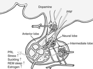
FIGURE 9-2. Control of pituitary secretion of prolactin (PRL). Dopamine from the hypothalamus is the predominant inhibitory regulator of pituitary PRL secretion. Multiple factors act as PRL-releasing factors (PRF; see text), and these come from both the hypothalamus and the posterior pituitary. Physiologic states that stimulate PRL release are listed on the figure. REM, Rapid eye movement.
Dopamine is synthesized by a two-step reaction in which tyrosine conversion to levodopa is catalyzed by tyrosine hydroxylase, and levodopa is converted to DA by the action of aromatic amine decarboxylase. As is the case for catecholamine synthesis in other cells, the momentary rate of DA synthesis in the TIDA neurons is determined by the activity of tyrosine hydroxylase. The negative feedback mechanism for controlling PRL release is to increase tyrosine hydroxylase activity in the TIDA neurons, thereby increasing the amount of DA available for release from the median eminence. PRL receptors are located in both the arcuate nucleus (site of the TIDA perikarya) and the median eminence.55 Therefore, circulating PRL may feed back on TIDA neurons at their terminals, which lay outside the blood-brain barrier, or systemic PRL may enter the cerebrospinal fluid via the choroid plexus. The choroid plexus expresses high levels of a short isoform of the PRL-R, which may serve to transport PRL across the blood-brain barrier. Levels of PRL in the cerebrospinal fluid reflect changes in PRL in the systemic circulation.56 Isolated PRL deficiency resulting from targeted gene disruption in the mouse causes decreased DA in the median eminence but does not affect DA levels in other regions of the hypothalamus.57
Activation of D2 receptors in lactotrophs has at least two main actions that result in inhibition of PRL. D2 receptors are members of the heptahelical G protein–coupled receptor superfamily, and they activate the αi subunits, which leads to inhibition of cyclic adenosine monophosphate (cAMP) synthesis.48 In addition, D2 receptors activate a G protein–coupled, inwardly rectifying potassium channel, which instantaneously causes hyperpolarization of the lactotroph membrane and closes voltage-gated calcium channels.58 Cytoplasmic calcium levels fall because of decreased influx of extracellular calcium, and the reduction in cytosolic free calcium decreases the exocytosis of secretory vesicles.
Dopamine-induced membrane hyperpolarization opposes the actions of some stimulatory factors such as thyrotropin-releasing hormone (TRH), which acts predominantly to increase influx of extracellular calcium by depolarizing the lactotroph membrane. Inhibition of cAMP by DA also opposes the actions of stimulatory factors such as vasoactive intestinal peptide (VIP), which acts via a positive effect on cAMP. This action decreases PRL release in the short to intermediate term. Second, because cAMP is mitogenic in lactotrophs, as well as in other pituitary cells, activation of Gi signaling by DA is antimitogenic. Lactotroph proliferation is important for physiologic elevation of PRL release during lactation. The proliferative action of cAMP on lactotrophs is understood to be an important promoter of pituitary tumor growth, thereby contributing to pathologic hyperprolactinemia.35
Other hypothalamic factors, as well as DA, and local pituitary peptides can inhibit PRL secretion. Somatostatin inhibits PRL secretion and acts through both cAMP-dependent and cAMP-independent mechanisms.59 Calcitonin has been shown to inhibit PRL secretion and may be secreted from the hypothalamus.60 Endothelin-1 is produced by lactotrophs and inhibits PRL secretion; transforming growth factor-β1 can act as a paracrine inhibitor of PRL.61,62 The biologic significance of these factors in pituitary development and physiology has not yet been established.
PROLACTIN-RELEASING FACTORS
A wide variety of stimulatory PRL secretagogues have been identified over the years, and it is likely that additional PRFs will be identified in the future. Known stimulators of PRL secretion include, but are not limited to, steroids (estrogen63), hypothalamic peptides (TRH, oxytocin, VIP,64,65 pituitary adenylate cyclase activating peptide [PACAP],66 and galanin67), and local pituitary factors (growth factors such as EGF68 and FGF-2,69 angiotensin II,70 and, again, PACAP71 and galanin43).
TRH is a potent and rapid stimulator of PRL release in vitro via a set of calcium-mediated pathways activated by a Gq-coupled receptor. However, the relative contribution of TRH to physiologic control of lactotrophs is not clear. VIP acts through cAMP to stimulate PRL synthesis and release on an intermediate to long-term basis. The importance of VIP as a positive lactotrophic factor is supported by two types of evidence. With the use of antibodies against VIP, the secretion of PRL can be inhibited to a very low level.47 In addition, VIP appears to be the primary PRF in birds and other nonmammals,72,73 suggesting that this positive mechanism may have been in place before the evolution of the dopaminergic inhibitory system in mammals. Oxytocin secretion is tightly coupled with PRL secretion during lactation, and both are secreted in response to nipple stimulation. The potential role of oxytocin as a PRF, given that it can reach the anterior pituitary through the short portal system, has remained controversial. Oxytocin antagonism partially suppresses PRL secretion,48 so this peptide is likely to provide some portion of the physiologic stimulus for PRL release. PACAP stimulates PRL synthesis and release. Galanin is synthesized in both the pituitary and the hypothalamus. In the pituitary, it colocalizes with PRL in lactotroph secretory granules and acts by autocrine and paracrine mechanisms to stimulate lactotrophs.43
A putative PRL-releasing peptide (PrRP) from the hypothalamus was identified by searching for ligands that activate an orphan pituitary G protein–coupled receptor. The mature peptide that was identified from bovine hypothalamus is a 20 amino acid molecule that originally was reported to cause rapid secretion of PRL from isolated pituitary cells.74 However, subsequent studies have failed to confirm that PrRP acts on lactotrophs to stimulate PRL release.75 Rather, PrRP may act within the hypothalamus to indirectly elevate PRL by inhibiting DA release. Antagonists of serotonin or opioid receptors inhibit PRL secretion under physiologically meaningful stimuli. Conversely, antidepressants that inhibit serotonin reuptake (fluoxetine [Prozac], etc.) may increase PRL secretion in humans and in laboratory animals. Serotonin and opioids are important indirect regulators of PRL by virtue of their actions on DA and releasing factor secretion in the hypothalamus.
Lactotrophs display a large degree of functional heterogeneity within the anterior pituitary. This heterogeneity is manifested as differences in morphology (i.e., secretory granule size and density), basal hormone release, electrical activity, and response to releasing and inhibiting factors. Assay of hormone release from single cells has revealed not only substantial cell-to-cell variations in function, but also marked temporal variations in a single cell.76
Transcription regulators that control the development of the anterior pituitary lactotrophs also participate in controlling PRL synthesis during adult life. Prominent among these factors is the Pit-1 protein. Pit-1 binds to two regions of the human PRL gene, the proximal promoter (within 250 bp of the transcription start) and a distal enhancer (beyond −1300 bp) (see Fig. 9-1). Multiple Pit-1–binding sites are present in each of these regions. Transcription regulators such as cAMP and estrogen receptors can control PRL gene expression by influencing Pit-1 activity.39,63
Pathophysiology of Prolactin Secretion
Normal plasma PRL concentrations in women who are neither pregnant nor lactating range from 4 to <20 ng/mL. In men, the values, on average, are several units lower. Late pregnancy and lactational levels normally range from 100 to 200 ng/mL, and the highest levels occur following active bouts of nursing. PRL is normally measured by radioimmunoassay (RIA). Although glycosylation and other chemical modifications of PRL can affect its immunoreactivity and therefore can lead to aberrant RIA results,26 pathologic levels generally are readily detected by RIA. The original method for bioassay of PRL involved measuring the growth of the pigeon crop sac mucosal epithelium.77 This method still is used occasionally and serves as the basis for the international standardization of PRL bioactivity. However, the method has been largely supplanted by a simpler bioassay that takes advantage of the ability of PRL to stimulate the proliferation of rat Nb2 lymphoma cells in culture.78
PROLACTIN DEFICIENCY
When PRL deficiency occurs, it is normally one component of a combined pituitary hormone deficiency. However, a few cases of PRL deficiency without evidence of other pituitary defects have been reported in women. Isolated PRL deficiency results in lactational failure and reproductive difficulty with no other obvious problems.79–82 No cases of isolated PRL deficiency have been reported in men. These results in a few humans are consistent with the phenotype of mice in which the PRL gene has been disrupted by a targeted mutation. In mice with disruptions of either PRL or its receptor genes, mammary gland development is defective; the females fail to reproduce, but the males do not have any overt symptoms.57,84,85 The concordance of these results from humans and mice is remarkable, given the possible differences between PRL physiology in humans and rodents. One important difference is that progesterone secretion in the rodent corpus luteum requires PRL, but in the human, it does not. This difference in luteal control probably explains why women who have isolated PRL deficiency are merely subfertile,79–82 whereas PRL-deficient mouse females are completely infertile.84,85
Mice with a targeted mutation of the PRL gene develop pituitary hyperplasia57 and adenomas86 that are more severe in females. Mice with targeted disruption of the PRL receptor gene also exhibit this lactotroph hyperplasia and prolactinoma development.87 Loss of PRL feedback in both of these genetic models leads to decreased hypothalamic DA, and the deficiency of DA leads to poorly restrained pituitary growth. Some forms of combined pituitary hormone deficiency have been identified in which PRL, GH, and TSH are hyposecreted as a consequence of mutations in important developmental factors. Familial inheritance of defects in the Pit-1 gene or PROP-1 results in individuals who fail to develop lactotrophs, somatotrophs, and thyrotrophs, and consequently are dwarfed and hypothyroid, as well as PRL deficient. Two spontaneous mutations that cause dwarfism in mice have been shown to correspond to these human conditions. In Snell dwarf mice, a mutation of the Pit-1 gene occurs, and in Ames dwarfs, the PROP-1 gene is mutated.35,88,89
HYPERPROLACTINEMIA
Hypersecretion of PRL is among the most common of pituitary disorders. Medications that elevate PRL secretion and may cause hyperprolactinemia include commonly used antiemetics, antipsychotics, antidepressants, and narcotics. These medications alter PRL secretion by antagonizing DA action, or by elevating serotonin or endorphin bioactivity. Reserpine and methyldopa increase PRL secretion as a result of DA depletion. DA receptor antagonists, such as haloperidol and phenylthiazines, increase PRL secretion. Serotonin reuptake inhibitors, such as fluoxetine, elevate serum PRL.48 It is uncommon for any of these medications to cause clinical signs of hyperprolactinemia, because the levels of PRL seldom reach more than 30 to 50 ng/mL with these drugs. One might imagine that subtle hormonal effects may be noted after long-term treatment.
Hyperprolactinemia that manifests clinical symptoms is most commonly a consequence of a lactotroph adenoma (see Chapter 25). These tumors may secrete high levels of PRL alone or of both PRL and GH. Any intracranial mass or trauma that causes compression or disruption of the pituitary stalk can cause hyperprolactinemia because of the loss of dopaminergic tone from the hypothalamus. Pituitary adenomas have been discovered to be much more common than was once believed, with more than 20% of individuals harboring tumors measuring at least 3 mm at autopsy.35 Tumors that do not hypersecrete hormones are usually of gonadotroph or lactotroph origin. Some symptoms of prolactinomas may be caused by tumor mass effects. These include visual field defects, associated with pressure on the medial aspect of the optic chiasm, and alterations in temperature regulation, feeding patterns, or other effects caused by hypothalamic compression. However, effects associated with the physiologic actions of the hormone are the more common presenting symptoms.
Galactorrhea (breast milk secretion in an individual who is not postpartum) and amenorrhea are the result of PRL actions directly on the breast and the hypothalamic-pituitary-ovarian axis. In men, galactorrhea and impotence are the most common presenting symptoms of a hypersecreting prolactinoma. The causes of impotence in hyperprolactinemia, whether hormonal or neurogenic, are unclear. Hyperprolactinemia is treated medically by administration of DA agonists, including bromocriptine and cabergoline, or surgically by resection of the tumor tissue.
Prolactin Receptors and Signal Transduction
RECEPTORS
The PRL-R is a member of the type 1 cytokine receptor family,90 and its nearest relative is the GH receptor. Several hematopoietic cytokine receptors, such as those for erythropoietin, most interleukins, and granulocyte-macrophage colony-stimulating factor, are also very similar to PRL and GH receptors. Other receptors, such as those for the interferons, are members of a broader superfamily of proteins that includes cell adhesion proteins. Features that define the type 1 cytokine receptor family include two signature motifs in the extracellular domain and one in the intracellular domain. Four cysteine residues in the extracellular domain are absolutely conserved among all of the type 1 cytokine receptors, and they form two disulfide bridges that are essential for the proper tertiary folding of the ligand-binding domain. A short sequence, which includes a tandem repeat of tryptophan-serine interrupted by a single amino acid (the WSXWS motif), is the second signature motif in the extracellular domain. This sequence is highly conserved near the base of the extracellular domain, but the function of these residues has not yet been proven with any degree of certainty. The structure of the PRL-R extracellular domain, like that of the GH and other cytokine receptors, has been analyzed extensively by x-ray crystallography, as well as by biochemical methods.91 This domain comprises two 100 amino acid subdomains, which are structurally related to the type III repeats of fibronectin. Each of the type III subdomains includes a conserved series of seven β-strands folded into two β-sheets that run in an antiparallel orientation. These type III subdomains are connected by a short, flexible hinge peptide, and residues that contact the ligand span this connector to include amino acids in each of the type III subdomains. Across the vertebrate lineages, substantial conservation of the major features of the PRL-Rs is evident, with some notable exceptions. In birds (pigeons, chickens), the extracellular domain has duplicated and diverged, and in cattle, the distal C-terminus has been truncated, thus eliminating a tyrosine residue that is conserved in other lineages (Fig. 9-3). Neither of these evolutionary changes appears to have functional significance.92,93
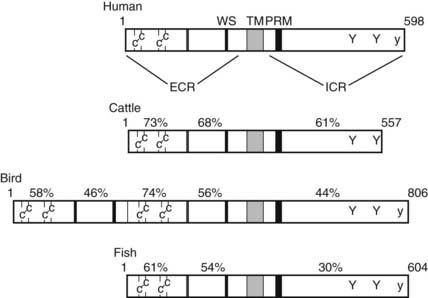
FIGURE 9-3. Prolactin receptor structure and function. Schematic diagram of the linear sequences of representative prolactin receptors. Pertinent structural features are two pairs of cysteines, the flexible hinge (double line), and the WS × WS repeat in the extracellular region (ECR). The transmembrane-spanning sequence (TM) marks the separation between the ECR and the intracellular region (ICR). In the ICR, the conserved motifs are the proline rich box 1 motif (PRM) and conserved tyrosine residues. The uppercase Y indicates ubiquitously conserved tyrosines, and the lowercase y is a tyrosine that is conserved in all known species except cattle. The percentage of identical amino acid residues in each region, compared with the human receptor, is labeled above each receptor.
Related posts:

Stay updated, free articles. Join our Telegram channel

Full access? Get Clinical Tree


