Transfusion Medicine


Transfusion Medicine


Susan A. Galel

Magali J. Fontaine

Maurene K. Viele

Christopher L. Gonzalez

Lawrence T. Goodnough


The first documented transfusion of blood in humans occurred in 1667, but it was not until almost 300 years later that transfusion become a therapeutic practicality.1 Landsteiner’s landmark discovery in the early 1900s of blood groups and agglutinating antibodies was the key that unlocked this therapeutic pathway. The development of anticoagulants, blood preservatives, and sterile collection sets in the middle of the 20th century made blood banking possible by enabling the collection and preservation of donor blood for later use.

In the past few decades, the complexity of blood banking and blood component therapy has virtually exploded. The recognition of both infectious and noninfectious complications of transfusion led to numerous practice changes involving blood donor screening, component production and modification, compatibility testing, and blood utilization. The menu of blood component options and therapeutic services has progressively expanded, along with efforts to establish evidence-based guidelines for their optimal use. Specialized recommendations have been developed for supporting specific patient populations such as immunosuppressed patients, chronic transfusion recipients, hematopoietic cell transplant recipients, and neonates.2,3 In the United States, all blood establishments (blood banks and transfusion services) are regulated by the US Food and Drug Administration (FDA).4 FDA regulations govern all aspects of blood collection, component processing, storage, compatibility testing, and administration. The FDA requires blood establishments to comply with highly stringent quality assurance standards that ensure control of processes and restrict variability. The AABB (formerly known as the American Association of Blood Banks) issues accreditation standards and recommendations that further establish the standard of practice in the US.5

Today, Transfusion Medicine is itself a Board-recognized clinical specialty. In addition to overseeing the complex donor center and transfusion service operations, Transfusion Medicine physicians are increasingly important participants in the clinical care team. Transfusion Medicine specialists can guide the selection of therapeutic options to best support a patient’s medical needs, and can coordinate the supply and delivery of these blood components and therapeutic services.6 This chapter serves as an introduction to blood components and transfusion services available in the United States.


BLOOD DONATION AND COLLECTION


Donor Selection

In the United States (US), donor eligibility criteria are established and enforced by the FDA and to a lesser degree by the American Association of Blood Banks (AABB). Donor selection is undertaken with two goals in mind: to protect the health of the donor by ensuring that a donation does not place the donor at risk, and to protect the recipient by ensuring that the donor meets all health and screening criteria so that the risk of transmitting infectious agents or causing other adverse events is minimized.2 In the US, most fresh blood products are from unpaid volunteers. These donors have decreased risk of transmitting infectious agents, especially in the “window period,” when screening tests fail to detect infection.7,8 Paying donors for fresh transfusion products was stopped after studies in the 1970s showed that paid donors had a much higher prevalence of hepatitis.8,9,10,11 and 12 Commercial plasma-derivative manufacturers still use paid donors, but comply with FDA-approved donor eligibility criteria and employ pathogen reduction processes during manufacturing that reduce infectious risks, as discussed later in this chapter.


Donor Identification and Registration

Donor registration must accurately identify the potential donor, including name, birthdate, address, and phone number, so the donor can be traced if needed. Records must link donors to all prior donations and test results and be kept for at least 10 years.


Donor Information

The donor must be given educational material describing signs and symptoms of AIDS and activities associated with increased risk of acquiring human immunodeficiency virus (HIV). The donor must be informed that testing may not detect all infections, and that individuals who have engaged in risk behavior should not donate.2


Donor Health History

Information to be elicited is defined by the FDA and the AABB. The medical history is obtained by a trained interviewer, or donors may complete an FDA-approved self-administered questionnaire on paper or a computer. Responses are then reviewed with the donor by qualified staff. 2 The history includes a review of current health, ensuring that the donor feels well, is free of signs of infection, and that his or her cardiovascular status can tolerate an acute blood volume loss of 10% to 15%. The donor is questioned about recent exposures to blood, potential exposure to HIV or hepatitis, sexual contact with individuals at risk for HIV, needle sharing by the donor or sexual partners, travel to or residence in areas endemic for malaria or variant Creutzfeldt-Jakob disease, medications, and immunizations.

The donor must meet certain requirements of age and vital signs. The hemoglobin must be at least 12.5 g/dl. Donor weight must be sufficient so that the donation constitutes no more than a 15% loss of blood volume. There must be no arm stigmata of parenteral drug abuse.5


Informed Consent

The donor must sign the health history form verifying that all questions were answered truthfully, that the donation process is understood, and that he consents to testing for infectious agents transmitted by transfusion, including HIV and hepatitis.2


Additional Donor Criteria for Specific Components

Donors may be eligible to donate some blood components but not others.


Medications That Impact Therapeutic Effectiveness of Particular Blood Components

Acetylsalicylic acid (ASA) irreversibly acetylates platelet cyclooxygenase and inhibits platelet aggregation.13 Platelet-mediated
hemostasis is restored, however, if ASA-inhibited platelets are mixed with untreated platelets.14 After a single dose of aspirin, ASA-exposed platelets are inhibited for the rest of their lifespan, but platelets produced after clearance of the drug restore hemostatic function. Therefore, platelets from donors who have taken ASA are acceptable as long as 48 hours have elapsed from the last dose. Platelets donated within 2 days of ASA ingestion are acceptable for use if mixed with platelets from unexposed donors.5

Nonsteroidal anti-inflammatory drugs (NSAIDs) may impair platelet function, but the effects of many are reversible13—that is, platelet function is restored once the platelets are removed from the offending drug.15 Therefore, individuals taking reversible NSAIDs can donate platelets for transfusion. Individuals who are taking irreversible NSAIDs, however, or other irreversible antiplatelet agents (e.g., ticlopidine) are not eligible to donate platelets for transfusion.5

Warfarin reduces levels of functional blood clotting factors (see Chapter 55). Transfusable plasma units or cryoprecipitate cannot be made from donors taking this medication.5


TRALI Mitigation Strategies for “High Plasma Volume” Components

Studies in the early 2000s indicated that blood products containing a large volume of plasma (e.g., plasma and apheresis platelet units) were associated with a higher per unit risk of transfusionrelated acute lung injury (TRALI) compared to components containing small amounts of plasma (e.g., red cell units and cryoprecipitate). Furthermore, plasma or platelet units containing donor antibodies to white blood cell (HLA and neutrophil) antigens were implicated in a substantial proportion of TRALI cases.16,17 In an attempt to reduce the frequency of TRALI, the AABB recommended in 2006 that blood collection agencies take steps to reduce production of high plasma volume products from donors with an increased likelihood of having WBC antibodies.18 Women with a history of pregnancy are at highest risk of being immunized to WBC antigens. Therefore, in response to AABB recommendations, many blood collection facilities have stopped making transfusable plasma components from women with a history of pregnancy or from all women. Whole blood collections are still accepted from these women, but the plasma portion of their collections is used only for cryoprecipitate production and/or manufacturing into plasma derivatives. It has been more challenging to exclude women with a history of pregnancy from plateletpheresis donations, as the platelet supply is difficult to sustain. Some blood collection facilities exclude multiparous women as platelet donors or screen them for the presence of HLA antibodies.


Directed Donations

A directed donation (DD) is a donation in which the donor directs his/her donated blood product to a specific designated patient.2 The donation must usually be ordered by the recipient’s doctor. DD programs exist primarily for emotional reasons, although these programs are required to be offered in certain states. In these programs, patients anticipating the need for blood can select who their donors will be. The donor is often a family member or acquaintance of the recipient. DDs are not of medical benefit where an established blood supply exists but can be useful in rare circumstances when specific characteristics of the donor’s blood are medically needed (e.g., for rare HLA or blood types obtained from family members). Directed donors must meet all regular donation criteria; however, exceptions can be made if rare types are needed. Data show DDs are no more safe than regular community donations from an infectious disease perspective. Directed donors are more likely to be first-time donors, who have a higher incidence of HIV and HCV than repeat blood donors.19 DDs theoretically may have higher risk of causing transfusion complications such as (1) hemolytic disease of the newborn in future pregnancies after a woman has received blood from her husband or his relatives,20 (2) TRALI in a mother to child transfusion, or (3) transfusion-associated graft-versus-host disease (TA-GVHD) from a related family donation.21 Directed donations from blood relatives are irradiated to prevent TA-GVHD.


BLOOD COLLECTION PROCESS

Phlebotomy and collection of blood proceed only if the donor is deemed suitable after pre-donation screening. Blood is collected in accordance with established standards 5 and is collected either manually or with an automated collection device.


Whole Blood (Manual) Collection

The phlebotomy site is swabbed with a disinfectant.22 Blood is collected in a primary plastic collection bag with a large bore needle allowing rapid flow and mixture with anticoagulant. The volume drawn is standardized for the collection bag used (either 450 or 500 ml). The blood draw is controlled by scales that discontinue flow when the desired weight is collected. Blood and anticoagulant are mixed gently during the collection. Specimen tubes for testing are also procured. Because most whole blood units will be separated into components, the primary bag has one to three attached satellite bags allowing separation of components in a sterile closed system. The collection tubing is heat-sealed into segments that are left attached to provide samples for crossmatching. The whole blood is transported to a laboratory where it can be separated into its components (red cells, plasma, platelets).


Blood Component Separation

Whole blood is centrifuged to sediment the red blood cells (RBCs) (Fig. 21.1). Most of the supernatant “platelet-rich plasma” is pushed off into an attached sterile satellite bag. The bag containing platelet-rich plasma may then be centrifuged at a higher rate to sediment platelets. Most of the plasma is then removed into a third satellite bag. This leaves behind a platelet pellet which is then resuspended in 40 to 70 ml of residual plasma resulting in a platelet concentrate. If platelet concentrates are not going to be produced from the donation, the initial centrifugation of the whole blood is done at high speed, and the plasma is removed directly from the red blood cells and frozen. Further processing of plasma and red cell components is discussed later in this chapter.


Terminology

The whole blood in the original collection bag (450 or 500 ml) is referred to as one whole blood unit, and each component made from that unit is defined as one “unit” of that component. Because a whole blood unit constitutes approximately 10% of a donor’s blood volume, each component can be considered roughly 10% replacement therapy for an adult patient.


“Closed” versus “Open” Systems

If blood component manipulation is done without opening the system to air (closed system), all components may be stored to the limit of their viability. If the bag or tubing is entered, however, the system is considered potentially open to air/bacteria, and the product outdates in 4 hours if stored at room temperature or 24 hours if refrigerated. Devices using high-temperature welds can sterilely attach additional containers or tubing to the original unit in a way that prevents entry of bacteria. With these “sterile connection devices,” blood components may be split into aliquots, filtered, or otherwise manipulated without loss of shelf life.2


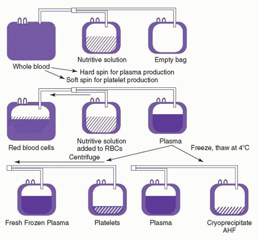
FIGURE 21.1. Preparation of components from a whole blood donation. AHF, antihemophilic factor; RBC, red blood cell. (Reprinted from Jeter EK, Spivey MA. Blood components and their use. In: Jeter EK, ed. Introduction to transfusion medicine: a case study approach. Bethesda, MD: American Association of Blood Banks Press, 1996:5, with permission.)


Automated Blood Collection and Separation Devices: Apheresis

The word apheresis is derived from a Greek word that means to separate or to take away. Initially, it referred to a manual process in which whole blood was withdrawn from the donor and centrifuged, the plasma retained, and the red cells returned to the donor. In 1914, Abel experimentally removed plasma from anephric dogs, and replaced it with crystalloid.23 It was not until the advent of plastic bags that manual apheresis could be used routinely in humans. Between 1950 and 1980, manual apheresis was the primary source of plasma for fractionation.

In the 1970s and 1980s automated cell separator devices changed the approach to apheresis. This technology has become the routine collection method for many blood components and is used in the treatment of many diseases.2,24

In discontinuous centrifugal devices, anticoagulated blood is collected into a spinning disposable bowl. Centrifugal force causes red cells to move to the outside of the bowl, and platelet-rich plasma moves to the inside. White blood cells (WBCs) (buffy coat) settle in between. With optical detectors, the desired component is pumped into an attached plastic blood bag. The remaining components are returned to the donor.

Continuous flow devices continuously subject incoming blood to a centrifugal force, establishing a standing cell gradient. The fraction(s) to be removed is pumped into a bag, and the rest is reinfused continuously. The extracorporeal blood volume is lower than with the discontinuous technique, and the procedure is faster. An increasing proportion of blood components are being collected using automated cell separation. These devices can collect multiple unit-equivalents of platelets, plasma, or red blood cells from one donation. Apheresis is the main source of plasma for fractionation because multiple units of plasma may be obtained without red cells and possible resultant iron deficiency. Apheresis technology is now used to collect the majority of platelets in the US and in many regions apheresis platelets have entirely replaced platelets derived from whole blood units. Apheresis platelets have several advantages. Up to three therapeutic platelets doses (equivalent to 18 whole blood-derived platelet units) can be obtained from one apheresis donation. This minimizes possible recipient exposure to infectious agents. If a donor is large enough, it is possible to obtain two RBC unit-equivalents from one apheresis donation, returning plasma, platelets, and saline to the donor to minimize volume loss. Some apheresis devices can be programmed to collect any desired combination of red cells, plasma, or platelet products from the same donation. Apheresis technology is also used for therapeutic plasma exchange (TPE) and for collection of peripheral blood hematopoietic progenitor cells (HPCs) or donor lymphocytes, as discussed later in this chapter and in Chapter 102.2

Membrane filtration can be combined with apheresis to collect plasma. Blood is pumped over a membrane with a specific pore size that permits passage of plasma but not cells. Such devices have been used to collect plasma for fractionation or to perform TPE.


Complications of Donation

Most people easily tolerate blood donation but occasional problems arise, the most common of which are from venipuncture, consisting of bruises, soreness, and hematoma. These complications may be striking but usually resolve spontaneously. Nerve irritation and/or injury (0.02% to 0.9%) and arterial puncture (0.003% to 0.01%) are less common. Donors normally compensate for volume loss by increasing heart rate and vascular resistance, but 2% to 7% of donors experience vasovagal reactions, with syncope occurring in 0.1% to 0.3%. Fatigue (8%), nausea, and vomiting (1.1%) are also seen.25,26,27,28 and 29 Apheresis donors can develop transient symptomatic hypocalcemia (tingling or muscle cramps) from the citrate infused when anticoagulated blood components are returned to them. These symptoms are treated by slowing the flow of the device and or giving the donor oral calcium supplements.30 Intravenous calcium infusions are needed only during prolonged apheresis procedures such as those for HPC collection.24


Donor Testing

Every blood donation undergoes a series of tests to determine its suitability for transfusion. In the United States, the following tests must be performed on every unit collected: ABO group and Rh type, red cell antibody screen, serologic tests for infectious markers including hepatitis B surface antigen (HBsAg), antibodies to hepatitis B core (HBc) antigen, hepatitis C, HIV-1 and -2, human T-cell lymphotropic virus (HTLV)-I and -II, and syphilis, and nucleic acid tests for HIV-1 RNA, HCV RNA, and West Nile virus RNA.5 In 2010 the FDA recommended one-time testing of allogeneic donors for antibodies to T. cruzi (the causative agent of Chagas disease).31 Donor testing is discussed more fully later in this chapter.

Blood components are not released for transfusion unless all donor tests for infectious markers are negative.5 This extensive testing results in a delay of 24 to 48 hours from the time of collection until an RBC component can be released for transfusion. Platelet products are typically not available until 36 to 48 hours after collection because of additional bacterial testing (see Bacterial Contamination).


RED BLOOD CELL PRESERVATION AND STORAGE

RBCs are collected and stored in solutions that maintain their viability.


Anticoagulant/Preservative Solutions

Blood banking was not practical until anticoagulant and preservative solutions capable of preserving red cells in viable form were developed.

Citrate anticoagulant is used for essentially all transfusable blood components today. It chelates ionized calcium in blood, blocking calcium-dependent coagulation steps. After transfusion, citrate is readily metabolized by the liver.32

During World War I, a sodium citrate and glucose solution was developed that permitted blood storage for several days. This citrated blood was used to treat shock in British and American soldiers.32 During World War II, it was found that the addition of citric acid (acid citrate dextrose [ACD]) preserved RBCs for 21 days. Continued efforts resulted in citrate phosphate dextrose (CPD) solution,33 a modified ACD, with added NaH2PO4 and a higher pH. Stored in CPD, red cell phosphate and 2,3-diphosphoglycerate (2,3-DPG) are maintained at higher levels than in ACD; however, RBC viability was still only 21 days.

In the 1950s, it was noted that red cells that had lost their adenosine triphosphate (ATP) did not survive well during storage.34 Nakao 35 showed that ATP content and posttransfusion viability of aged red cells could be improved by the addition of adenine, which allows the RBCs to maintain the adenine nucleotide pool. Simon et al. 36 described the maintenance of red cell ATP levels using preservatives containing glucose and low concentrations of adenine, leading to the adenine-supplemented anticoagulants now in use.32,37

CPDA-1, which is CPD fortified with adenine, became available in the United States in 1978. It had been used extensively in Europe for several years before then. Initial concerns about potential toxicity of adenine proved to be unfounded. CPDA-1 is now a common anticoagulant-preservative and allows storage of RBC concentrates for 35 days.5


Packed Red Cells versus Red Cells in Additive Solutions

After whole blood is collected into CPD or CPDA-1, the red cells may be centrifuged, allowing removal of most of the plasma (“packed RBCs”). Approximately 20% of the anticoagulant-containing plasma must be left to provide metabolic substrate for RBCs during storage. Another approach to red cell preservation involves more complete removal of the anticoagulated plasma from the red cells (“dry pack”), followed by resuspension of the red cells in 100 ml of an additive solution. Such additive solutions contain saline, adenine, and glucose, with or without mannitol to decrease hemolysis. The storage of RBCs in additive solutions is extended to 42 days.2,32 RBCs in additive solution are now the most common preparations for transfusion. RBCs collected in any of the anticoagulants and preservatives must be stored at 1° to 6°C to maintain optimum function.5


Changes in Red Cells during Storage

Stored liquid RBCs undergo biochemical and structural changes that have major influences on their viability and function after transfusion.32,38,39,40 and 41


Structural Changes

A number of red cell changes contribute to decreased cell viability after storage.38,40,41 RBCs are normally disc shaped. Soon after storage they become spherical with surface projections (spheroechinocytosis). Later defects include loss of membrane lipids and protein, as well as alterations in structural proteins. Loss of membrane deformability correlates with viability.39 The more severe membrane changes are irreversible and probably contribute to decreased posttransfusion RBC survival.38,40

Blood bag plasticizers appear to influence membrane stability. Red cells are stored in polyvinyl chloride (PVC) bags that contain the plasticizer di-2-ethylhexylphthalate (DEHP). Morphologic deterioration is greater in RBCs stored in containers that do not have DEHP, with increased hemolysis and loss of deformability, suggesting that DEHP has a direct membrane stabilizing effect.42,43 Adding DEHP can both prevent and repair deterioration of stored red cells, with many of the spherical cells reverting to normal discoid morphology.44 However, concerns have been raised about potential toxic effects of DEHP. Efforts are being made to find alternatives to DEHP.45


Biochemical Changes

During storage, red cells metabolize glucose, producing lactic and pyruvic acid, resulting in lower pH and decreased glycolysis. As glycolysis slows, RBC ATP content falls. Because human RBCs contain no enzymes to synthesize adenine or other purines de novo, the nucleotide pool gradually becomes exhausted. In the presence of adenine, ATP may be regenerated. Understanding this has led to prolonged RBC storage by the addition of exogenous adenine and inorganic phosphate, both of which improve the cells’ ability to regenerate ATP.32

Red cells lose potassium and gain sodium during storage. This is because the Na+-K+ gradient is normally maintained by a Na+-K+ ATPase that does not function well at 4°C. Gamma irradiation of red cells to prevent graft-versus-host disease (GVHD) (see section GVHD) doubles the rate of potassium leakage.46,47 Red cells reabsorb potassium after transfusion.48


Red Cell 2,3-Diphosphoglycerate

Another significant change in stored RBCs is 2,3-DPG depletion, which decreases RBC oxygen delivery.32,49,50 In blood preserved in ACD, 2,3-DPG drops to below 50% within 48 hours. In CPD, CPDA-1, and additive solution-stored red cells, 2,3-DPG is better maintained but is still depleted after about 2 weeks. Studies show that if RBCs are cooled down rapidly to 17 to 18 degrees C within 1 hour after collection, the fall of 2,3-DPG can be delayed
significantly.51 2,3-DPG levels improve rapidly within the first 6 hours after transfusion, and return to near-normal levels within 24 hours.48,52

The clinical implications of transfusion of blood with decreased 2,3-DPG content is controversial 32,53 The oxygen dissociation curve of cells that are 2,3-DPG-depleted is shifted to the left, resulting in increased hemoglobin oxygen affinity and decreased tissue oxygenation. These changes are thought to be of limited clinical significance because the stored red cells rapidly regain their 2,3-DPG in the circulation. In select patients, such as those in shock, lower RBC 2,3-DPG levels may have a negative effect. However, the acidosis that may be present in such patients shifts the oxygen dissociation curve to the right. Because of such compensatory mechanisms, the need for blood specifically altered to preserve or reconstitute red cell 2,3-DPG has not been demonstrated.


Rejuvenating Solutions

A number of chemical agents—dihydroxyacetone,54 pyruvate,55 phosphoenolpyruvate,56 and inosine 57—are capable of maintaining near-normal red cell 2,3-DPG content during storage, or of replenishing 2,3-DPG after storage. Although none of these chemicals are likely to be used in transfusion because of their side effects, studies with these agents have resulted in the development of rejuvenating solutions.

Rejuvenating solutions contain pyruvate, inosine, glucose, phosphate, and adenine, and may be added to red cells up to 3 days after the expiration date. Treatment with rejuvenating solutions corrects the metabolic defects of the red cell, with a return to normal levels of ATP and 2,3-DPG. These rejuvenated red cells may either be washed and transfused within 24 hours or frozen for later use.2 Such rejuvenated red cells have a normal survival and oxygen affinity.58,59 and 60 In practice, rejuvenation is rarely performed.


In Vivo Recovery of Stored Red Cells

After transfusion of stored blood, red cells that have developed lethal degrees of damage are removed promptly from the circulation of the recipient. Red cells that survive the first 24 hours after transfusion have normal survival thereafter.61,62 Therefore, the criterion by which the adequacy for transfusion of banked blood is assessed is the proportion of transfused red cells that remain in circulation at 24 hours after transfusion. Generally, 75% survival at 24 hours is considered evidence of adequate viability; the anticoagulant systems in current use readily achieve this goal. Work to develop optimal additive solutions capable of maintaining red cell ATP and 2,3-DPG levels and to prolong red cell storage time continues.


Clinical Implications of Stored Blood

Whether the age of transfused blood affects clinical outcomes is highly controversial, with many studies coming to different conclusions 63,64 For a variety of complex methodologic reasons, the ability of even well-designed randomized controlled trials to demonstrate significant clinical differences based on the age of transfused blood has been questioned.65


Frozen Red Cells

Red cell freezing is a labor-intensive process that is used primarily for storing rare blood types or prolonged storage of autologous red cells in the event of planned or postponed surgery. Glycerol is gradually added to the red cells as a cryoprotectant to a final concentration of 40% (weight/volume). The cells are then frozen at -65°C or colder for up to 10 years. Immediately after thawing, an automated cell processor must be used to wash the glycerol from the cells. The washed cells are resuspended in isotonic saline and glucose. In most cases, postthaw storage is limited to 24 hours because an open system is used to process the cells.2 However a closed system maintaining sterility has been developed and RBCs processed in this manner can be stored in a refrigerator for up to 2 weeks after thawing.66,67

Potential new technologies for RBC biopreservation are now under investigation, such as hypothermic storage and lyophilization. Hypothermic red cell storage potentially maintains higher levels of 2,3-DPG but still requires improvements. Lyophilization of red cells potentially would allow stable, indefinite, room temperature storage and would be ideal for remote storage and military applications. These methods have still not been sufficiently developed.68


PLATELET PREPARATION AND STORAGE


Preparation of Platelet Concentrates

Platelets, like erythrocytes, are actively metabolizing cells and require specific conditions for their preparation and storage to optimally maintain viability and function.69,70 They are prepared for transfusion either as platelet concentrates from whole blood or by apheresis. Although the preparation differs, both products are stored under the same conditions.

Platelet concentrates may be prepared from whole blood collected into bags with satellites (Fig. 21.1). The anticoagulants in current use, CPD and CPDA-1, are satisfactory for preparation of platelet concentrates. The whole blood is kept at room temperature and must be processed within 8 hours of collection. The unit of whole blood is centrifuged at low speed at room temperature, and the supernatant, platelet-rich plasma is expressed into a satellite bag. The supernatant is centrifuged again to further concentrate the platelets. Most of the supernatant platelet-poor plasma is expressed, and after 1 hour, the platelets are gently resuspended in the remaining plasma (50 to 60 ml). Approximately 60% to 75% of the donor platelets, or a minimum of 5.5 × 10¹⁰ platelets/unit, are recovered.71

In Europe and Canada, whole blood platelet concentrates are prepared from the buffy coat.2 Briefly, the whole blood unit is spun inverted at high speed, and the platelet-poor plasma and the buffy coat are withdrawn, each into its own satellite bag. The buffy coat is then centrifuged at low speed to separate the platelets from the red and white cells. The functional quality of these platelets is comparable to those prepared by the American method.71 Platelet concentrates prepared from buffy coat contain fewer white cells than those prepared from platelet-rich plasma, although filtration is necessary to meet the European standards for leukoreduced products. In some European countries, platelet concentrates are pooled, resuspended in an additive solution, and filtered before storage.72 In the United States and Canada, the pooling of platelets before storage has been evaluated73,74,75; currently, only one system has been FDA-approved for this use in the US.76


Apheresis (Pheresis) Platelets

In the US the majority of platelets are collected from single donors by apheresis using the automated collection devices described earlier.2 Platelets collected in this way must contain at least 3 × 10¹¹ platelets in approximately 300 ml of plasma by AABB standards. This is equal to about six whole blood platelet concentrates. With improved apheresis technology many platelets collections are so abundant that they can be split into multiple products that still meet the 3 × 10¹¹ requirement. Apheresis platelets are leukocyte-reduced by the collection technology. Because the procedure
is carried out in a closed system, the platelets can be stored for 5 days in the appropriate plastic bag.2,5,77 Apheresis platelets are usually collected from random donors but can also be collected from HLA-compatible donors to support alloimmunized patients. There is no significant functional difference between apheresis platelets and those from whole blood donations.75 However, pheresis platelets typically contain fewer WBCs and, therefore, are less likely to cause febrile transfusion reactions than whole blood-derived platelet concentrates,78,79 unless these concentrates have been leukoreduced prior to storage.80,81 The use of apheresis platelets also results in fewer donor exposures for recipients.


Platelet Storage and Functional Integrity

Platelet products must be kept under specific conditions to ensure optimal recovery and function. Unlike red cells, platelets stored at 4°C undergo shape changes and lose their viability. Platelet survival and function are optimized by storage at room temperature (20° to 24°C).82,83 During storage, platelets metabolize glucose to lactate and hydrogen, which are buffered by bicarbonate present in the plasma, resulting in a release of CO2. The nature of the bag in which platelet concentrates are stored is important. The plastics used in the early era of platelet transfusion were made of PVC and plasticizers and were not permeable to O2 and CO2. In these bags, stored platelets become depleted of oxygen, resulting in a shift from oxidative to glycolytic metabolism, with increased lactate generation, decreased pH, and decreased platelet viability. As the pH decreases below 6.2, platelets undergo shape change, are damaged, and show reduced in vivo recovery. The platelets could not be stored for longer than 72 hours in the PVC bags. The plastic blood bags currently in use are more gas permeable. This permits continued oxidative metabolism and prolongation of storage time.84,85 and 86 Although platelet viability is maintained for up to 7 days in the new plastic containers,87,88 storage of platelet concentrates at room temperature is currently approved for only 5 days in the US because of the risk of bacterial growth.89,90 Viability is best preserved if the platelets are gently agitated during storage. Platelets must be stored in sufficient plasma to maintain a pH greater than or equal to 6.2.5

Even under optimum storage conditions, platelets, like red cells, develop a storage lesion.91 After storage at room temperature, the changes that occur in the platelets include decreased aggregation in response to single platelet agonists such as adenosine diphosphate, and reduction in adenosine diphosphate and ATP content both in granules and in the metabolic pool. Beta-thromboglobulin and platelet factor-4 are released, and both dense and alpha-granules are depleted. There is increased surface expression of P-selectin (CD62), a molecule derived from the alpha-granule membrane of the resting platelet. Platelets may develop morphologic changes and impaired responses to hypotonic shock. It has been difficult, however, to correlate the clinical response to platelet transfusions with specific in vitro findings.

The in vivo effectiveness of stored platelets is dependent on the recovery of transfused platelets in the circulation of recipients. This has been assessed through platelet recovery and survival studies in normal volunteers using autologous radiolabeled platelets.92 Platelet recovery is the percentage of transfused platelets that are found in circulation immediately after transfusion. Even when fresh platelets are transfused, only about two thirds of the transfused platelets are recovered in the circulation93; the remaining 30% to 40% are pooled in the spleen.94 At the end of storage, mean platelet recovery is approximately 40% to 50%.86,95 After the initial recovery, there is little difference in survival of fresh versus stored platelets; both show a half-life of 3 to 5 days in healthy adults.83 In patients, however, the observed recovery and survival of transfused platelets are often substantially lower than these figures (as discussed in the section Dosage and Expected Response).

The in vivo hemostatic efficacy of transfused platelets is difficult to assess. The use of bleeding times in thrombocytopenic patients as an indicator of function is not of value because of great variability in technique and lack of reproducibility of results.96 Function is best assessed through clinical assessment of hemostasis.97 The clinical response generally correlates with the posttransfusion platelet increment.


Platelet Additive Solutions

Platelet additive solution (PAS) use is less developed than additive solutions for RBCs, especially in the US. Removal of variable amounts of plasma and storage of platelet in PAS has many potential advantages, including optimized survival, decreased adverse reactions (e.g., allergic, febrile, TRALI), and pathogen inactivation.98

PAS generally contain acetate, which functions both as a metabolic substrate and as a buffer. Platelets stored in one of these solutions (platelet additive solution-2) showed increased P-selectin expression and decreased in vivo recovery compared to platelets stored in plasma 72,99; moreover, platelets stored in the additive solution resulted in lower transfusion responses.99,100 Another solution, platelet additive solution-3, however, appears to show suitable preservation of platelet function for 7 days and is now in use in both Europe and the US. Platelet additive solutions are included in the pathogen reduction systems for platelets that are approved in Europe. These systems include exposure of the platelets to pathogen reduction agents and UV light, as discussed later in this chapter. This processing appears to be associated with some loss of platelet product potency, but the clinical importance of these changes is unclear.101,102


Frozen Platelets

Frozen storage of platelets has been investigated and a potential role of such preparations has been established only for patients with alloantibodies for whom satisfactory donors cannot be found. Autologous platelets may be frozen using dimethyl sulfoxide as the cryoprotective agent. In vitro recovery of frozen and thawed platelets and posttransfusion increments may be satisfactory for clinical use, although only approximately half of the platelets survive the freeze-thaw process.103,104 This recovery may be improved by combining a platelet additive solution with a reduced concentration of dimethyl sulfoxide.105 Theoretically, autologous frozen platelet transfusions may permit support of highly alloimmunized patients through periods of chemotherapy-induced myelosuppression.104,106 Currently, frozen platelets are not available for clinical use; however, studies are beginning to evaluate preserving platelets by freeze-drying with trehalose.107,108


PREPARATION OF PLASMA COMPONENTS

Plasma is obtained from whole blood donations and apheresis collections. The plasma may be used for transfusion, further processed by the blood center into cryoprecipitate, or sent to commercial facilities for manufacturing into plasma derivatives.


Plasma Components for Transfusion

Plasma components such as FFP and cryoprecipitate are produced from individual volunteer blood donations and are briefly described here. As noted earlier, transfusable plasma units are not typically made from donors with an increased likelihood of having antibodies to WBC (e.g., women or women with a history of pregnancy); this strategy reduces the TRALI risk associated with plasma transfusion.


Fresh Frozen Plasma, Plasma Frozen within 24 Hours (FP24)

Fresh frozen plasma (FFP) is prepared by separating citrated plasma from whole blood and freezing it within 8 hours of collection or by freezing citrated apheresis plasma within 6 hours of collection. Each unit of FFP prepared from whole blood contains approximately 200 ml of plasma. Apheresis plasma may be packaged into 200- or 400-ml bags. FFP may be stored at -18°C or below for up to 1 year. Under these conditions, there is minimal loss of activity of the labile coagulation factors V and VIII. One milliliter of FFP contains approximately one unit of coagulation factor activity. After thawing, FFP may be stored in the refrigerator for up to 24 hours before use.

A product called “Plasma frozen within 24 hours after phlebotomy” (FP24) has largely replaced FFP production in the United States. As discussed earlier, TRALI mitigation strategies have restricted the donations from which transfusable plasma units can be made. In order to maintain a sufficient supply of plasma components for transfusion, most blood collection facilities in the US are labeling their plasma units as “Plasma frozen within 24 hours after phlebotomy” rather than “FFP.” This labeling permits blood centers to make transfusable plasma units from donations that reach the component processing facility more than 8 hours after collection. The content of FP24 is identical to that of FFP except that the Factor VIII content may be reduced to 80% that of FFP.109,110,111 This Factor VIII content is more than sufficient for the therapeutic applications in which plasma is utilized, e.g., trauma, liver disease, warfarin reversal, etc. Therefore, FFP and FP24 are typically used interchangeably.112


Liquid Plasma, Thawed Plasma

Liquid plasma is a term for plasma that is separated from whole blood and stored at 1°C to 6°C without freezing. Thawed plasma is a term for FFP or FP24 that is thawed and stored in the refrigerator for up to 5 days after thawing. The only significant difference between these products and FFP/FP24 is the content of the labile coagulation factors (V and VIII).

After 5 days of refrigerated storage, Factor VIII and V levels are at about 65% to 75% of their original levels. After 28 days’ storage, Factor VIII activity is approximately 40% of normal and Factor V levels approximately 50% to 60% of normal. Other coagulation factors, including fibrinogen, ADAMTS 13, and factors II, VII, IX, X, and XIII, are generally stable under refrigerated storage conditions.112,113,114,115 and 116,117

Liquid plasma or thawed plasma should not be used for clotting factor replacement in patients who have specific deficiencies of factor V or VIII. However, these products can be used for plasma replacement in massively bleeding patients, as these products will still maintain clinically hemostatic factor levels in the patient (i.e., 30% of normal). Because Vitamin K-related factors are not depleted during refrigerated storage, these prolonged storage products would be equivalent to FFP or FP24 for warfarin reversal.


Plasma, Cryoprecipitate Reduced

This is the supernatant remaining after removal of cryoprecipitate from FFP (see “Cryoprecipitated Antihemophilic Factor”). Storage conditions are the same as for FFP. This product is deficient in fibrinogen, Factor VIII, von Willebrand factor (vWF), and factor XIII. Cryoprecipitate-reduced plasma was initially thought to be superior to FFP for treatment of TTP because of its lower vWF content. Randomized studies have verified that this fluid is therapeutically effective for TTP; however, it is not superior to FFP.118


Cryoprecipitated Antihemophilic Factor

Cryoprecipitated AHF, or cryoprecipitate, is an extract of FFP that is enriched in high-molecular-weight plasma proteins. It is prepared by thawing one unit of FFP at 1°C to 6°C. Under these conditions, the high-molecular-weight proteins remain as a precipitate. The precipitated protein is concentrated by centrifugation, and all except approximately 15 ml of supernatant is removed. The remaining 15 ml and the precipitate are refrozen. Each unit of this cryoprecipitate contains approximately 80 to 120 units of Factor VIII and at least 150 mg of fibrinogen. It also contains factor XIII, fibronectin, and the high-molecular-weight multimers of vWF.

Cryoprecipitate was originally developed for the treatment of hemophilia A. It is no longer the treatment of choice for that disorder, because less infectious alternatives are available. At the present time, cryoprecipitate is most often used for correction of hypofibrinogenemia (<100 mg/dl) in bleeding patients. Cryoprecipitate has also been used topically, along with thrombin and calcium, as a “fibrin glue.” However, commercial products that are much more effective as topical hemostatic or sealant agents are now available. A commercial fibrinogen concentrate for intravenous infusion is available, but as of April 2012 this product was approved in the US only for treatment of congenital fibrinogen deficiency.

The typical dose of cryoprecipitate of one unit per 5 to 10 kg can be expected to raise the recipient’s fibrinogen level by approximately 70 mg/dl.2,3 Multiple units of cryoprecipitate are often pooled before administration.


Commercial Plasma Derivatives

Commercial plasma derivatives are made from pooled plasma collected from hundreds or thousands of donors. In the United States, paid plasmapheresis donors provide most of the plasma derivatives, but excess (“recovered”) plasma from volunteer whole blood donations is used also. Plasma units are pooled and fractionated into a number of purified proteins. The most commonly used fractionation procedure is based on Cohn’s cold ethanol fractionation process, developed in the 1940s.119 As the temperature, ionic strength, pH, and ethanol concentration are varied, plasma can be separated into several fractions. Fraction I contains Factor VIII and fibrinogen, fraction II contains the immunoglobulins, and fraction V contains albumin. Fractions III and IV contain a number of other coagulation factors and proteins. Although other approaches such as ion-exchange chromatography have been applied to the preparation of certain plasma products, Cohn’s method remains the standard.

Because the plasma pools used for the production of plasma derivatives are derived from many donors, contamination with infectious agents is common. All plasma derivatives are treated by methods demonstrated to inactivate HIV, HCV, and hepatitis B virus (HBV), such as prolonged heat and treatment with organic solvents and detergents, which inactivates lipid-coated viruses. Pooled plasma products, however, could still transmit infectious agents that lack a lipid coat and that are resistant to heat. Human parvovirus B19 is one such agent. The FDA requires screening for, and exclusion of, donations that contain high titers of the B19 virus,120 but low levels of B19 virus can still be present in the pools. Many plasma derivatives undergo additional purification steps such as affinity chromatography, precipitation, or nanofiltration that would further reduce their contamination by infectious agents.119,121

There are many commercial plasma derivatives available. Some examples are described in the following.


Solvent/Detergent-treated Plasma

Solvent/detergent-treated plasma is made from hundreds or thousands of units of FFP that have been thawed, pooled, subjected to treatment with organic solvents and detergents, filtered, and refrozen.122 The product was developed to reduce the risk
of transmitting enveloped viruses, such as HIV, HCV, and HBV. It appears to be therapeutically equivalent to FFP.122 A version of this product introduced in the US in the late 1990s was withdrawn after reports of thromboembolic complications. European versions of this product, however, are in current use and have a good safety record. In contrast to individual plasma units, this pooled plasma product has not been associated with transfusion-related acute lung injury (TRALI), presumably because of dilution of antibodies from individual donors during plasma pooling.


Coagulation Factor Concentrates

The coagulation factor concentrates, both the recombinant products and those made from plasma, are discussed in detail elsewhere as part of the management of inherited or acquired coagulation disorders (see Chapters 53 and 54). Recombinant coagulation products are used when available; these include Factors VIII, IX, and VIIa. Some factors are available only as plasma-derived concentrates, including fibrinogen and protein C.


Immunoglobulins

Intramuscular immune globulin preparations are prepared from pooled plasma by cold ethanol fractionation. They contain dimeric and polymeric IgG, artifacts of the fractionation procedure, which are capable of nonspecifically activating complement by both the classic and alternative pathways. This mechanism probably explains the major adverse effects that occur if these products are administered intravenously.119 Products labeled for intramuscular use must therefore not be given intravenously.

Intravenous immune globulin preparations (IVIGs) are produced by various chemical modifications designed to decrease the aggregation of IgG. Nonspecific complement activation is reduced, whereas the ability of the Ig molecules to interact with pathogenic organisms and complement is retained. Many of these products may also be administered subcutaneously.

Nonspecific immune globulin preparations contain a broad spectrum of antibodies naturally present in the donor population. They are most often used for treatment of primary immunodeficiency or as immune modulators.123 Immune globulins against a particular target are derived from the plasma of donors selected for high concentrations of antibodies to that target. Such preparations include Rh immune globulin, hepatitis B immune globulin, and Varicella zoster immune globulin.

IVIGs have been associated with some adverse reactions, including renal failure, hemolysis, and thrombotic events. Both IM and IV immune globulin products should be used with caution in patients with IgA deficiency, because they may contain small quantities of IgA.


BLOOD COMPONENT MODIFICATION


Leukocyte Reduction

When whole blood is separated by centrifugation, WBCs sediment at the interface between red cells and platelet-rich plasma. Therefore, WBCs typically contaminate both red cell and platelet components, with concentrations of WBCs approximately 109/product. WBCs in blood components can mediate febrile transfusion reactions, stimulate HLA alloimmunization in transfusion recipients, and transmit some cell-associated pathogens such as cytomegalovirus (CMV).124 Therefore, it is desirable to remove WBCs from transfusable blood components.

Historically, several methods have been used to reduce the number of WBCs in transfusable blood components. Relatively nonspecific methods were used initially, including saline washing of red cells or physical separation of the WBC layer (buffy coat) from the RBCs.124 Later, microaggregate filters were used to remove WBCs after centrifugation.125 These methods resulted in white cell reduction of 70% to 90% and were effective in preventing most febrile reactions to red cells. Freezing and deglycerolization of red cells have also been used to remove WBCs and result in approximately 2-log WBC removal. Ultimately selective leukoreduction filters were developed that can reduce WBCs from blood components by 3 or more logs. These synthetic fiber filters remove WBCs by a combination of mesh density, chemical attraction, and active adhesion.124 All leukocyte-reduced red cells are now produced using these special filters and, by FDA criteria, have less than 5 × 10⁶ WBCs/unit and at least 85% of the original RBC component.

Leukofiltration of RBC components may be performed at the blood collection center, in the hospital transfusion service, or at the bedside. Filtration prior to storage reduces WBC breakdown products in the blood component and there is some evidence that RBC viability is better preserved. The clinical importance of these benefits has not been demonstrated.124 In practice, most RBC leukofiltration is performed by blood collection centers within the first few days after collection.

Apheresis platelets usually contain very few WBCs and usually qualify as leukoreduced (<5 × 10⁶ WBCs) without the need for filtration. In contrast, whole blood-derived platelet concentrates contain large numbers of WBCs, and many of the febrile transfusion reactions to these products appear to be due to cytokines produced by the WBCs in these products during storage.126,127,128 and 129 Therefore, removal of WBCs from whole blood-derived platelet concentrates before storage is beneficial. One system is approved in the US that allows prestorage leukocyte reduction and pooling of whole blood platelets.

The use of leukoreduced products has evolved over the last decade or two. Initially these products were indicated for patients with a history of febrile, nonhemolytic transfusion reactions, to reduce the risk of HLA alloimmunization, and as an alternative to cytomegalovirus (CMV) antibody screening of donors to reduce the risk of transfusion-transmitted CMV. Now the use of leukoreduced products has become nearly universal, although the medical necessity of universal leukoreduction remains somewhat controversial.130


Washed Products

Saline washing with automated cell washers can be used to reduce the amount of plasma in cellular blood products. These washers are capable of removing approximately 99% of plasma proteins from red cell products.2 Although cell washing was previously also used for leukocyte reduction, it is no longer used for this purpose. Today, washing is primarily used to reduce incompatible plasma and also prevent severe allergic reactions (which are thought to be triggered by donor plasma proteins). Washing is also used to reduce RBC supernatant potassium, which may be required prior to massive or rapid infusion of stored RBC to neonates. Washing on an automated cell processor takes 30 to 45 minutes/unit. Because the washing procedure is usually performed in an “open” system, the red cells have only a 24-hour shelf life after washing. A closed processing system has been developed that may permit longer storage of washed cells.66 Although many facilities perform red cell washing, few offer washed platelets. Use of automated cell washers to wash platelets has been described.2 However, in practice, it may be difficult to ensure adequate platelet recovery and viability after washing.


Irradiation of Blood Products

Gamma irradiation of cellular blood components is used to prevent transfusion-related GVHD by impairing the proliferative capacity of lymphocytes in the blood component. The recommended dose
for the irradiation of blood and blood products is 2500 cGy at the center of the irradiation field, with a minimum dose of 1500 cGy at any point in the field.5,131 This dose of radiation has no significant adverse effect on red cell, platelet, or granulocyte function. However, there are changes in the red cell membrane that result in an increased loss of potassium from the cell, limiting the storage time of red cell concentrates to 28 days.46,47 The amount of accumulated free potassium in the supernatant of irradiated red cells may be clinically important in massive transfusion, especially in the neonate.132 It may be desirable to irradiate proximate to transfusion, or wash stored irradiated RBCs if massive transfusion of irradiated products is required for a patient at risk for hyperkalemia. The dose of irradiation used for cellular blood components is not sufficient to inactivate pathogens.133 The irradiation doses required for pathogen inactivation would irreparably damage blood components.


ALTERNATIVES TO ALLOGENEIC DONOR BLOOD


Autologous Blood

Autologous blood donation is blood donated by a patient, intended for transfusion back into the same patient. Blood collected for autologous use is not released to other patients.

Use of a patient’s own (autologous) blood may reduce or eliminate the need for allogeneic blood. There are three types of autologous blood collection procedures. In preoperative autologous donation (PAD), patients donate one or more units of blood to a blood bank during the weeks preceding an elective procedure. In acute normovolemic hemodilution (ANH), blood units are collected in the operating room immediately prior to surgery. In autologous blood cell salvage, blood lost during or after a surgical procedure is salvaged for reinfusion.


Preoperative Autologous Donation

Preoperative blood donation (PAD) is most often used for patients who are expected to require transfusion during elective surgery. It is also used in patients for whom crossmatch-compatible blood cannot otherwise be made available, as in patients with rare blood groups or with multiple alloantibodies. For autologous collections, the donor eligibility criteria are not as stringent as for allogeneic donors. The key consideration is whether the patient can tolerate the acute withdrawal of a unit of whole blood representing 10% to 15% of their blood volume. Patients with significant cerebral or cardiac disease should be evaluated before they are enrolled in a PAD program. Children are also eligible for autologous blood donation, but the volume of blood collected and anticoagulant used must be adjusted to body weight.

An autologous donor may donate blood every 3 days as long as the donor’s hemoglobin remains at or above 11 g/dl. An “aggressive” donation schedule stimulates a more substantial endogenous erythropoietin response, with the potential for more autologous units collected or a higher patient hemoglobin at surgery.134 In most instances, the units of blood are stored in the liquid state for up to 35 to 42 days. They may be frozen if a longer interval between donation and surgery is required, but this significantly increases the cost and is not routinely recommended.135,136

All autologous collections must be tested for ABO group and Rh type. The units must be labeled For Autologous Use Only. If autologous blood is to be transfused at an institution that is not the collecting facility, the blood unit must be tested for transfusiontransmitted infectious diseases.5 Units with reactive infectious disease tests must be labeled with biohazard labels. Regulators permit the use of autologous units with positive infectious disease tests. However, some hospitals do not accept such units because of concerns related to the risk of accidental transfusion of the unit into the wrong patient.

PAD is not necessarily beneficial to the patient. There is a reported 12-fold increase in the number of autologous donors hospitalized after a donation compared to allogeneic donors, with an increased risk in the elderly.137 Iron supplementation is recommended, but may not be sufficient to prevent anemia, especially if the last unit of autologous blood is collected <7 days preoperatively. This anemia may increase the patient’s likelihood of requiring transfusion.138 PAD is expensive and the logistics of collecting and shipping the blood in the desired timeframe are complex. Autologous blood may be wasted or transfused unnecessarily, particularly if collected for procedures in which transfusion is rarely needed.139,140

Transfusion complications such as bacterial contamination, febrile nonhemolytic transfusion reactions (FNHTRs), allergic reactions, and volume overload can occur with autologous transfusion.141 The possibility of an accident or error such as the transfusion of the wrong unit or an allogeneic unit into the autologous donor/patient has been reported to be as high as 1.2%.142 The cost-effectiveness of PAD is has been questioned, given its high cost, risks, and limited benefit.143


Acute Normovolemic Hemodilution

The second approach to autologous blood procurement involves the withdrawal of blood immediately before the surgical procedure and replacing the blood with crystalloid, colloid, or both, thereby acutely lowering the patient’s hematocrit.139,144 The blood lost during surgery is therefore relatively dilute, reducing total red cell loss. The higher hematocrit blood withdrawn immediately prior to surgery is used for transfusion. Patients most likely to benefit from this maneuver are those with anticipated large surgical blood losses who can tolerate low intraoperative hematocrits.145,146

Units collected by ANH can be stored at room temperature for up to 8 hours or at 1°C to 6°C for up to 24 hours.147 The blood so collected does not undergo any storage-related changes. The relative efficacy of ANH in comparison to other blood conservation techniques has been debated.146,148,149 The cost of ANH is significantly less than that of PAD because there is little incremental cost associated with collecting the blood and no required testing.149


Intraoperative and Postoperative Salvage and Reinfusion

A third approach to autologous transfusion is the collection and retransfusion of blood lost during or after surgery.2,150 Perioperative salvage has been shown to be effective in reducing the need for allogenic blood in a variety of surgical procedures. The AABB publishes Standards for Perioperative Autologous Blood Collection and Administration, which provide guidance for use of these blood conservation options.147

There are two basic techniques available. Intraoperatively, an anticoagulated vacuum suction device can be used to collect blood from the surgical field and deliver it to a centrifuge- like device that washes the shed blood with saline before it is reinfused. Only red cells are salvaged by this method (platelets and plasma are lost). There has been concern about the safety of reinfusing materials suctioned from obstetric, cancerous, or contaminated surgical fields. Published experience to date, however, suggests that reinfusion of salvaged cells after processing with leukoreduction filters may be safe in these settings.151 Postoperatively, blood shed into joints or body cavities can be collected into sterile containers. The salvaged material must be filtered to remove fat and particles, and may then be reinfused either directly or after washing. The shed fluid contains red cell stroma, free hemoglobin, activated clotting factors, and fibrin degradation products.
It appears that there is not an increased risk associated with infusion of unwashed shed blood if the volume reinfused is limited to approximately 1 L. There is little value to salvaging shed blood in settings where the volume of fluid drained from the surgical site is small or has a low hematocrit. Because these devices are expensive, patient selection is important.152


Erythropoiesis Stimulating Agents

Erythropoiesis stimulating agents (ESA) have been used to stimulate red cell production in patients, either to increase the number of units that can be collected preoperatively 134,153,154 or to increase preoperative red cell mass.155 The use of epoietin alfa has been approved to facilitate autologous blood donation in the European Union, Canada, and Japan, but not in the United States.156 Epoietin alfa has also been approved for perisurgical use in anemic patients (Hgb ≤13 g/dl) in the US and Canada.


Artificial Oxygen Carriers

Some aspects that make the artificial oxygen carriers particularly appealing include: the prospect of being free of most or all of the infectious risks of allogeneic blood; no need to perform blood grouping and cross match; extended shelf life and possibility of storage at room temperature; the potential for a virtually unlimited supply; and the possibility of development of homogeneous and standardized products with controlled characteristics optimized to achieve the goal of oxygen delivery without raising all other complexities of allogeneic blood.157,158,159 and 160 No artificial oxygen carrier is currently approved by the FDA for clinical human use in the US.

Perfluorocarbon (PFC) emulsions boost oxygen delivery by increasing the amount of dissolved oxygen. Use of these oxygen carriers must be coupled with oxygen and increased FiO2 to further increase the amount of dissolved oxygen. The PFCs that reached clinical trials include Fluosol-DA (Alpha Therapeutics, Los Angeles, CA) and Oxygent (Alliance Pharmaceutical Corporation, San Diego, CA). Fluosol-DA was initially used to improve oxygen delivery to the heart muscle during percutaneous transluminal coronary angiography, but it was subsequently withdrawn because of difficulties in storage and preparation, and lack of utilization in angioplasty.

Hemoglobin-based oxygen carriers (HBOCs) increase the oxygen delivery by increasing total hemoglobin. Despite being an effective oxygen carrier, vasoconstriction (initially attributed to extravasation of the HBOC to interstitial space and scavenging nitric oxide)161, hypertension, and renal, pancreatic, and liver injury have been described as complications. Approaches such as purification, polymerization, cross-linking, conjugating with other macromolecules, and encapsulating in vesicles or other nanoparticles have been pursued to minimize toxicity and associated complications in subsequent generations of HBOCs.162

A 2008 meta-analysis of 16 trials on 5 different HBOCs indicated that regardless of the individual product or indication studied, HBOCs are associated with significantly higher risk of death (relative risk of 1.30) and myocardial infarction (relative risk of 2.71) compared to the controls.163,164,165 A new generation of products is in development as “oxygen bridges” until anemia management via compensatory erythropoiesis with production of cellular hemoglobin can be achieved with the use of ESA and intravenous iron therapy.


USE OF BLOOD COMPONENTS

Table 21.1 lists the blood components available for clinical use and briefly summarizes the indications for the use of each. The use of each component is discussed in detail in the following sections.


Patient Informed Consent

Although approximately 14 million units of RBCs are transfused in the United States each year, their efficacy has never been demonstrated in well-designed trials. For purposes of obtaining the patient’s informed consent, the treating physicians and patient must understand that not all is known about the relative risks and benefits of blood transfusion.

The elements of transfusion consent comprise: a discussion of blood transfusion risks 166,167 and benefits; alternatives to blood; an opportunity to ask questions; and patient consent.168 Consent should occur as far in advance of transfusion as possible, so that alternatives to allogeneic blood such as autologous blood can be made available. Some states have legislated that alternatives to allogeneic blood be offered to patients whenever there is a reasonable possibility that a blood transfusion may be necessary. It should also be noted that blood transfusion has been legislated to be a medical service not subject to commerce and trade laws, thus excluding the principle of implied warranty and granting blood banks immunity from strict product liability.169


Patient Blood Management

Of the estimated 39 million discharges in the US in 2004, 5.8% (2.3 million) were associated with blood transfusion.170 The rate of blood transfusion more than doubled from 1997 to 2009. Increased provider awareness of the costs associated with blood transfusion171 and recognition of the potential negative outcomes have stimulated initiatives in Patient Blood Management.172 Blood Management has been defined as the appropriate use of blood and blood components, with a goal of minimizing their use. This goal has been motivated historically by: 1) known blood risks; 2) unknown blood risks; 3) preservation of the national blood inventory; and 4) constraints from escalating costs.172

Patient-focused blood management173 is an evidence-based approach that is multidisciplinary (transfusion medicine specialists, surgeons, anesthesiologists, and critical care specialists) and multiprofessional (physicians, nurses, pump technologists, and pharmacists). Preventative strategies are emphasized to identify, evaluate, and manage anemia174,175 (e.g., pharmacologic therapy139 and reduced iatrogenic blood losses from diagnostic testing)176; to optimize hemostasis (e.g., pharmacologic therapy177 and point of care testing) 178; and to establish decision thresholds (e.g., guidelines) for the appropriate administration of blood therapy (the impact of these activities on blood transfusion outcomes is illustrated in Figure 21.2.172,179,180

In the US, The Joint Commission (TJC) developed Patient Blood Management Performance Measures which have been placed in their Topic Library. These are available to be used as additional patient safety activities and/or quality improvement projects by provider institutions.181

Patient Blood Management strategies for patients undergoing cardiac surgery have been shown to be safe and effective in reducing transfusion, while at the same time delivering high-quality outcomes. One institution reported that only 11% of patients undergoing cardiac surgeries received blood transfusions; this program ranked first in their state for lowest risk-adjusted mortality.182


RED CELL TRANSFUSION

The main indication for RBC transfusion is inadequate oxygen delivery as a result of anemia and, in some cases, hypovolemia. Wound healing is not impaired in the presence of anemia, and transfusion does not improve wound healing. Patients with chronic diseases should never be transfused simply because of mild asymptomatic anemia or as part of supportive care.


TABLE 21.1 BLOOD COMPONENTS AND INDICATIONS FOR USE


Component


Composition


Volume


Indications and Expected Benefit


Whole blood


RBC and plasma (approx. Hct, 40%); WBCs; plateletsa


500 ml


To increase red cell mass and plasma volume (plasma deficient in labile clotting factors V and VIII); for hypovolemic anemia, massive transfusion, or exchange transfusion in neonates


Packed RBCs


RBC and reduced plasma (approx. Hct, 75%); WBCs; plateletsa


250 ml


To increase red cell mass in symptomatic anemia; 10 ml/kg raises Hct by 10%


RBCs, adenine-saline added


RBC and 100 ml of additive solution (approx. Hct, 60%); WBCs; plateletsa; little plasma


330 ml


To increase red cell mass in symptomatic anemia; 10 ml/kg raises Hct by 8%


RBCs, leukocytes reduced (prepared by filtration)


>85% original volume of RBCs; <5 × 10⁶ WBCs


>85% of original volume


To increase red cell mass; <5 × 10⁶ WBCs to decrease the likelihood of febrile reactions, immunization to leukocytes (HLA antigens), or CMV transmission


RBCs, washed


RBCs (approx. Hct, 75%); reduced WBCs; no plasma


225 ml


To increase red cell mass; reduce risk of allergic reactions to plasma proteins; or reduce free potassium dose


RBCs, frozen


RBCs (approx. Hct, 75%);


225 ml


To increase red cell mass; minimize febrile or allergic transfusion reactions; use for prolonged RBC blood storage


RBCs, deglycerolized


<5 × 10⁶ WBCs; no platelets; no plasma


Granulocytes, pheresis


Granulocytes (>1.0 × 10¹⁰ polymorphonuclear cells/unit); lymphocytes; platelets (>2.0 × 10¹¹/unit); some RBCs


220 ml


To provide granulocytes for selected patients with sepsis and severe neutropenia (<0.5 × 10⁹/L)


Platelet concentrates


Platelets (>5.5 × 10¹⁰/unit); RBCs; WBCs; plasma


50 ml


Bleeding due to thrombocytopenia or thrombocytopathy; 1 unit/10 kg raises platelet count by 17-50 × 109/L


Platelets, pheresis


Platelets (>3 × 10¹¹/unit); RBCs; WBCs; plasma


300 ml


Same as platelets; sometimes HLA-matched; benefit is equivalent to 6 platelet concentrates


Platelets, leukocytes reduced


Platelets (as above); <5 × 10⁶ WBCs/final dose of pooled or pheresis platelets


300 ml


Same as platelets; <5 × 10⁶ WBCs to decrease the likelihood of febrile reactions, alloimmunization to leukocytes (HLA antigens), or CMV transmission


FFP; thawed plasma


FFP: all coagulation factors; thawed plasma: reduced factors V and VIII


200 ml


Treatment of some coagulation disorders; 10 ml/kg of FFP raises factor levels by approximately 10%


Cryoprecipitated antihemophilic factor


Fibrinogen; factors VIII and XIII; von Willebrand factor


15 ml


Deficiency of fibrinogen, 1 unit/5 kg raises fibrinogen 70 mg/dl; also used for factor XIII replacement; not first-choice therapy for hemophilia A, von Willebrand disease, topical fibrin sealant


approx., approximate; CMV, cytomegalovirus; FFP, fresh frozen plasma; Hct, hematocrit; RBC, red blood cell; WBC, white blood cell.


a WBCs and platelets are nonfunctional.


Modified from Triulzi DJ, ed. Blood transfusion therapy: a physician’s handbook, 7th ed. Bethesda, MD: American Association of Blood Banks, 2002.


Indications for Red Blood Cell Therapy

The therapeutic goal of a blood transfusion is to improve oxygen delivery according to the physiologic need of the recipient. The usual response to an acute reduction in hemoglobin in the normovolemic state is to increase cardiac output to maintain adequate oxygen delivery.183 The heart is the principal organ at risk in acute anemia. In a normal heart, increased lactate production and an oxygen extraction ratio of 50% occur at a hemoglobin of approximately 3.5 to 4 g/dl.184 In a model of coronary stenosis, the anaerobic state occurs at a hemoglobin of approximately 6 to 7 g/dl.185 No single number, either extraction ratio or hemoglobin, can serve as an absolute indicator of transfusion need. However, the use of a physiologic value in conjunction with clinical assessment of the patient status would permit a rational decision regarding the appropriateness of transfusion before the onset of hypoxia or ischemia.186

Literature on mortality in surgical patients refusing transfusion provides insight into the circumstances in which transfusion may be of benefit. In a review of 16 reports of the surgical outcomes in Jehovah’s Witness patients who underwent major surgery without blood transfusion, mortality associated with anemia occurred in only 1.4% of the 1,404 operations.187 A more detailed analysis of 61 studies of Jehovah’s Witness patients found that, with the exception of three patients who died after cardiac surgery, all deaths attributed to anemia occurred in patients with hemoglobin ≤5 g/dl.188 In one large study of surgical patients refusing transfusion, the risk of death was found to be higher in patients with cardiovascular disease than in those without.189 A subsequent analysis190 found that although the risk of death was low in patients with postoperative hemoglobin levels of 7.1 to 8.0 g/dl, morbidity occurred in 9.4%; the odds of death in patients with a postoperative hemoglobin level ≤8 g/dl increased 2.5 times for each gram decrease in hemoglobin level.190

A large retrospective study of elderly patients who underwent surgical repair of hip fracture found that transfusion of patients with hemoglobin levels ≥ 8 g/dl did not influence 30-day or
90-day mortality.191 This was confirmed by a subsequent192 randomized, prospective study. Prospective, randomized trials in patients undergoing cardiac surgery193 and receiving critical care194 have each demonstrated that such patients can tolerate anemia without transfusion to hemoglobin levels between 7 and 8 g/dl, with equivalent clinical outcomes comparable to patients maintained at hemoglobin levels of greater than 10 g/dl. A Cochrane meta-analysis of prospective randomized trials comparing “high” versus “low” hemoglobin thresholds on more than 3700 patients195 found that a hemoglobin of 7 g/dl was sufficient for the majority of patients.


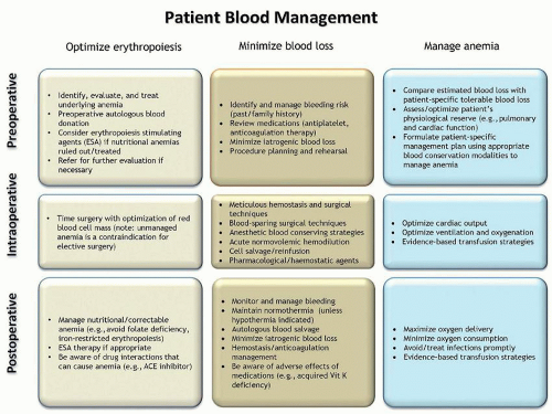
FIGURE 21.2. Patient blood management. These principles applied in the perisurgical period enable treating physicians to have the time and tools to provide patient-centered evidenced-based patient blood mangement in order to minimize allogeneic blood transfusions. (From Goodnough LT, Shander A. Patient blood management. Anesthesiology 2012;116:1367-1376, with permission.)

AABB clinical practice guidelines suggest that patients should not be transfused with red blood cells, in the absence of symptoms/signs of anemia, unless the Hgb concentration is less than 7 to 8 g/dl, or less than 8 g/dl for patients with symptoms or known to have cardiovascular disease.196 Comprehensive blood management guidelines from the Societies of Thoracic Surgeons and Cardiovascular Anesthesiologists contain similar recommendations, suggesting that RBC transfusion may be life-saving when hemoglobin is less than 6 and is reasonable in most patients when hemoglobin is less than 7.197 However, it is unlikely that a hemoglobin value alone should serve as a “transfusion trigger;” patients should be managed, rather than laboratory values.198


Red Cell Transfusion in Specific Settings


Massive Hemorrhage

One of the major indications for blood transfusion is the restoration of circulating blood elements after the loss of large amounts of blood (Table 21.2).199 In general, adults who lose <20% of their blood volume (or approximately 1 L) do well without red cell transfusion, providing that fluid resuscitation is adequate to maintain the circulating blood volume and that further blood loss is avoided. Young healthy patients can sustain losses of up to 30% to 40% of blood volume as long as intravascular blood volume is adequately maintained with intravenous (IV) fluids.

Massive hemorrhage is generally defined as transfusion of more than 10 U of RBC (one complete blood volume replacement) within 24 hours. Massive hemorrhage is a common complication in a number of clinical settings. In traumatic injury, hemorrhage is a major cause of morbidity and is responsible for almost 50% of deaths occurring within 24 hours of injury and up to 80% of intraoperative trauma mortalities.200,201 and 202 In addition, cardiovascular and hepatobiliary procedures can frequently result in massive bleeding203,204; postpartum hemorrhage events can complicate labor and delivery; and diverticulosis or varices can lead to significant gastrointestinal bleeding.204,205,206 and 207 Blood component support before and after control of massive hemorrhage is critical in these
scenarios.206 Despite the need for consensus on the management of patients with massive bleeding, currently no such consensus exists.


TABLE 21.2 CLINICAL SIGNS OF BLOOD LOSS


Volume Lost


ml


% of Blood Volume


Clinical Signs


500


10


None; occasionally vasovagal syncope


1,000


20


At rest, there may be no clinical evidence of volume loss; a slight postural drop in blood pressure may be seen; tachycardia with exercise


1,500


30


Resting supine blood pressure and pulse may be normal; neck veins flat when supine; postural hypotension


2,000


40


Central venous pressure, cardiac output, systolic blood pressure below normal even when supine and at rest; air hunger, cold clammy skin; tachycardia


2,500


50


Signs of shock; tachycardia, hypotension, oliguria, drowsiness, or coma


An estimated 10% of military trauma patients and 3% to 5% of civilian trauma patients receive massive transfusions (MTs).205,208,209 Within the standard definition of massive hemorrhage there appears to be an important subset of patients who may benefit from blood components in addition to RBCs, specifically, those with rapid massive bleeding.210 Both Moore et al.210 and Holcomb et al. 205 demonstrated that patients receiving 10 U of RBCs in the first 6 hours after injury had a higher rate of mortality than those receiving the same quantity of RBCs over a 24-hour period. Early identification of this patient population and specific massive transfusion (MT) support strategies involving multiple blood components have been associated with improved survival.211

In response, transfusion services have implemented protocols to quickly and efficiently provide packages of blood products to patients with massive hemorrhage. There are a number of criteria available to help evaluate the effectiveness of different strategies used by transfusion services. Evaluation of the effectiveness of these protocols should include several parameters: clinical outcomes (survival, length of hospital stay, multisystem organ failure, infection rate, etc); post-resuscitation laboratory parameters (hemoglobin, prothrombin time [PT], partial thromboplastin time [PTT], fibrinogen, and PLT count); and 24-hour blood component and crystalloid use. A recent review summarizes the literature to date, highlights some of the limitations of the available evidence, and identifies areas in need of additional study.212

Recent data from the US Army’s Institute of Surgical Research have shown improvement in outcomes when soldiers requiring MTs received resuscitations with ratios of component types that were similar to whole blood transfusions.213,214 Subsequent reports, primarily in the military literature, further support a component therapy transfusion in ratios of 1 U RBC/1 U plasma/1 random donor unit of PLTs.205,215,216 and 217 However, these are retrospective studies, with a survivorship bias.205,215,216 and 217 The military data also lack adjustment for confounding variables such as injury severity.205,215,216 and 217 Nevertheless, these data have led to widespread support of this ratio, particularly the 1:1 ratio of RBC/plasma, although considerable debate remains on this topic.218,219 and 220

Civilian trauma studies have also evaluated the impact of more aggressive ratios and noted an association with improved survival with use of MT protocols.221,222,223 and 224 Similar to the military data, such studies also support using a more aggressive RBC/plasma ratio, reporting a significant reduction (41% vs 62%) in 30-day mortality as compared with those that received less plasma.225 This was independent of age and Trauma Related Injury Severity Score, which by themselves were independent predictors of mortality.225

Several studies have called into question the benefit of higher plasma ratios.223,226 Nevertheless, the cumulative data appear to support early, proactive support with high ratios of plasma to RBC along with additional support with platelets.218,219 Not all studies showed a mortality benefit, and in the absence of randomized trials, data to convincingly support a particular ratio or formula are needed. However, the existing data suggest that a well-organized MT protocol that is activated in a timely fashion is likely to demonstrate improved patient outcomes and result in less overall blood product usage in large trauma centers.218,219


Elective Surgery

In preparation for surgery, preoperative requests for typed and crossmatched blood should be based on the predetermined likelihood of a procedure requiring a blood transfusion. “Maximum surgical blood order schedules” or “standard blood orders” specify the number of units that should be crossmatched for a variety of procedures and include guidelines for pretransfusion assessment.227,228 A pretransfusion request for type and crossmatch should be sent to the blood bank if it is likely (≥10% probability) that blood will be required for a specific surgical procedure. The request should be for a type and screen if it is unlikely (<10%) that the patient will require blood. As discussed previously under patient blood management, preoperative evaluation and management of anemia to correct deficits in red cell mass is the single most important determinant of the likelihood of perioperative blood transfusions.172


Nutritional Deficiencies and Other Anemic States

RBCs are often transfused in the management of various types of anemia. Published guidelines (see above) advise transfusing on the basis of clinical indications. Physiologic adaptations to anemia, including elevated red cell 2,3-DPG content and increased cardiac output, compensate to preserve oxygen transport and delivery to a significant extent in chronic anemias. Transfusion is rarely indicated in these patients if there is time to correct anemia with alternative therapies.

Patients who are anemic solely because of deficiency of iron, folate, or B12 rarely require transfusion. Patients with “lifethreatening” anemia (Hgb <6.5 g/dl) by WHO (World Health Organization) and NCI (National Cancer Institute) criteria may require transfusion.229 Elderly patients who present with pernicious or severe iron deficiency anemia may require red cell transfusion, particularly when angina or congestive heart failure has been the cause of the patient seeking medical attention. Iron-deficient patients who are also bleeding actively (e.g., from the gastrointestinal tract) may also require red cell transfusion. In such situations, the goal of transfusion is not to correct the patient’s hemoglobin concentration, but to raise it sufficiently to stabilize the patient until specific therapy can be administered.


Hemolytic Anemias

Patients with acute or chronic hemolytic anemias may require red cell transfusion; often, this need arises at the time of a hemolytic or aplastic crisis. Such patients are often critically ill, and safe transfusion requires careful clinical attention. In autoimmune hemolytic anemia,230,231 the clinician may be faced with a severely anemic patient for whom crossmatch-compatible blood cannot be obtained. These patients produce an antibody that
reacts with all RBCs including their own, and the transfusion of serologically incompatible red cells may be necessary. At a hemoglobin <6 g/dl, most patients require transfusion. In these cases, withholding transfusion in the absence of “compatible” RBCs places the patient in needless danger. Although “incompatible” cells will have a shorter than normal lifespan, transfusion reactions are infrequent. The risk of complications is increased if the patient has brisk hemolysis and a large volume of blood is infused, or if the patient has an undetected alloantibody in addition to the autoantibody. If time allows, special techniques should be used to evaluate these patients for alloantibodies prior to transfusion (see “Autoagglutination”). Consultation with a transfusion medicine specialist is recommended in these cases.


Hypoproliferative Anemias

In aplastic and sideroblastic anemias, myelodysplastic states, and myelofibrosis, patients often depend on regular transfusion of red cells and may die of transfusion-induced iron overload after several years of such support unless precautions are taken to remove iron. The development and use of erythropoiesis stimulating agents have reduced the need for transfusion in many patients with hypoproliferative anemia, such as those with end-stage kidney disease (see Chapter 41) and in patients with chemotherapyinduced anemia.232


Hereditary Red Cell Disorders

In children with thalassemia (see Chapter 34), bone marrow hyperplasia with its undesirable effects on the skeleton may be ameliorated, and iron absorption decreased, by regular transfusions to maintain a near-normal hemoglobin concentration.233 Such a program is possible only in conjunction with an aggressive iron chelation program, as the iron load otherwise leads to fatal hemosiderosis. In patients with sickle cell disease and vaso-occlusive crises (see Chapter 33), the adverse microvascular effects of sickle cells can be relieved temporarily by hydration with crystalloid therapy to restore intravascular volume, rather than with RBC transfusions (to avoid chronically unnecessary risks of alloimmunization and iron overload). Red cell exchange may be indicated when impaired oxygenation leads to <90% O2 saturation, in order to address the generation of sickle cells in acute chest syndromes. A multicenter trial (STOP trial) showed a significant decrease in the incidence of stroke in patients with abnormal transcranial Doppler studies who were treated with simple or exchange transfusions to maintain their hemoglobin S concentrations <30%, compared to patients who remained on standard supportive care with transfusion only when clinically indicated.234


Pretransfusion Testing of Red Cells


Compatibility Testing Process

The process of selecting red cells for transfusion involves three stages. Blood grouping involves determination of the ABO group and Rh type of both recipient and donor specimens. The recipient’s serum or plasma is screened for the presence of unexpected red cell antibodies. Crossmatching, either serologically or electronically, after selection of a donor unit of the appropriate group and type determines whether the donor cells are compatible with the recipient’s plasma.

Properly selected blood products will be compatible with the recipient, indicating that transfusion should not result in hemolysis of donor red cells. Because only the ABO and Rh(D) red cell antigens are prophylactically matched in routine transfusion practice, there are always significant antigenic differences between donor and recipient, both for red cells and for the accompanying leukocytes, platelets, and plasma. Repeated exposure to foreign antigens with chronic transfusion or pregnancy may result in antibody formation in the recipient.

There is no room for error in the provision of blood for transfusion. If clerical or laboratory error results in donor and recipient being mismatched for the ABO group, transfusion of even a few milliliters of red cells may lead to a life-threatening transfusion reaction with shock, intravascular coagulation, and acute renal failure. Such reactions are uncommon because of rigid adherence to a routine designed to maximize safety at all levels of the transfusion process; however, with the decrease in the risk of transfusion-transmitted infections, such reactions are becoming one of the leading risks of transfusion.166,235 Careful identification of the patient for whom the blood is ordered, including complete labeling of the specimen at the bedside of that patient, is essential. Careful ABO blood grouping along with comparing results of ABO testing with historical records for each patient adds to the level of safety. Ensuring positive identification of crossmatched units of blood and verifying that the information identifying the unit with the intended recipient is reviewed in the presence of that recipient before the administration of the blood are crucial.2,5


Blood Grouping

The presence of ABO antigens is determined by testing the unknown RBCs with anti-A and anti-B sera by one of a variety of methods including slide, tube, gel, or microplate tests.2 Identifying which ABO antigens are on the surface of an individual’s RBCs is called the forward grouping or forward typing. Cells agglutinated only with anti-A serum are group A; those reacting only with anti-B are group B. Those reacting with both antisera are group AB, and red cells that fail to agglutinate with either anti-A or anti-B are group O.

“Reverse grouping” or “reverse typing” should be performed to confirm the reaction obtained by the forward grouping test. This involves testing the reactions of the serum or plasma from the person of unknown type with reagent red cells of known A and B type. Agglutination of the red cells indicates the presence of anti-A or anti-B in the individual’s serum. The conclusions of forward and reverse tests should agree as shown in Table 21.3.

The antisera used in blood group antigen detection can be obtained from donors with naturally occurring high levels of antibodies, from people or animals specifically stimulated to produce antibodies against blood groups, or from monoclonal antibodies of mouse or human origin. The advantages of monoclonal antibodies include their high quality and stability and their ease of production in large quantities. Antisera must be of known specificity and potency, and control testing must be done on a routine basis.2

The Rh type of red cells is determined by examining the cells’ reaction with anti-D serum from commercial sources. Commercial
antisera may be modified by the manufacturer with the addition of high concentrations of protein or by chemically altering the immunoglobulin (Ig) G molecules in such a way that they perform as direct agglutinins in the laboratory. This permits rapid, reliable testing to determine the D antigen status of the cells. However, these high-protein reagents may cause false positive reactions because of spontaneous aggregation of some red cells in their presence. If this happens, an Rh-negative patient could be typed as Rh-positive if the recommended Rh control is not tested simultaneously. This problem has led to the development of low-protein, saline-reactive monoclonal reagents. Monoclonal anti-D reagents contain both human IgM and polyclonal IgG antibodies and are currently most widely used. They can also be used in the antiglobulin test for weak D. As with all reagents, the manufacturers’ instructions must be followed.2


TABLE 21.3 ABO GROUPING


Expected Reactions of


Patient’s Cells with


Patient’s Serum with


Patient Blood Group


Anti-A


Anti-B


A Cells


B Cells


O


None


None


+


+


A


+


None


None


+


B


None


+


+


None


AB


+


+


None


None


Red cells reacting weakly with anti-D reagents are called weak D. If donor blood is being tested, the absence of D must be confirmed. If the initial test for D is negative, a second, more sensitive test must be performed using a method that detects weak D. If D is detected by either method, the unit is labeled Rh-positive.2,5 In patients, testing for weak D is not required. Patients who are typed as Rh-negative, but who are really weak D-positive, will not be adversely affected by the transfusion of Rh-negative products. Patients known to be weak D-positive, however, may be given Rh-positive donor blood. Before concluding that a patient is weak D-positive, care must be taken to ensure that the patient has not recently been transfused with Rh-positive red cells or experienced a large fetal-maternal hemorrhage.


Testing for Red Cell Antibodies

Antibodies in potential blood transfusion recipients fall into several categories. The most common blood group antibodies that are clinically significant and may be implicated in hemolytic transfusion reactions or hemolytic disease of the newborn are shown in Table 21.4.


TABLE 21.4 SIGNIFICANCE OF CERTAIN BLOOD GROUP ANTIBODIES


Clinical Significance


Blood Group System


Antibody


Relative Frequency in Antibody Screening


Hemolytic Transfusion Reaction


Hemolytic Disease of the Newborn


ABO


Anti-A


All group B and O


Yes


Yes


Anti-B


All group A and O


Yes


Yes


Rhesus


Anti-D


Common


Yes


Yes


Anti-c


Common


Yes


Yes


Anti-E


Common


Yes


Yes


Anti-C


Common


Yes


Yes


Anti-e


Uncommon


Yes


Yes


Kell


Anti-K


Common


Yes


Yes


Anti-k


Rare


Yes


Yes


Kidd


Anti-Jka


Common


Yes


Yes


Anti-Jkb


Rare


Yes


Yes


Duffy


Anti-Fya


Common


Yes


Yes


Anti-Fyb


Uncommon


Yes


Yes


MNS


Anti-M


Common


Rare


Rare


Anti-N


Rare


Rare


Rare


Anti-S


Uncommon


Yes


Yes


Anti-s


Rare


Yes


Yes


Lewis


Anti-Lea


Common


Yes


No


Anti-Leb


Uncommon


No


No


P


Anti-P1


Common


Rare


No


Ii


Anti-I


Uncommon


No


No


All human plasmas contain naturally occurring antibodies that react with the complementary antigens of the ABH system. These are of great importance in transfusion, as they are complementfixing IgM antibodies; transfusion of incompatible red cells leads to immediate hemolytic reactions. Many people also have naturally occurring antibodies (usually low-titer IgM antibodies reacting at or below room temperature) that react with some antigens of the Lewis, P, Ii, MN, or other systems; these are rarely active above room temperature and are only occasionally important in transfusion. Finally, people exposed to foreign red cells by prior transfusion or pregnancy may produce IgG antibodies to antigens of certain other systems, primarily Rh (C, c, D, E, e), Kell, Duffy, Kidd, and Ss, but many less common possibilities exist. These red cell antibodies are clinically significant. They do not often lead to intravascular hemolytic reactions, but transfused incompatible red cells may exhibit decreased survival caused by increased clearance in the reticuloendothelial system. Many of these IgG antibodies can also cause hemolytic disease of the fetus and newborn.2,236

There are two major classes of antibodies that react with red cells. Complete or saline antibodies agglutinate red cells suspended in saline solution; these are usually IgM. Antibodies that do not react visibly in saline and are capable of producing agglutination reactions only with special techniques to make their interaction with red cells detectable are called incomplete agglutinins; these are generally IgG antibodies.

The best example of a room temperature saline agglutination test is that used in ABO grouping. Other red cell antibodies that are readily detected in saline suspension are those belonging to
the Lewis, MN, P, and Ii blood group systems. With the important exception of ABO system antibodies, many of the others detected with this test are of no clinical significance, as they are not reactive at 37°C.

The best examples of incomplete agglutinins, or IgG antibodies, are those that react with antigens of the Rh system. If such antibodies are not detected in the recipient, immediate hemolysis of transfused, incompatible red cells is extremely rare. However, their presence may lead to a significantly decreased survival of transfused cells and the development of an extravascular hemolytic syndrome (delayed hemolytic transfusion reaction [DHTR]).


Antiglobulin Test

The antiglobulin (Coombs) test (Figs. 21.3 and 21.4) is based on the reaction between an antihuman globulin (AHG) reagent and antibody- or complement-coated red cells. AHG reagents are commercially available and are prepared either by the injection of an animal with human globulin or through hybridoma technology. AHG reagents may be polyspecific or monospecific. The polyspecific reagents contain antibodies with both antihuman IgG and anticomplement activity. Monospecific AHG reagents, anti-IgG, anti-C3b, and anti-C3d, are used to determine which protein is responsible for a positive direct antiglobulin test.2

The direct antiglobulin test is performed by washing the patient’s cells with saline, adding polyspecific AHG, and observing for agglutination (Fig. 21.3). Positive reactions (agglutination) suggest the presence of IgG antibodies or complement bound to the red cell.2 The indirect antiglobulin test is used to determine the presence of red cell antibodies in serum or plasma (Fig. 21.4). Reagent red cells are incubated with the patient’s serum or plasma, washed to remove unbound immunoglobulins, mixed with AHG (usually monospecific anti-IgG), and then centrifuged briefly. The cell button is gently resuspended and examined for agglutination. A positive reaction suggests that IgG antibodies in the patient’s plasma have bound to the reagent cells. A positive indirect antiglobulin test therefore indicates the presence of antibodies capable of reacting with red cells and possibly capable of hemolyzing such cells if they were transfused.

Direct and indirect antiglobulin tests are the simplest approaches to the detection of IgG anti-red cell antibodies. Because many of these serologic reactions are rather weak, the addition of various media has been used to enhance the agglutination reaction. These tests involve procedures that diminish the mutually repulsive electrostatic forces between red cells, permitting visible agglutination by IgG antibodies.2 Antigens that often require such enhancing tests include those of the Kidd (Jka and Jkb), Rh (D, C, E, c, e), Kell, and Duffy (Fya and Fyb) systems.


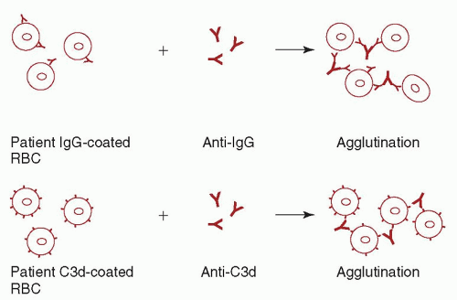
FIGURE 21.3. Direct antiglobulin test with anti-immunoglobulin G (IgG) and anti-C3d. RBC, red blood cell. (Reprinted from Jeter EK, Spivey MA. Pretransfusion testing. In: Jeter EK, ed. Introduction to transfusion medicine: a case study approach. Bethesda, MD: American Association of Blood Banks Press, 1996;56, with permission.)


Media That Enhance Agglutination

Adding albumin, low-ionic-strength saline (LISS) or polyethylene glycol (PEG) solutions to antibody identification tests can enhance the sensitivity of the test system.237 These solutions augment the antibody-antigen interaction in a variety of ways, enhancing the detection of weak or otherwise undetectable antibodies. Treating reagent red cells with proteolytic enzymes such as papain or ficin also increases the sensitivity for some antibodies such as those reacting with Rh and Kidd system antigens. These enzyme reagents weaken or destroy other red cell antigens (M,N,Fya, Fyb, and, in some cases, S, s)—a trait that can be helpful in the identification of multiple red cell antibodies in a single serum sample.2


Other Antibody Identification Tests

Sera containing several antibodies may be analyzed by absorbing with one or more selected red cells.2,236 Antibodies so adsorbed may be eluted from the cells, and their specificity may be determined. Alternatively, the specificity of antibodies not absorbed and remaining in the supernatant can be identified. When necessary, the identity of certain antibodies may be confirmed by their inhibition by soluble antigens, such as A, B, and Lewis substances present in the saliva of secretors. Neonatal (cord) red cells exhibit a number of antigens very weakly and may be used to investigate antibody specificity.


Selection of Red Cells for Transfusion

A series of serologic tests is used to select donor blood for patients. Although individual transfusion services prefer different specific methods, the general principles of compatibility testing are the same.

A properly labeled, fresh sample of patient blood must be provided. If the patient has been transfused or has been pregnant within the preceding 3 months, the specimen must be obtained within 3 days of the anticipated transfusion.5


Donor

The ABO group of the donor unit must be confirmed. The donor unit Rh typing must also be repeated if the unit is labeled as Rh-negative. These tests are performed to confirm the blood group and to ensure that the unit has not been mislabeled.


Recipient

The recipient’s ABO group and Rh type must be determined. The recipient’s serum is screened for the presence of antibodies that may have been induced by prior pregnancy or transfusion. A set of commercially prepared group O red cells, expressing 18 clinically relevant antigens (D, C, E, c, e, M, N, S, s, P1,Lea, Leb, K, k, Fya, Fyb, Jka, and Jkb), is used in this test in accordance with FDA rules. The use of group O reagent red cells avoids agglutination by anti-A or anti-B. These reagent cells are incubated with the patient’s serum and tested with the indirect antiglobulin test for reactions indicating the presence of antibody in the serum.2

If such screening reactions are positive, the antibody specificity can be determined by reaction of the serum with a commercially prepared panel of reagent red cells of known antigenic composition. If an antibody has been found on the screen and the patient’s clinical status allows, it is best to withhold transfusion until identification is complete. The incidence of unexpected RBC antibodies in patients requiring transfusion is low.238,239


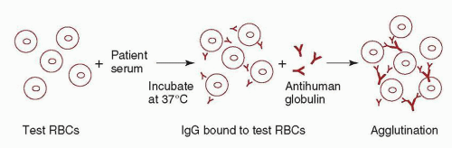
FIGURE 21.4. Indirect antiglobulin test. IgG, immunoglobulin G; RBC, red blood cell. (Reprinted from Jeter EK, Spivey MA. Pretransfusion testing. In: Jeter EK, ed. Introduction to transfusion medicine: a case study approach. Bethesda, MD: American Association of Blood Banks Press, 1996;56, with permission.)


Type and Screen

If it is unlikely that blood will be required, for example, for a surgical procedure with <10% likelihood of transfusion, a “type and screen” rather than a crossmatch should be requested. In this instance, the blood bank types the patient’s blood and screens for unexpected antibodies; if antibodies are not found, the blood bank ensures that blood of the appropriate group is available for transfusion if necessary. In such an event, a telephone call can trigger a rapid crossmatch test and blood will be available with minimal delay. The appropriate use of type and screen improves the efficiency of the blood bank. It assists in inventory control by not segregating blood for patients who are unlikely to require it and is therefore more cost-effective.


Crossmatch

If no antibody has been detected on the screen and there is no record of the previous presence of a clinically significant antibody, only verification of ABO compatibility between the donor unit and recipient is required before transfusion. This can be done either by an immediate spin crossmatch or a computer crossmatch.

The immediate spin crossmatch consists of mixing the patient’s serum with donor saline-suspended red cells at room temperature, spinning the tube, and reading the results immediately. The purpose of this test is to detect ABO incompatibility due to the presence of anti-A, anti-B, or both, in the patient’s serum.2

The conditions for computer or “electronic” crossmatch are outlined in the AABB standards.5 Briefly, the computer system must be validated to prevent release of ABO-incompatible blood. This computer crossmatch can be used only for patients who do not have a record of clinically significant antibodies. The recipient’s ABO blood group must have been determined on at least two separate tests. The system must contain complete information on the donor unit and the recipient, including ABO group and Rh type. Data entered must be verified as correct before the release of blood. The system must contain logic to alert the user to discrepancies for either the donor unit or the recipient, including unit labeling, blood grouping, and ABO incompatibilities.

If a clinically significant red cell antibody has been found when a patient’s plasma or serum is screened for unexpected antibodies, antibody identification should be performed. Once the antibody specificity has been identified, donor units that lack the corresponding antigen should be selected, and a crossmatch using an indirect antiglobulin test should be performed on each unit to ensure compatibility. The physician should also be advised about the nature of the problem, as well as the potential for delays if further units are required.

Once a unit of blood is crossmatched for a patient, there must be positive identification of the patient and the blood product both in the laboratory before release of a blood product to the nursing ward as well as at the patient’s bedside by the transfusionist. Before every transfusion, the requisition, the label on the blood product, and the patient’s identification must be checked. These aspects of patient and blood product identification are critical safety steps and must be documented.5


Uncrossmatched Blood for Emergency Transfusion

For patients in hemorrhagic shock, it is necessary to transfuse blood immediately, and no blood bank testing should be attempted before emergency transfusion. The risk of transfusing group O “uncrossmatched” red cells is extremely low and is certainly much lower than the risk of the patient’s death if blood transfusion is delayed. If a patient is to be given uncrossmatched blood, a specimen of the patient’s blood should be obtained prior to transfusion so that typing and screening can be performed while the transfusion is proceeding.

Once the patient’s blood group has been determined, ABO group-compatible uncrossmatched blood may be used.5 Until the patient’s Rh type is determined, uncrossmatched blood should be Rh(D)-negative when used in women of childbearing age, in whom sensitization to D would be undesirable. As Rh-negative blood is often in limited supply, Rh-positive blood is often used for emergency transfusion of older females and of males of unknown blood group. In such cases, sensitization may occur, but the risk of an immediate hemolytic reaction is low.236

Despite the lack of a crossmatch, transfusion of group-specific blood under emergency situations is safe. The incidence of red cell alloantibodies in healthy people is low, and most such antibodies do not cause dangerous acute intravascular hemolytic transfusion reactions. However, the decision to use uncrossmatched blood is the responsibility of the attending physician, who must weigh the risks against the expected benefits and document in the patient’s record the need for the uncrossmatched blood.

Table 21.5 outlines the selection of blood and plasma by ABO type. If the blood group is known, group-compatible red cells and plasma can be selected. If the blood group is not known, group O
red cells should be used; if plasma is required, group AB plasma should be used because it contains no anti-A or anti-B.


TABLE 21.5 SELECTION OF BLOOD AND PLASMA BY ABO TYPE


Selection of Blood Component


Component


Recipient ABO Type


Preferred


Alternate


Red blood cells


O


O


None


A


A


O


B


B


O


AB


AB


A, B, O


Fresh frozen plasma


O


O


A, B, AB


A


A


AB


B


B


AB


AB


AB


None


Modified from Jeter EK, Spivey MA. Introduction to transfusion medicine: a case study approach. Bethesda, MD: American Association of Blood Banks Press, 1996.

Only gold members can continue reading. Log In or Register to continue

Stay updated, free articles. Join our Telegram channel

Oct 21, 2016 | Posted by in HEMATOLOGY | Comments Off on Transfusion Medicine

Full access? Get Clinical Tree

Get Clinical Tree app for offline access