Gene Therapy for Hematologic Disorders


Gene Therapy for Hematologic Disorders


Andre Larochelle

Cynthia E. Dunbar

John Tisdale


INTRODUCTION

The molecular characterization of congenital and acquired human disease over the past several decades has stimulated scientists and clinicians to envision genetic therapy as a new and exciting possibility.1 By the 1990s, gene transfer technologies had progressed sufficiently to offer real hope for successful widespread clinical application.2, 3 and 4 The first replication-defective retroviral vector was described in 1983, offering a safe and feasible route for transfer of exogenous genes to nontransformed human cells.5 By 1989-1990, clinical studies using gene transfer began.6,7,8 Hundreds of clinical trials have been completed or are in progress (Fig. 71.1). Although surrounded by expectations, none of the early trials had even minimal evidence for clinical benefit, and in 1996 an expert panel convened by the director of the National Institutes of Health was critical of the premature initiation of clinical gene therapy trials and the subsequent overselling of results by investigators and the media.9 At the time of the report, no clinical gene “therapy” trial had shown clinical efficacy. The focus thus turned to rationally designed, small-scale clinical trials in diseases for which the pathophysiology is well understood, as well as the more important need for continued basic science investigations into vector systems and target cell biology.10, 11 and 12


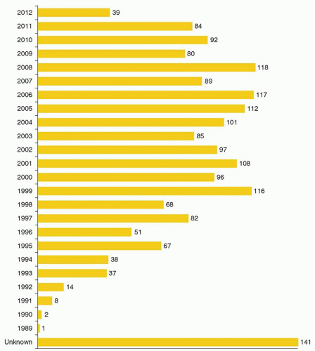
FIGURE 71.1. Number of gene therapy clinical trials approved worldwide from 1989 through part of 2012. (From The Journal of Gene Medicine, © 2012 John Wiley and Sons Ltd, www.wiley.co.uk/genemed/clinical. Reproduced with permission of John Wiley and Sons, Inc.)

Hematopoietic stem cells (HSCs) and lymphocytes have remained central target cell populations for two major reasons: first, they are relatively easy to manipulate ex vivo, and, second, many acquired and congenital diseases are potentially curable by their genetic correction.13,14 A significant fraction of ongoing human clinical trials target hematopoietic cells or are designed to treat congenital or acquired diseases of the hematopoietic or immune system (Fig. 71.2). Moreover, many important experimental advances in our understanding of hematologic physiology have resulted from the application of gene transfer techniques in vitro or in animal models.15,16,17, 18, 19 and 20,21 For instance, retroviral tagging of HSCs has allowed tracking and quantitative analysis of murine and nonhuman primate stem cell behavior, and experiments overexpressing oncogenes or cytokines in primary hematopoietic
cells using similar techniques has helped elucidate the in vivo role of these proteins. Significant advances in gene transfer technology have occurred through systematic preclinical testing in large animal models, and clinically relevant gene transfer levels have now been achieved in these models. These results predicted success in humans, at least for disorders for which modest transfer rates of genes not requiring complex regulation could be curative, and unequivocal efficacy has since been realized.22,23 These longanticipated results represented a significant advance for the field, providing not only restoration of immunity in children with severe combined immunodeficiency (SCID), but proof of principle in the context of a human disorder, and a great deal of enthusiasm for returning to the clinic for these and other disorders quickly developed. However, serious adverse events arising as a direct result of insertional mutagenesis have since been reported and efforts to understand these events more fully, develop additional safeguards, and adjust the risk-benefit assessment have now become necessary. Thus, it is particularly important for hematologists to have a general understanding of the field, even if widespread clinical applications may be a decade or more in the future. This chapter reviews the fundamental features of gene transfer technologies and their applications in preclinical and early clinical trials. The pace of the field is rapid, and many details may become obsolete, but the central concepts should remain relevant to any future gene therapy applications.


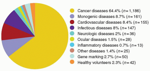
FIGURE 71.2. Indications for gene therapy clinical trials. (From The Journal of Gene Medicine, © 2012 John Wiley and Sons Ltd, www.wiley.co.uk/genemed/clinical. Reproduced with permission of John Wiley and Sons, Inc.)


OVERVIEW OF BASIC CONCEPTS

Gene therapy can be defined as the transfer of a gene or genetic material (DNA or RNA) into a cell with therapeutic intent (Fig. 71.3). The genotype of the cell is thus altered, with subsequent gene expression altering the phenotype of the cell. The therapeutic agent is the gene product, generally a protein, or less frequently RNA, for example, ribozymes or antisense molecules. This is in contrast to conventional therapies that act by directly altering the phenotype, even if the congenital or acquired defect is a genetic one. Three examples illustrate the conventional approach to underlying genetic disorders. Dietary avoidance of phenylalanine can prevent the consequences of phenylketonuria by circumventing the genetic deficiency of phenylhydroxylase. Cancer chemotherapy acts by preferentially killing tumor cells based on their cell cycle characteristics, thus removing cells with an acquired genetic defect. And factor replacement in hemophilia directly replaces the defective or missing gene product by infusion of an exogenously manufactured or isolated protein. Gene therapy strategies instead attempt to alter the underlying genetic abnormality to circumvent the need for these therapies.

The identification and cloning (isolation) of genes responsible for many congenital disorders, as well as of the cellular genes mutated in acquired disorders such as cancer has led to the concept of genetic correction of affected cell populations. Ideally, actual substitution of a defective gene with a therapeutic gene would be the most desirable method for returning target cells to a normal genotype and phenotype (gene replacement). However, this goal requires homologous recombination, a complex and inefficient process, and current gene transfer methods predominantly rely on either insertion of new genes into the chromosomes, or on extrachromosomal (episomal) maintenance of a newly introduced gene (gene addition).24, 25 and 26 To date, efforts have focused on somatic (nongerm cell) therapy, with genotypic alteration of only the diseased target tissue. Manipulation of germ cells, with transmission of altered genetic material to subsequent generations, is not yet feasible in humans, but the profound ethical and societal implications need to be addressed through the political process before the technology progresses much further.27 The recent development of induced pluripotent stem cells (iPS cells), whose behavior is analogous to embryonic stem cells (ESCs) but can be derived from somatic cells, has introduced the possibility of genetic manipulation of autologous pluripotent stem cells, circumventing the ethical issues surrounding ESCs.

The vehicle for transferring new genetic material into a target cell is called a vector. At a minimum, a vector contains the gene or genes of interest along with regulatory elements such as promoters or enhancers that govern expression of the gene product.
A vector may be a simple particle consisting of a fragment of DNA encapsulated within a liposome or conjugated to proteins that facilitate uptake into cells, or may be a more complex viral vector, capitalizing on the ability of viruses to enter cells easily and express genes robustly. Characteristics of the major vector systems are summarized in Fig. 71.4 and Table 71.1, and are detailed in subsequent sections. The successful interaction of a vector with a target cell, leading to an alteration in that cell’s genotype, is termed transduction.


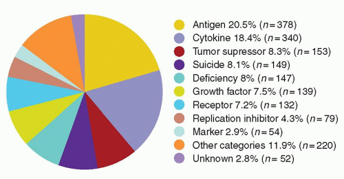
FIGURE 71.3. Gene types transferred in gene therapy clinical trials. (From The Journal of Gene Medicine, © 2012 John Wiley and Sons Ltd, www.wiley.co.uk/genemed/clinical. Reproduced with permission of John Wiley and Sons, Inc.)

Vector production procedures are unique to each system, but a number of considerations are common to all, especially those being developed for actual clinical use. A clinical vector must be feasible and practical to produce safely at pharmaceutical grade.28,29 To prevent indiscriminate spread of viral genomes, most viral vectors designed for clinical applications must be rendered replication-defective meaning that once a viral vector enters a target cell, the cell will produce no new viral particles. High-titer vector preparations, containing a high concentration of functional vector particles, are also very important, allowing exposure of target cells to the highest possible multiplicity of infection (MOI), defined as the ratio of vector particles to target cells; this increases the probability of successful vector-cell interaction.

Exposure of target cells to vectors can occur either ex vivo or in vivo. Autologous hematopoietic targets such as stem cells or lymphocytes are generally transduced ex vivo, because these cells can be easily collected, manipulated in culture, and then re-infused intravenously. Ex vivo transduction allows for a controlled exposure of only the desired target cells to large concentrations of vector, and is less likely to generate an inflammatory or immune response or be hindered by vector inactivation by complement. Other cellular targets for ex vivo transduction have included hepatocytes, keratinocytes, tumor cells, and muscle progenitor cells.

In vivo gene transfer has been used for applications involving cells that cannot successfully or easily be harvested and manipulated ex vivo, such as airway epithelium, vascular endothelium, differentiated muscle cells, retinal pigment epithelium, and neurons. The ideal in vivo system would allow intravenous injection of a vector followed by rapid and safe specific transduction of target cells around the body.

A number of important steps must occur between exposure of a target cell to a vector and successful transduction of that cell, with persistence of the transferred genetic material in the correct cellular compartment and expression of the gene of interest or transgene. The vector must cross the plasma membrane efficiently and without damaging the cell. Most viral vectors enter cells via specific cell-surface receptors, and an important consideration is the number of functional receptors on the proposed target cell for the vector being used.30 A process called pseudotyping can be used to redirect viral vectors to different cell-surface receptors by substituting alternative viral envelope proteins during the vector production process.31,32,33,34 Nonviral vectors may cross the plasma membrane without need for a specific receptor.

After crossing the plasma membrane, the vector must then travel through the cytoplasm and cross the nuclear membrane in order to enter the nucleus and utilize the cell’s transcriptional machinery for expression of the transgene. Nuclear entry of some vectors may be dependent on mitosis, with temporary breakdown of the nuclear membrane; others carry nuclear localization determinants that result in specific conveyance across the intact membrane. The transferred genetic material may integrate permanently into the target cell’s own chromosomal DNA, ensuring passage of the new gene to all daughter cells with every cell division. The need for integration depends on the target cell and therapeutic application: it is absolutely required for gene transfer into HSCs where the transgene must be transmitted to all progeny cells, but is superfluous for cellular targets such as neurons or muscle cells that are not mitotic.


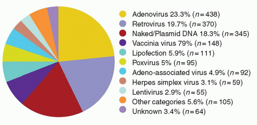
FIGURE 71.4. Vectors used in gene therapy clinical trials. (From The Journal of Gene Medicine, © 2012 John Wiley and Sons Ltd, www.wiley.co.uk/genemed/clinical. Reproduced with permission of John Wiley and Sons, Inc.)

Alternatively, the gene may remain episomal, or nonintegrated. Some vectors are very stable as nuclear episomes, with prolonged persistence of transgene expression as long as the cell does not undergo mitosis. Unless the episome can reproduce itself, cell division will eventually dilute out episomal DNA, limiting the application of nonintegrating vectors to nonmitotic tissues or to situations requiring only transient expression.

The level of transgene expression and the ability to restrict expression to specific target cell types are also important factors to consider. Expression of the transduced gene is dependent on both vector and target cell determinants. Transcriptional promoters must be included along with the actual transgene protein coding sequences in the vector, and often lineage or tissue-specific promoter and enhancer elements are used to limit expression to a particular cell lineage derived from a target cell population.35, 36 and 37 For instance, hemoglobin gene regulatory sequences are required to drive transgene expression specifically in erythroid cells in strategies targeting the globin disorders. In other situations, constitutive control elements that can drive transcription continuously in most cell types can be used. Genetic control elements that are inducible, or turned on by some exogenous manipulation such as the administration of an antibiotic, are also under development for inclusion in gene transfer vectors.38,39 Endogenous cellular factors may shut off expression of transferred genes in some situations.40,41 These factors have not been fully elucidated, and vary from vector to vector. Silencing of transferred genes via methylation of vector sequences is one possible mechanism.42 The level of expression necessary for the desired therapeutic effect is very important to determine during in vitro and animal experiments and varies greatly depending on the target cell type and the proposed clinical application.


Nonviral Vectors

The simplest approach to gene transfer is to use only the DNA of the transgene, with the necessary control sequences, as the vector.29,43 Recombinant DNA production in bacteria can result in purified plasmids containing the gene(s) of interest along with regulatory elements. For over two decades, scientists have introduced purified DNA into target cells ex vivo by a variety of physical and chemical means. The least complex technique is direct microinjection into individual cells, which has little clinical
utility due to the impossibility of injecting enough cells to produce most of the desired effects. One exception is the observation that direct DNA injection into muscle or skin can stimulate a very potent immune response against antigens introduced as plasmid transgenes.44,45 This observation led to preclinical and clinical development of genetic vaccination strategies against bacterial and viral pathogens.46


TABLE 71.1 GENE TRANSFER VECTOR SYSTEMS


Vector System


Integration


Cell Cycle Dependence


Insert Size Limit


Clinical Experience


Advantages


Disadvantages


Major Applications


Murine retrovirus


Yes


Yes


8-10 kb


Extensive


Stable producer lines No viral genes in vector Low immunogenicity Well-understood biology Efficient entry and integration in many cell types Proven clinical safety


Low titer, fragile vector Requirement for cycling Erratic expression Insertional mutagenesis


Ex vivo—stem cells, lymphocytes, tumor cells, hepatocytes, myoblasts In vivo—producer cell or vector injection into tumors


HIV-based lentivirus


Yes


No


None


Faithful delivery of complex genes


Well understood


Efficient entry and integration


Pseudotyping allows broad tissue range


Production labor-intensive Erratic expression


Insertional mutagenesis Recombination with wildtype HIV


Ex vivo—stem cells, lymphocytes, tumor cells, nondividing cells


Adenovirus


No


No


8-10 kb


Moderate


High titer, stable vector


High-level transgene expression


Efficient entry into many cell types


No stable producer lines Potential for recombination and replication-competent virus


Multiple viral genes expressed from vector


High immunogenicity (may be advantage as a vaccine vector!)


Pre-existing immunity


Inflammatory responses


In vivo—pulmonary epithelium, tumor cells, muscle, liver


AAV


Yes—inefficient


Yes—controversial


4.5 kb


Minimal


Stable vector, extra- and intracellularly


High titer


High-level transgene expression


No expressed viral genes in vector


No stable producer cell lines High percentage of defective particles


Requirement for helper adenovirus during production Very limited insert size


Pre-existing immunity


Undefined


Naked DNA


No


No


No limit


Moderate


Ease of production


High level of safety


No extraneous expressed vector genes


No immunogenicity of vector


Inefficient cell entry, uptake into nucleus Poor stability within cell Low-level expression


In vivo—tissues accessible to injection, for transient expression or vaccination


Facilitated DNA (liposomes, polylysine conjugates, inactivated adenovirus, etc.)


No


No


No limit


Moderate


Same as naked DNA plus: Can be targeted to specific cell types


More efficient uptake and intracellular stability


No mechanism for persistence


Same as naked DNA, plus in vivo tumor cells, vascular endothelium


AAV, adeno-associated virus, kb, kilobases.


A number of other methods have been explored for getting plasmid DNA into cells. Liposomes, composed of phospholipid bilayers enclosing an aqueous space loaded with DNA, can directly fuse with the plasma membrane, releasing DNA into the cytoplasm.47 The gene gun technique involves bombardment of the cell membrane with gold microparticles complexed to DNA.48,49, 50, 51 and 52 Electroporation and calcium phosphate precipitation are generally too toxic and inefficient for use in gene transfer strategies aimed at primary human cells.53, 54 and 55

All of these methods lack mechanisms to stabilize intracellular vector DNA, nor do they allow transport into the nucleus; they therefore rarely result in chromosomal integration or long-term persistence and expression. The development of cationic liposomes has improved cellular uptake of plasmid DNA and has circumvented cytoplasmic degradation.56,57 In aqueous solution these positively charged liposomes bind with up to 100% of negatively charged DNA without size restrictions and can deliver DNA to the cell nucleus, albeit inefficiently, where it remains primarily episomal.58 If administered in vivo, liposomes demonstrate no target cell tropism and are rapidly cleared by the reticuloendothelial system. However, after intravenous injection of cationic liposomes, long-term low-level persistence of vector sequences in many murine organs has been demonstrated.59,60

Nonviral plasmid DNA transfer has been improved by conjugation of vector DNA to substances that improve cellular transport
and allow target cell specificity via cell-surface receptors.61,62 For example, adenoviral capsid elements (especially the penton base protein) help disrupt endosomes, releasing DNA more efficiently into the cytoplasm, and inclusion of transferrin or other ligands in DNA-polylysine conjugates allows specific uptake via cell-surface receptors.63, 64, 65, 66 and 67

Manufacturing nonviral vectors is much simpler than manufacturing viral vectors, and cannot generate potentially dangerous replication-competent infectious particles.29,68 Transduction is not dependent on target cell cycling, and no viral proteins are present to induce an antivector immune response. There are no size constraints. Limitations include a generally lower transduction efficiency than with viral systems. Most important, transgene integration is poor and persistent expression rare, limiting utility to situations allowing transient transgene expression.


Viral Vectors

Viral vectors are Trojan horses, taking advantage of the viral capsid or envelope and of the viral machinery to deliver nonviral transgene sequences efficiently to target cells.69 In general, the external capsid or envelope of the virus is unaltered in a viral vector, but the genome of the virus is in large part replaced by a transgene or genes. This strategy is limited by the space available in a viral particle for new genetic material. Viral vectors also depend on the presence of a specific viral receptor on target cells. The efficiency of gene transfer utilizing viral vectors is the main advantage when compared to some of the physical and chemical cell entry methods described above. Furthermore, the relatively low toxicity of transduction of cells with certain viral vectors (e.g., retroviruses, lentiviruses, and adeno-associated viruses [AAV]) is another advantage when compared to physical or chemical methods. In order to modify a virus into a vector system, detailed understanding of the viral genome and life cycle is necessary to retain viral genome sequences required for packaging of vector nucleic acids into viral particles and for appropriate trafficking in the target cell, while removing sequences that might allow production of replication-competent viral particles.


Retroviral Vectors

The murine retroviruses exemplified by the Moloney murine leukemia virus (MLV) were the basis of the first practical viral vector system.70, 71 and 72 These retroviruses consist of two single strands of linear viral RNA bound to protein core and coated by a lipid envelope that is acquired from the plasma membrane of the infected cell upon viral release. The linear RNA genome can contain 2 to 9 kilobases (kb) of coding and regulatory sequences, flanked on each end by sequences termed long terminal repeats (LTRs) that permit integration into chromosomes and contain strong promoter/enhancer elements that normally drive expression of full-length viral RNA genomes, or via alternative splicing, the individual retroviral genes. These simple murine retroviruses contain only three genes necessary for viral replication and packaging: gag, pol, and env. The retrovirus enters a cell after binding to a specific cell-surface receptor via the env gene product. These receptors are large, widely expressed proteins involved in phosphate transport and other cellular homeostatic functions.30 The amphotropic receptor is found on both rodent and primate cells, and is the entry site for most murine retroviral vectors directed at human cells.73, 74 and 75

After cell entry, the viral RNA is reverse transcribed via the pol gene product into proviral cDNA and enters the nucleus. The LTR sequences allow random integration of the viral cDNA into the host chromosomes. The integrated retroviral genome, or provirus, relies on the host cell’s transcriptional machinery for expression of proviral genes and production of full-length viral RNA. The gag, pol, and env gene products are packaged along with the new viral RNA into viral particles, dependent on the presence of a packaging (ψ) sequence in the viral RNA, and viral particles are released from the cell via budding through the plasma membrane, without damaging the infected cell.

Recombinant retroviral vectors are constructed by removing the gag, pol, and env gene sequences from the viral nucleic acid backbone and replacing them with up to 7 to 8 kb of a gene or genes of interest, retaining only the LTRs and the packaging signal (ψ).5 The resulting recombinant viral vector can integrate and express the gene or genes of interest, but cannot replicate and produce new retrovirus once within a target cell, because of the lack of gag, pol, and env genes within its genome. Thus it is termed replication defective. Figure 71.5 diagrams the steps involved in making a retroviral vector. A packaging cell line is created by introducing a plasmid containing gag, pol, and env sequences but no ψ sequence into an immortalized cell line such as NIH3T3. The lack of the ψ sequence prevents these helper genes from being packaged into viral particles. A second plasmid containing the recombinant vector sequences (LTRs flanking the transgene or genes) is then introduced into these cells to create a producer cell line. Full-length vector RNA is transcribed from the vector plasmid sequences and packaged into viral particles using the gag, pol, and env proteins encoded by helper plasmid sequences. In this way, producer cell lines release helper-free replication-defective vector particles containing the recombinant genome into cell culture media at a titer of up to 107 particles/ml. These particles contain the full-length vector RNA, consisting of the viral LTRs flanking the transgene or genes, and the env, gag, and pol proteins, but because they do not contain any actual gag, pol, or env viral gene sequences, no further infectious virus can be made once the vector infects the target cell.

The potential for generation of replication-competent virus through recombination events between the vector and helper sequences in the producer cell line is a significant safety concern.28,76 In order to allow packaging of replication-competent viral particles these recombination events must result in the transfer of an intact ψ packaging sequence being transferred into the helper sequences containing the gag, pol, and env genes. The presence of replication-competent virus could allow spread of vector and helper particles indiscriminately to nontarget cells in vivo, thus greatly increasing the risk of insertional mutagenesis by repeated infection of susceptible cell populations.76,77 The absolute need for avoidance of replication-competent viral particles in clinical vector preparations was inadvertently demonstrated when high-grade lymphomas occurred in rhesus monkeys transplanted with HSCs transduced with a vector preparation contaminated with high levels of replication-competent virus.78 Over the next several years, a number of modifications in the organization of genetic sequences included in the packaging cell lines has greatly decreased the risk of recombination events, and sensitive systems for detecting replication-competent virus have been developed.28,79,80,81 A number of investigators have used packaging cell lines derived from human instead of murine cells to make producer cell lines, in hopes that lower levels of endogenous retroviral sequences in human cells would also decrease recombination events and thus replication-competent viral contamination.82,83

As described below, retroviral vectors are capable of stable integrated transduction of a large number of cell types, including repopulating stem cells, but have a number of important limitations. Stable transduction and integration requires passage of the target cell through the S phase of the cell cycle, preventing transduction of quiescent cells.84 The amphotropic receptor density on certain target cell types may be too low to allow efficient transduction.85 Thus, redirection of receptor specificity via pseudotyping with alternative envelope proteins has been explored, with some success.32,86, 87, 88, 89 and 90

Biophysical considerations may also limit vector-target interactions. Vector particles are very fragile, and degrade quickly in

solution or within cells if cell division allowing nuclear entry and integration does not occur. A number of investigators have tried to increase the likelihood of successful transduction by flowing vector solution continuously over target cells or co-localizing vector and target cells using culture dishes coated with fibronectin fragments.91,92 New methods of concentrating and stabilizing vector preparations are also under development.33,87,93


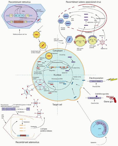
FIGURE 71.5. Schematic representation of transduction of a generic target cell by the four major gene transfer vector systems. The same “gene of interest” is shown being transferred with each vector. AAV, adeno-associated virus; Ad, adenovirus; ITR, inverted terminal repeat; LTR, long terminal repeat.


Lentiviral Vectors

There has been real progress toward the development of alternative retroviral systems that may overcome some of the limitations of the MLV vectors. HIV-derived lentivirus vectors have been most actively pursued. Relatively high-titer vectors have been reported using production strategies with numerous safeguards against recombination events that could result in generation of wildtype HIV.94,95,96 Vectors retaining the HIV gp120 envelope protein could be used to transduce CD4+ T-cells, monocyte/macrophages, or glial cells, and pseudotyping with alternative envelope proteins such as vesicular stomatitis virus (VSV) or amphotropic leukemia virus envelope proteins could be used for transduction of a wider range of cells. Lentiviral vectors can traverse an intact nuclear membrane, requiring the HIV gag protein and an accessory VpR protein, thus allowing transduction of nondividing cells.97,98 The promise for more efficient HSC transduction using lentiviral vectors remains an attractive feature, however, other features of HIV-based vectors have also stimulated interest. Specifically, the faithful delivery of nonrearranged genes with complex regulatory elements appears superior, and success in delivering the human β-globin gene along with large portions of the locus control region has finally been achieved with HIV-based vectors.99 A number of additional safety modifications have been employed, including deletions in the 3′ LTR reducing the risk of insertional mutagenesis by self-inactivation of the integrated LTRs. Indeed, the first clinical trial employing lentiviral vectors for the treatment of HIV have been carried out,100,101 and subsequent trials have demonstrated clear efficacy in adrenoleukodystrophy (ALD)102 and thalassemia.103


Human Foamy Viral Vectors

The other member of the retroviridae under consideration as a gene transfer vector is the human foamy virus (HFV). HFV, a poorly characterized retrovirus, has three potential advantages. It has never been associated with pathology in animals or humans, it infects a wide variety of primate cell types, and it has the capacity to package longer transgene(s).104 It does not appear, however, to transduce nondividing cells, although it may be more stable than conventional retroviruses within a target cell, tolerating a more prolonged period before cell division and then integration.105 Virologists are trying to define the packaging signal and other important elements necessary for engineering a replication-incompetent HFV vector,106,107 and a method for the production of helper-free vector stocks has recently been described.108 Third-generation vectors have now been developed, and these foamy virus vectors appear efficient for transducing HSCs, with high gene transfer rates observed in human cord blood (CB) in vitro, and in murine BM (BM) in vivo after a single, overnight vector exposure.109 Recently, HFV vectors have proven highly efficient in a canine model of leukocyte adhesion deficiency (LAD).110


Adenoviral Vectors

Adenoviruses are nonenveloped double-stranded large DNA viruses.111 The linear adenovirus genome contains 36 kb with an inverted terminal repeat (ITR) of 100 to 165 base pairs at each terminus. A set of early genes encodes for regulatory proteins that serve to initiate cell proliferation, DNA replication, and down-modulation of host immune defenses, whereas the late genes encode for structural proteins. Adenovirus readily crosses the plasma membrane of many cell types, whether replicating or not, via receptor-mediated endocytosis112 through the receptor, common to two distinct viral pathogens, coxsackie B and adenovirus 2 and 5, termed the coxsackie and adenovirus receptor.113 Adenovirus escapes the endosome by altering the pH and then enters the nucleus where it remains as a linear episome. In permissive cells, adenovirus replicates and then enters a lytic cycle, destroying the host cell and releasing daughter viral particles. Of the 42 adenovirus serotypes, most are known to cause mild respiratory, gastrointestinal, and conjunctival infections in immunocompetent humans; no associated malignancies in humans have been reported, although some serotypes can transform cells in culture. The human embryonic kidney cell line HEK293 was, in fact, immortalized by transfection of kidney cells with sheared adenovirus serotype 5 DNA; the E1 gene that is integrated into the cellular genomic DNA is apparently responsible for the immortalization of the cell line.

Recombinant adenovirus vectors have been engineered from adenovirus (usually serotype 5) by the removal of the E1 and E3 genes (regulating replication and immune recognition) and replacement by the gene or genes of interest, with space for 7 to 8 kb of new genetic material.114,115,116,117,118 High-titer adenovirus vectors, up to 1012 plaque-forming units (pfu) per ml, can be reliably packaged through the use of a human embryonic kidney transformed cell line,293 which provide the helper or replication E1 genes, followed by purification and concentrating procedures. The final product is a replication-defective adenovirus vector which is free of helper or wildtype virus that can efficiently transduce nondividing cells.116 These vectors do not integrate into the target cell genome, avoiding insertional mutagenesis and resulting in only transient transgene expression in proliferative tissues. Because of the tropism of adenovirus for epithelial cells, these vectors were initially investigated for the treatment of pulmonary diseases and diseases in which liver gene transfer is desirable.119,120

Transient transgene expression also may result from host cellular and humoral immune responses directed at either transgene-encoded antigens or adenovirus proteins expressed from the large portions of the adenovirus genome retained in these vectors.121,122 Another concern is inflammation resulting from in vivo transduction of certain cell types, especially airway epithelium.123 In vivo use may also be compromised by pre-existing or new antiviral neutralizing antibodies, limiting the efficacy of repeated dosing, which may be required for applications directed at mitotic tissues.124 Nonessential adenoviral sequences are being gradually eliminated from the vectors.125,126 The use of adenovirus vectors in which essentially all viral genes are removed is the ultimate goal of such a strategy. Unfortunately, although these vectors have little except the capsid proteins to mark themselves as foreign, there is evidence that they can trigger innate immunity by recognition through the toll-like receptor 9 pathway in target cells.127 Further technical issues that are important include the decreased efficiency of vector production as more viral genes are deleted, inasmuch as these functions need to be provided by accessory plasmids through transfection of vector-producing cells. Hence, these highly deleted vectors are often called “helper-dependent.” Specific dosage schedules (e.g., neonatal or embryonic exposure) or co-administration of immune modulators such as cyclosporine or IL12 may also prevent sensitization.128, 129, 130, 131 and 132 Another strategy being explored is use of different vectors based on adenovirus serotypes other than adenovirus 5. One survey of unusual group B and group D adenovirus serotypes that might be considered for use as HIV vaccines indicates that in a target population in sub-Saharan Africa the existing immunity to adenovirus serotypes 11, 35, and 50 from group B and adenovirus serotypes 26, 48, and 49 from group D are substantially less than that of the commonly studied adenovirus 5 serotype from group C.133 By employing alternatives to adenovirus 5, it might be possible to get around the barrier of pre-existing antivector antibodies for vaccination
efforts. Furthermore, it might be possible to alternate the vector type with subsequent vaccinations to thwart neutralizing antiadenovirus immunity to the vaccine vector. Recently, investigators developing HIV vaccination strategies have reported an adenovirus 41 serotype vector that can express HIV envelope protein.134 The adenovirus 41 serotype virus is an enterotropic pathogen that causes diarrhea in its wildtype form; the use of a gene transfer vector with tropism for a mucosal surface may present advantages for vaccination against HIV, a pathogen that is normally encountered by the host in the mucosa. This vector has not yet been clinically tested.

In initial clinical trials, adenovirus vectors were used to transfer a normal cystic fibrosis transmembrane conductance regulator (CFTR) gene into airway cells (bronchial or nasal) of patients with cystic fibrosis.135, 136 and 137 Despite the demonstration that airway cells could be transduced with these vectors in vivo with correction of the chloride transport defect, clinical utility has been precluded by the harmful immune and inflammatory responses noted above. However, the active immune response induced by adenoviral vectors is also being explored as a possible advantage for adenovirus vectors when they are used to transduce tumor cells with cytokines or other immune modulators for tumor vaccine protocols.138, 139 and 140 The fact that most adenoviral gene transfer results in transient transgene expression (in contrast to retroviral-mediated gene transfer) has limited their use for congenital diseases in which permanent expression of a missing protein is desirable, such as hemophilia, ornithine transcarbamylase (OTC)-deficiency, or α-1-antitrypsin inhibitor deficiency. The use of alternating serotypes for repeated administration can only increase the number of administrations so much before immunity to the vectors neutralizes further administration. The powerful and often toxic immune reaction against viral vectors makes their use for vaccination or antitumor therapy more likely than for correction of metabolic disorders.141 The tragic death of a subject who received the OTC-adenovirus vector was mediated in a large part by the “cytokine storm” and resultant systemic immune response that led to widespread capillary leak multiorgan failure.141


Adeno-Associated Viral Vectors

AAV are small nonenveloped single-stranded DNA viruses in the parvovirus family, dependovirus subfamily, that require a helper virus (typically a double-stranded DNA virus such as adenovirus or herpes simplex virus) for production of new viral particles.142, 143 and 144 The linear AAV genome is approximately 4.7 kb long and consists of two homologous ITRs of 145 bp flanking two groups of genes: the rep or nonstructural genes and the cap or structural genes. There are at least eleven serotypes of AAV that differ mainly on the basis of their external capsid proteins. AAV-2 is the best characterized serotype. AAV-2 enters host cells primarily through interaction with the heparin sulfate proteoglycan protein on the cell-surface, and by interaction with the αVβ5 integrin and fibroblast growth-factor receptor proteins. AAV has oncoprotective and HIV-suppressive properties, and is not known to cause disease in humans or other animals.145, 146 and 147 Prior infection with AAV-2 in humans is common: seroepidemiologic studies demonstrate that 80% of the adult population has antibodies.143,148

After cell entry mediated by the capsid protein, wildtype AAV integrates within the host chromosome.149 Integration of multiple copies in tandem occurs in a site-specific manner within a relatively small area on chromosome 19: this site specificity appears to require rep protein.150,151 Specifics of replication and integration are less well understood than for retroviruses.

Recombinant AAV vector DNA contains the AAV ITRs flanking a gene of interest replacing the rep and cap genes. This plasmid is introduced into a cell line permissive for adenovirus, along with a helper plasmid containing the AAV rep and cap genes but no ITRs. Upon exposure to adenovirus or transfection with adenovirus genes such as E4 (particularly open reading frame 6), the cell line packages the recombinant vector sequences using the rep and cap gene products produced by the helper plasmid, and recombinant vector particles are released as the producer cell lyses.152,153, 154, 155 and 156 Recombinant AAV vectors have size constraints: vector sequences longer than 115% of the wildtype length are not packaged or encapsidated efficiently. Very high titers of recombinant AAV particles can be produced, but often a significant percentage of capsids are empty or otherwise defective.

The need for replication-competent adenovirus during AAV vector production complicates the manufacturing and safety issues, inasmuch as the adenovirus must be inactivated or (preferably) removed before use. Some progress is being made toward introducing only the specific adenovirus genes necessary for AAV replication into producer cell lines, avoiding live adenovirus. The inability to harvest AAV vector without actual lysis and death of the producer cells also complicates production of pure and defined vector preparations. Generation of stable packaging cell lines is also hindered because the AAV rep gene product harms most cell types; but recently producer cell lines expressing rep from an inducible promoter have been isolated.157,158 Another approach to avoid the issue of helper-virus contamination is to transfect cells with multiple plasmids that separately encode the desired transgene nucleic acid as well as the structural and replication proteins.159,160

Integration of the recombinant vectors into target cell chromosomes appears to be very inefficient, and thus these vectors prove more useful in situations not requiring integration, inasmuch as they have been shown to be stable as episomes for long periods.156,161,162 Site-specific integration in chromosome 19, very desirable to avoid random insertional mutagenesis, does not occur with the recombinant AAV vectors, presumably due to lack of rep protein in the vector particle.162 Many cell types can be efficiently transduced, including nondividing cells such as neurons, but integration and increased efficiency of transduction still appears to depend on cell division or other DNA-disrupting events, although this conclusion is very controversial.163, 164, 165, 166 and 167

Recently, there has been a great deal of interest in the use of vectors based on serotypes other than AAV-2, especially AAV-6, AAV8, and AAV-9, although the work thus far has consisted of in vitro vector production development160 and preclinical animal studies.168, 169 and 170 It is possible that a number of other parvoviruses can be developed as vectors, including AAV-3, nonpathogenic strains of B19, or novel autonomous parvoviruses isolated from nonhuman primates or other animal species.171, 172 and 173 These viruses are similar to AAV, but some, such as B19, never integrate. Self-complementary AAV vectors based upon serotypes with high-efficiency delivery to liver cells have been utilized recently to deliver factor IX as a potential treatment for hemophilia B in the canine model with correction of the bleeding abnormalities.174,175 These results supported the first successful clinical trials in humans described in detail below.176


Miscellaneous Other Vectors

Herpes viruses are large DNA viruses with marked neurotropism, generating intense interest in their potential as vectors targeted at the nervous system.177, 178, 179 and 180 They can accommodate very large DNA sequences (up to 30 kb). More recently, these vectors have been reported to transduce some types of hematopoietic cells, including monocytes, leukemic blasts, and progenitor cells.181,182 However, these vectors result in only transient expression in dividing cells, and cause cytotoxicity, limiting clinical utility, at least for hematologic applications.

Vaccinia virus, a large DNA virus that replicates cytoplasmically, has also been considered for gene therapy applications.183,184 It can accommodate very large transgenes (up to 30 kb) and expresses these genes at very high levels, but expression is
transient, and production of replication-incompetent vectors has not yet been possible. Very high immunogenicity limits most clinical applications, but may be advantageous for in vivo vaccination with vaccinia-transduced tumor cells.184, 185, 186 and 187


Genome Editing

Current vectors and protocols have attempted phenotypic correction or modification by gene addition, generally randomly in the genome. A different process, gene correction by homologous recombination, in hematopoietic tissues has been proposed as an alternative.188 Correction of the genetic defect in lymphoblastoid cell lines derived from patients with sickle cell disease was described189 using a RNA-DNA oligonucleotide, yet the applicability of this approach remains elusive.

An alternative corrective approach for some disorders has recently been described. Constructs targeting aberrant splice sites in the form of oligonucleotides, morpholinos, or U7 snRNAs lead to increased levels of correctly spliced β-globin mRNA by effectively blocking the aberrant splice site through the use of sequences complementary to the corrected sequence.190, 191 and 192 A major problem with this approach has remained the difficulty in delivering such constructs in vivo with sufficient efficiency to correct the phenotype and recently, lentiviral vectors designed to deliver modified U7 snRNAs permanently have been tested in cell lines and in primary cells from individuals with thalassemia with encouraging early results.193

Targeted DNA double-strand breaks through the use of artificial nucleases was recently shown to increase the efficiency of homologous recombination by several logs, renewing enthusiasm for this approach. The development of zinc finger nucleases (ZFNs) that allow cleavage of DNA in a sequence-specific manner allows the potential for genome editing at virtually any site. These artificial endonucleases mediate DNA cleavage at targeted sites and have been utilized successfully in a broad range of experimental systems including plants, flies, worms, rodents, ESCs, and primary human cells.194 The ideal set of ZFNs would target double-strand DNA breaks at high efficiency, and more importantly, with high specificity. Specificity has thus far been difficult to achieve. ZFNs can also be utilized to modify normal genes, such as the CCR5 (required for HIV entry), and a clinical trial is now underway testing this approach as a potential therapy for HIV.195 Transcriptional activatorlike effector nucleases are a new class of nucleases that allow tailored genome editing in a variety of cell types with the potential for increased specificity.196 The advent of iPS cells that can be generated from individuals with genetic diseases raises the possibility of genome editing followed by differentiation to a cell type of interest as a form of therapy for a number of diseases, and proof of concept has already been achieved in a murine model of sickle cell disease in which iPS cells were corrected with homologous recombination and transplanted after differentiation into hematopoietic progenitors.197


HEMATOPOIETIC STEM AND PROGENITOR CELLS AS TARGETS FOR GENE TRANSFER

Since the development of helper-free retroviral gene transfer technology, now decades ago, the HSC has been a primary target for gene therapy applications. The curative potential of HSCs carrying corrective genes has been well established through the use of allogeneic bone marrow (BM) transplantation in genetic disorders whereby an individual carrying a normal genotype serves as the stem cell donor, yet procedural toxicities and finite donor availability limit this approach. The prospect of a curative, one-time therapy using genetically modified autologous stem cells for the treatment of a wide variety of congenital disorders such as hemoglobinopathies, immunodeficiencies, or metabolic storage diseases, and of a new weapon against malignancies and HIV infection, has proven irresistible.13,14,198, 199, 200 and 201 Gene therapy directed at HSCs must utilize an integrating vector, because ongoing self-renewal or proliferation/differentiation would rapidly dilute out an episomal vector in daughter cells. Initial preclinical studies in the 1980s reported the introduction of genetic markers and clinically relevant genes into mouse HSCs using integrating retroviral vectors, providing both conceptual and methodologic insights that led to first-, second-, and third-generation human gene therapy clinical trials (Fig. 71.6).


Gene Therapy Preclinical Evaluation Using Standard Mouse Transplantation Models

Retroviral gene transfer into murine hematopoietic repopulating stem cells has become routine in many laboratories.202,203,204 The isolation and availability of various hematopoietic growth factors has allowed ex vivo maintenance and increased retroviral gene transfer into hematopoietic target cells. Several different combinations of growth factors have been successful, including at least two or three cytokines that are active on primitive cells, such as interleukin-3 (IL-3), interleukin-6 (IL-6), and stem cell factor (SCF). Their mechanism of action probably includes both induction of cell cycling, a necessity for retroviral transduction and integration, and up-regulation of retroviral cell-surface receptors.85,205,206, 207, 208 and 209 Other manipulations that have been found to increase gene transfer efficiency to murine stem cells include co-culture of target cells directly on a layer of retroviral producer cells, use of high-titer (>105 viral particles/ml) vectors, induction of stem cell cycling by pre-treatment of donor mice with 5-fluorouracil, and co-localization of vector and target cells using fibronectin-coated culture dishes.92,210, 211, 212 and 213 Using these techniques, successful gene transfer can now be achieved in virtually all mice transplanted with transduced syngeneic BM stem cells, with long-term persistence of the vector sequences in 10% to 100% of cells from all hematopoietic lineages.206,210,211,213,214 The continued presence of vector sequences in short-lived granulocytes for the lifespan of the mouse and in multiple lineages of the blood of serial transplant recipients indicates that murine repopulating stem cells can be successfully modified with retroviral vectors.15,17,206,215,216,217 Documentation of the shared single-cell ancestry of gene-modified cells from different lineages by examining retroviral integration sites among transduced cell progeny has also supported this contention.

The ability of retroviral vectors to introduce marker genes permanently into murine cells with lifelong repopulating ability led investigators to test whether this approach was feasible when clinically relevant genes were used. Genetic disorders with high morbidity and mortality and with no effective conventional treatments were considered prime candidates for preclinical evaluation.


Severe Combined Immunodeficiency Disease

SCID resulting from adenosine deaminase (ADA) deficiency was initially chosen and stable expression of functional human ADA was demonstrated in all hematopoietic lineages at levels near endogenous murine levels in reconstituted murine transplants.202,218, 219, 220, 221 and 222 The efficiencies of gene transfer and ADA levels achieved in these preclinical models, together with the anticipated in vivo selective survival advantage of transduced T-cells, were thought to be predictive of successful correction of lymphoid dysfunction in patients with ADA deficiency.13 Retroviral vector-mediated gene transfer has also been used successfully to evaluate gene therapy strategies in several murine models of human immunodeficiencies, including those caused by deficiency
of the common γ-chain expressed in T-cell cytokine receptors (X-SCID),223 and 224,225 JAK3 kinase deficiency,226 deficiency in the ZAP70 protein,227 and recombination-activating gene-2 (RAG-2) deficiency.228


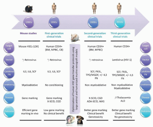
FIGURE 71.6. Timeline for the development of human gene therapy protocols targeting hematopoietic stem cells (HSCs). The main phases in the evolution of HSC gene therapy clinical trials are ordered chronologically. The six principal features defining each phase are outlined. ADA (adenosine deaminase); ALD (adrenoleukodystrophy); BM (bone marrow); CB (cord blood); CGD (chronic granulomatous disease); Flt3L (fms-related tyrosine kinase 3 ligand); FN (fibronectin); HIV (human immunodeficiency virus); IL3 (interleukin 3); IL6 (interleukin 6); LSK (Lin, Sca1+, Kit+); MGDF (megakaryocyte growth and development factor); MPBC (mobilized peripheral blood cells); SCF (stem cell factor); SCID (severe combined immunodeficiency disease); TPO (thrombopoietin); WAS (Wiskott-Aldrich syndrome).


Gaucher Disease

Gaucher disease is an inherited deficiency of the lysosomal enzyme glucocerebrosidase (GC) and the associated accumulation of glucocerebroside in the lysosomes of macrophages results in multisystem damage, including hepatosplenomegaly, gradual replacement of bone marrow (BM), skeletal deterioration, and neuropathology in some cases of Gaucher disease. Correction of enzyme deficiency in macrophages by HSC gene therapy has been considered an attractive therapeutic option for these patients. Mouse BM transplant models have been invaluable for initial evaluation of retrovirus-based gene transfer vectors developed for Gaucher disease.206,214,229, 230, 231 and 232 These studies established the feasibility of efficient transfer of the GC gene to normal mouse HSCs and long-term expression in their progeny after reconstitution, strengthening the rationale for gene therapy as a treatment option for Gaucher disease.


Hemoglobinopathies

Hemoglobinopathies, including β-thalassemia and sickle cell disease, were also among the first diseases selected as targets for genetically based therapeutic approaches. Transfer of the human β-globin gene into murine HSCs proved more challenging due to the requirement to include specific endogenous regulatory elements from the β-globin locus that were obligatory to achieve clinically relevant expression. Early preclinical studies demonstrated that retroviral vectors containing the β-globin gene and its promoter could transduce murine HSCs. However, human β-globin gene expression was either absent or very low, usually varying between 0% and 2% of mouse β-globin RNA levels.205,233, 234, 235, 236, 237 and 238 As outlined below, it is not until new vector designs became available that successful genetic correction of β-thalassemia was first demonstrated in murine preclinical models, laying the foundation for a recent clinical trial of gene therapy for β-thalassemia.103


Chronic Granulomatous Disease

Chronic Granulomatous Disease (CGD) results from mutations in any one of four genes encoding the essential subunits of the phagocytic antimicrobial system NADPH phagocyte oxidase (phox),239 including gp91phox, p22phox, p47phox, and p67phox, rendering individuals born with CGD particularly susceptible to bacterial and fungal micro-organisms. Retrovirus-based gene transfer vectors have been tested in two CGD knockout mouse models.240, 241 and 242 These studies indicated that gene transfer into murine HSCs is feasible and that partial reconstitution of NADPH oxidase activity achieved after retroviral gene transfer can improve host defense if an adequate number of phagocytes exhibit enzyme activity.


Wiskott-Aldrich Syndrome

Wiskott-Aldrich Syndrome (WAS) is an X-linked complex primary immunodeficiency disorder caused by mutations in the gene that encodes the WAS protein (WASP).243 It is characterized by an increased susceptibility to recurrent infections associated with adaptive and innate immune deficiency, thrombocytopenia, eczema, and autoimmunity.244,245 The generation of two Wasp-deficient mice has facilitated preclinical safety and efficacy studies of retroviral vectors for the treatment of WAS. In one study,246 vector-mediated WASP expression was shown to correct the T-cell defect in Wasp mice. In another study, investigators demonstrated rescue of T-cell signaling and amelioration of colitis upon transplantation of transduced WAS HSCs in mice, supporting the development of gene therapy approaches for WAS.247


First-Generation Human Gene Therapy Trials

Because of the absence of in vivo assays that measure the repopulating capacity of human HSCs, gene transfer protocols employed in early clinical trials were adapted from the murine studies described above, and initially tested using human in vitro colony-forming cell (CFC) assays that detect committed progenitor cells, and long-term BM culture assays that detect a cell capable of maintaining production of CFC for at least 5 weeks on a layer of stromal cells. Notwithstanding the fact that progenitors do not have the same biologic properties as stem cells, investigators were initially encouraged when high gene transfer efficiency (up to 100%) was observed in these in vitro progenitor assays using gene transfer vectors and transduction conditions comparable to those employed in the mouse.248,249,250 Demonstration of correction of GC and ADA deficiency after retroviral vector-mediated gene transfer into progenitors from patients with Gaucher disease251 and ADA-SCID,252 respectively, provided even further impetus to regulators and investigators to initiate a first generation of human HSC gene transfer clinical trials, including gene marking studies and gene therapy trials with therapeutic intent (see Table 71.2.


Genetic Marking Trials

The first human clinical gene transfer trials used retroviral vectors carrying nontherapeutic marker genes, and were critical for establishing proof of principle and allaying safety concerns (Table 71.2).7,253 Patients already undergoing autologous transplantation as therapy for an underlying malignancy received genetically marked hematopoietic stem and progenitor cells to determine whether re-infused tumor cells contribute to relapse after autologous transplantation and to establish if transduced HSCs could contribute to long-term hematopoietic reconstitution.254,255,256, 257 and 258 Genetically marked tumor cells were unequivocally demonstrated in several patients at the time of relapse, suggesting that the re-infused marrow had contributed to progression, and that investigation of therapeutic purging strategies was worthwhile. The presence of the marker gene was also followed in nonmalignant hematopoietic cells. Gene-marked cells contributed only 0.1% to 1% of the total BM but detection of the marker gene in T-cells and B-cells for as long as 18 months after transplantation was consistent with low-level transduction of primitive hematopoietic cells with multilineage capacity.

In a similar marking study, investigators determined that autologous BM used for transplantation in patients with chronic myelogenous leukemia following intensive therapy also contained cells that contributed to relapse.259 Other gene marking trials involving patients with multiple myeloma,260,261,262 breast cancer,261,262 follicular lymphoma,263 and AML264 failed to show stable levels of marked cells of more than 0.1% after transplantation with autologous gene-marked grafts, contrasting with the high-level marking observed in preclinical mouse models. Although valuable in the early stages, the perceived level of risk related to retroviral-marking clinical trials was altered dramatically after the first report of leukemia in a patient receiving retroviral gene therapy for X-SCID,265 and given the lack of any possible benefit to the patient related to use of marking vectors, this type of trial has been completely abandoned.


Gene Therapy Trials with Therapeutic Intent

Despite the low efficiencies of gene transfer into long-term repopulating stem cells achieved in early human marking trials, several Phase I/II clinical trials investigating the transfer of potentially therapeutic genes were initiated (Table 71.2). Important information was obtained on both the safety and feasibility of stem cell engraftment without ablation, and although these initial trials were largely disappointing, there were glimmers of hope regarding clinical benefit.


Severe Combined Immunodeficiency Disease

SCID due to ADA deficiency has been a prototype target disease for gene therapy since the initial development of retroviral vectors over a decade ago,266 and children with ADA-deficient SCID were the subjects of the first clinical trial utilizing a vector carrying a therapeutic gene directed at T-lymphocyte targets (see below). HSCs would be theoretically preferable to T-cells as genecorrection targets in this and other immunodeficiency disorders, because of the potential for permanent and complete reconstitution of the T-cell repertoire.

To address this hypothesis directly, two ADA-deficient children received autologous BM and mature lymphocytes transduced with two different retroviral vectors carrying the therapeutic ADA gene and the Neo gene and then repeatedly re-infused without conditioning.267 In the first year after initiation of these infusions, vector-containing T-cells originating from the transduced T-cells were observed, but with time, there was a shift to vector-containing T-cells originating from transduced BM cells. The proportion of gene-corrected clonable T-cells was 2% to 4%, and analysis of T-cell-receptor gene rearrangements indicated a wide repertoire of corrected clones. A surprisingly high number of marrow myeloid colonies resistant to neomycin were also reported, despite lack of conditioning, and an in vivo selective advantage for gene-corrected cells of all lineages was hypothesized.

For genetic disorders diagnosed in utero, the use of CB cells as targets for gene transduction represents an exciting alternative approach268,269 CB may contain greater numbers of primitive reconstituting cells with higher proliferative potential that may prove more susceptible to retroviral transduction. Moreover, early treatment is crucial in diseases that progress to irreversible damage before a child is old enough to allow collection of mobilized peripheral blood or BM cells.270 Three infants diagnosed in utero with ADA deficiency allowed the testing of this concept, and CB was collected at the time of delivery. The cells were CD34-enriched and transduced with an ADA/Neo


retroviral vector and infused in the absence of conditioning.271 Vector sequences were initially detected in circulating myeloid and lymphoid cells at low levels of <0.1% in all three children for >18 months. As expected if corrected lymphocytes have an in vivo advantage, 4 years after the newborns were given infusions of transduced autologous CB CD34+ cells, the frequency of gene-containing T lymphocytes rose to 1% to 10%, whereas the frequencies of other hematopoietic and lymphoid cells containing the gene remained <0.1%. Cessation of polyethylene glycol-conjugated ADA enzyme replacement in one subject led to a decline in immune function, despite the persistence of gene-containing T lymphocytes. Thus, despite the long-term engraftment of transduced HSCs and selective accumulation of gene-containing T lymphocytes, this study indicated that improved gene transfer and expression were needed to attain a therapeutic effect.267


TABLE 71.2 FIRST-, SECOND-, AND THIRD-GENERATION HSC GENE THERAPY CLINICAL TRIALS


First-Generation HSC Gene Therapy Clinical Trials


Transduction


Outcome


Reference


Indication


Gene


CD34+ cells


Cytokines and FN


Duration (MOI)


Vector


Conditioning


Gene Marking (No. Patients)


Genotoxicity (No. Patients)


Brenner MK et al. (1993)255


Gene marking (AML)


NeoR BM No cytokines


No FN


P: 0 h T: 6 h (MOI=10)


γ-RV Ampho env MLV-LTR promoter


Busulfan Cyclophosphamide


Low long-term gene marking


No


Brenner MK et al. (1993)254


Gene marking (AML, neuroblastoma)


NeoR


BM


No cytokines No FN


P: 0 h T: 6 h (MOI=10)


γ-RV Ampho env MLV-LTR promoter


Bu/Cy (AML) Carboplatin/Etoposide (neuroblastoma)


Low long-term gene marking


No


Rill DR et al. (1994)258


Gene marking (Neuroblastoma)


NeoR


BM MNC


No cytokines No FN


P: 0 h T: 6 h (MOI=10)


γ-RV Ampho env MLV-LTR promoter


Carboplatin Etoposide


Low long-term gene marking


No


Deisseroth AB et al. (1994)259


Gene marking (CML)


NeoR


BM


No cytokines No FN


P: 0 h T: 6 h (MOI=10)


γ-RV Ampho env MLV-LTR promoter


TBI Cyclophosphamide Etoposide


Low long-term gene marking


No


Dunbar CE et al. (1995)261


Gene marking (MM and BC)


NeoR


BM MPBC


IL3, SCF +/- IL6 No FN


P: 0 h T: 72 h


γ-RV Ampho env MLV-LTR promoter


Melphalan/TBI (MM) or ICE chemotherapy (BC)


Low long-term gene marking


No


Cornetta K et al. (1996)264


Gene marking (AML and ALL)


NeoR


BM


No cytokines No FN


P: 0 h T: 4 h


γ-RV Ampho env MLV-LTR promoter


Busulfan Cyclophosphamide


Low long-term gene marking


No


Emmons RV et al. (1997)262


Gene marking (MM and BC)


NeoR


BM MPBC


No cytokines +/- Stroma No FN


P: 0 h T: 6-72 h


γ-RV Ampho env MLV-LTR promoter


Melphalan/TBI (MM) or ICE chemotherapy (BC)


Low long-term gene marking


No


Bachier CR et al. (1999)263


Gene marking (NHL)


NeoR


BM MPBC


No cytokines No FN


P: 0 h T: 6 h (MOI=10)


γ-RV Ampho env MLV-LTR promoter


TBI Cyclophosphamide Etoposide


Low long-term gene marking


No


Alici E et al. (2007)260


Gene marking (MM)


NeoR


BM MPBC


IL3, IL6, SCF, bFGF No FN


P: 0 h T: 12 h (MOI=5)


γ-RV Ampho env MLV-LTR promoter


Melphalan


Low long-term gene marking


No


Bordignon C et al. (1995)439


ADA-SCID


ADA


BM


Co-culture No cytokines No FN


P: 0 h T: 72 h


γ-RV Ampho env MLV-LTR promoter


No conditioning


Low long-term gene marking


No


Kohn DB et al. (1995 1998)267,271


ADA-SCID


ADA


CB


IL3, IL6, SCF FN


P: 0 h T: 72 h


γ-RV Ampho env MLV-LTR promoter


No conditioning


Low long-term gene marking


No


Malech HL et al. (1997)278


CGD


P47phox


MPBC


PIXY321, G-CSF No FN


P: 18 h T: 72 h


γ-RV Ampho env MLV-LTR promoter


No conditioning


Low long-term gene marking


No


Hesdorffer C et al. (1998)633


Breast CA Ovarian CA Glioblastoma


MDR1


BM MPBC


IL3, IL6, SCF FN


P: 48 h T: 24 h


γ-RV Ampho env MLV-LTR promoter


High dose chemotherapy


Low long-term gene marking


No


Dunbar CE et al. (1998)277


Gaucher


GC


BM MPBC


IL3, IL6, SCF No FN


P: 0 h T: 72 h


γ-RV Ampho env MLV-LTR promoter


No conditioning


Low long-term gene marking


No


Kohn DB et al. (1999)634


HIV


RRED


BM


IL3, IL6, SCF +/- FN


P: 0 h T: 72 h


γ-RV Ampho env MLV-LTR promoter


No conditioning


Low long-term gene marking


No


Cowan KH et al. (1999)635


Breast CA


MDR1


BM MPBC


IL3, IL6, SCF No FN


P: 0 h T: 72 h


γ-RV Ampho env MLV-LTR promoter


ICE chemotherapy


Low long-term gene marking


No


Liu JM et al. (1999)276


FA


FANCC


BM MPBC


IL3, IL6, SCF No FN


P: 0 h T: 72 h


γ-RV Ampho env MLV-LTR promoter


No conditioning


Low long-term gene marking


No


Second-Generation HSC Gene Therapy Clinical Trials


Transduction


Outcome


Reference


Indication


Gene


CD34+ cells


Cytokines and FN


Duration (MOI)


Vector


Conditioning


Gene marking (No. Patients)


Genotoxicity (No. Patients)


Cavazzana-Calvo M et al. (2000)22, 450, 451


X-SCID


IL2Rγ


BM


SCF, Flt3L, MGDF, IL3 FN


P: 24 h T: 72 h


γ-RV Ampho env MLV-LTR promoter


No conditioning


Correction of X-SCID (9/10)


T-ALL (4/9) LMO2, CCND2, BMI1 gene insertions265, 450, 451


Abonour R et al. (2000)422


Germ cell tumors


MDR1


MPBC


SCF, IL6 or SCF,MGDF, G-CSF FN


P: 48 h T: 48 h


γ-RV Ampho env HaMSV-LTR promoter


Etoposide 2,250 mg/m2 Carboplatin 2,100 mg/m2


Low long-term gene marking (6/11)


No


Aiuti A et al. (2002)23


ADA-SCID


ADA


BM


SCF, Flt3L, TPO, IL3 FN


P: 24 h T: 72 h


γ-RV Ampho env MLV-LTR promoter


Busulfan 4 mg/Kg


Correction of ADA-SCID (2/2)


No


Amado RG et al. (2004)636,637


HIV (Phase I)


Anti-HIV1 ribozyme


MPBC


MGDF, SCF +/- FN


P: 18 h T: 72 h (MOI=5)


γ-RV Ampho env MLV-LTR promoter


No conditioning


Low long-term gene marking (10/10)


No


Gaspar HB et al. (2004)444, 452, 455


X-SCID


IL2Rγ


BM


SCF, Flt3L, TPO, IL3 FN


P: 40 h T: 56 h


γ-RV GALV env MLV-LTR promoter


No conditioning


Correction of X-SCID (10/10)


T-ALL (1/10) LMO2 gene insertion454


Thrasher AJ et al. (2005)456


X-SCID


IL2Rγ


BM


SCF, Flt3L, MGDF, IL3 FN


P: 24 h T: 72 h


γ-RV Ampho env MLV-LTR promoter


No conditioning


Failure to correct X-SCID in older patients (2/2)


No


Ott MG et al. (2006)446


X-CGD


gp91phox


MPBC


SCF, Flt3L, TPO, IL3 FN


P: 36 h T: 72 h


γ-RV Ampho env SFFV-LTR promoter


Busulfan 8 mg/Kg


Correction of X-CGD (2/2)


MDS (2/2) MDS1-EVI1, PRDM16, SETBP1 gene insertions457,458


Gaspar HB et al. (2006)441


ADA-SCID


ADA


BM


SCF, Flt3L, TPO, IL3 FN


P: 40 h T: 56 h


γ-RV GALV env SFFV-LTR promoter


Melphalan 140 mg/m2


Correction of ADA-SCID (1/1)


No


Chinen J et al. (2007)455


X-SCID


IL2Rγ


MPBC


SCF, Flt3L, TPO, IL3 FN


P: 16 h T: 96 h (MOI=1-2)


γ-RV GALV env MLV-LTR promoter


No conditioning


Clinical improvement (3/3)


No


Mitsuyasu RT et al. (2009)637,638


HIV (Phase II)


Anti-HIV1 ribozyme


MPBC


MGDF, SCF FN


P: 36 h T: 48 h (MOI=5)


γ-RV Ampho env MLV-LTR promoter


No conditioning


Lower HIV-1 viral load (10/10)


No


Aiuti A et al. (2009)440


ADA-SCID


ADA


BM


SCF, Flt3L, TPO, IL3 FN


P: 24 h T: 72 h


γ-RV Ampho env MLV-LTR promoter


Busulfan 4 mg/Kg


Correction of ADA-SCID (9/10)


No


Boztug K et al. (2010)447


WAS


WAS


MPBC


SCF, Flt3L, TPO, IL3 No FN


P: 48 h T: 48 h (MOI=5)


γ-RV GALV env MPSV promoter


Busulfan 8 mg/Kg


Correction of WAS (9/10)


T-ALL (4/9) LMO2 +/- other gene insertions


Kang EM et al. (2010)444


X-CGD


gp91phox


MPBC


SCF, Flt3L, TPO, IL3 FN


P: 18 h T: 96 h (MOI=2)


γ-RV Ampho env MLV-LTR promoter


Busulfan 10 mg/Kg


Long-term clinical benefits (2/3)


No


Kang HJ et al. (2011)445


X-CGD


gp91phox


MPBC


SCF, Flt3L, TPO, IL3 FN


P: 40 h T: 40 h (MOI=1-2)


γ-RV Ampho env MLV-LTR promoter


Fludarabine 120 mg/m2 + Busulfan 1.6 mg/Kg


Short-term clinical benefit (2/2)


No


Gaspar et al. (2011)443


ADA-SCID


ADA


BM


SCF, Flt3L, TPO, IL3 FN


P: 40 h T: 56 h


γ-RV GALV env SFFV-LTR promoter


Melphalan 140 mg/Kg or Busulfan 4 mg/Kg


Correction ADA-SCID (4/6)


No


Third-Generation HSC Gene Therapy Clinical Trials


Transduction


Outcome


Reference


Indication


Gene


CD34+ cells


Cytokines and FN


Duration (MOI)


Vector


Conditioning


Gene marking


Genotoxicity


Cartier N et al. (2009)102


X-ALD


ABCD1


MPBC


SCF, Flt3L, MGDF, IL3, FN


P: 19 h T: 17 h (MOI=25)


SIN-HIV-1 VSV-G env MND promoter


Busulfan 16 mg/Kg + Cyclophos 200 mg/Kg


Correction of X-ALD (2/2)


No


Cavazzana-Calvo et al. (2010)103


β-thalassemia


β-globin


BM


SCF, Flt3L, TPO, IL3 FN


P: 34 h T: 18 h


SIN-HIV-1 VSV-G env β-globin promoter β-LCR


Busulfex 12.8 mg/Kg


Correction of β-thalassemia (1/1)


Clonal dominance (1/1) HMGA2 insertion103


ADA, adenosine deaminase; ALD, adrenoleukodystrophy; ALL, acute lymphoblastic leukemia; AML, acute myelogenous leukemia; Ampho, amphotropic; BC, breast cancer; bFGF, basic fibroblast growth factor; BM, bone marrow; BMI1, B lymphoma MLV insertion region 1 homolog; Bu, busulfan; CA, cancer; CB, cord blood; CCND2, cyclin D2; CGD, chronic granulomatous disease; CML, chronic myelogenous leukemia; Cy, cyclophosphamide; Env, envelope; EVI1, ecotropic virus integration site 1 protein homolog; FA, Fanconi anemia; FANCC, Fanconi anemia complementation group C; Flt3L, fms-related tyrosine kinase 3 ligand; FN, fibronectin; GALV, gibbon ape leukemia virus; GC, glucocerebrosidase; G-CSF, granulocyte-colony stimulating factor; HaMSV, Harvey murine sarcoma virus; HIV, human immunodeficiency virus; HMGA2, high mobility group AT-hook 2; ICE, ifosfamide, carboplatin, etoposide; IL2Rγ, interleukin 2 receptor γ chain; ABCD1, ATP binding cassette subfamily D, member 1; IL3, interleukin 3; IL6, interleukin 6; LCR, locus control region; LMO2, LIM domain only 2; LTR, long terminal repeat; MDR1, multidrug resistance gene 1; MDS, myelodysplastic syndrome; MGDF, megakaryocyte growth and development factor; MLV, murine leukemia virus; MM, multiple myeloma; MNC, mononuclear cells; MND, myeloproliferative sarcoma virus enhancer, negative control region deleted, dl587rev primer binding site substituted; MOI, multiplicity of infection; MPBC, mobilized peripheral blood cells; MPSV, myeloproliferative sarcoma virus; NeoR, neomycin resistance; NHL, non-Hodgkin’s lymphoma; P, prestimulation; Phox, phagocyte oxidase; PRDM16, PR domain containing 16; RRED, rev-responsive element decoy; RV, retroviruses; SCF, stem cell factor; SCID severe combined immunodeficiency disease; SETBP1, SET binding protein 1; SFFV, spleen focus forming virus; SIN, self-inactivating; T, transduction; T-ALL, T-lineage acute lymphoblastic leukemia; TBI, total body irradiation; TPO, thrombopoietin; VSV-G, vesicular stomatitis virus-G protein; WAS, Wiskott-Aldrich syndrome.


Fanconi Anemia

Fanconi Anemia (FA) is another genetic disorder of the hematopoietic system that appears to be an excellent candidate for gene therapy. This congenital syndrome is characterized by BM failure, physical anomalies, and an increased susceptibility to marrow failure and leukemias. Cells from these patients are abnormally sensitive to chemically induced DNA cross-linking. Many of the complementation groups have been cloned and sequenced, making a genetic approach feasible for this disorder.272 Phenotypic correction of this abnormality in cells from one patient group was successful after transduction with viral vectors carrying the Fanconi anemia complementation group C gene (FACC).273,274 A possible in vivo survival advantage for gene-corrected primitive cells and their progeny has made FA an attractive candidate disease for stem cell gene therapy. Even a very low efficiency of transduction might result in gradual in vivo expansion of corrected progenitor and stem cell populations. A clinical trial testing this hypothesis using granulocyte-colony stimulating factor (G-CSF)-mobilized peripheral-blood CD34+ cells as targets yielded disappointing results, in part because of the very poor mobilization in these subjects,275 yet engraftment with corrected progenitor cells was demonstrated.276

Only gold members can continue reading. Log In or Register to continue

Stay updated, free articles. Join our Telegram channel

Oct 21, 2016 | Posted by in HEMATOLOGY | Comments Off on Gene Therapy for Hematologic Disorders

Full access? Get Clinical Tree

Get Clinical Tree app for offline access