Origin and Development of Blood Cells


Origin and Development of Blood Cells


Andrew Chow

Paul S. Frenette


BLOOD CELLS

The blood contains several different types of cells. Each of these cell types is quite distinct in appearance, and each has a specific biologic function. Erythrocytes are anucleate, biconcave discoid cells filled with hemoglobin, the major protein that binds oxygen. Erythrocytes transport the respiratory gases oxygen and carbon dioxide. Granulocytes and monocytes are cells that can exit from blood vessels and migrate into many tissues. These two cell types play key roles in inflammation and phagocytosis. Platelets are very small, anucleate cells that contain molecules required for hemostasis. In addition, platelets provide hemostasis through their abilities to adhere, aggregate, and provide a surface for coagulation reactions. Lymphocytes mediate highly specific immunity against microorganisms and other sources of foreign macromolecules. B-lymphocytes confer immunity through the production of specific, soluble antibodies, whereas T lymphocytes direct a large variety of immune functions, including killing cells that bear foreign molecules on their surface membranes. Despite these extreme structural and functional differences among the cells of the blood, strong evidence exists that the vast majority of blood cells are the progeny of a single type of cell: the hematopoietic stem cell (HSC). The processes involved in the production of all of the various cells of the blood from the HSCs are collectively called hematopoiesis. Hematopoiesis includes HSC self-renewal, HSC commitment to specific lineages, and maturation of lineage-committed progenitors into functional blood cells. Self-renewal may occur by symmetric HSC division, such as expansion of the HSC pool during fetal life or post-HSC transplantation. Other possible fates of HSC divisions include apoptosis or mobilization to the peripheral circulation following stress such as growth factor stimulation or depletion of marrow cells by irradiation or chemotherapy. During normal steady-state conditions, HSCs reside mainly in the marrow cavity, but under certain stress conditions, HSCs can migrate and colonize other organs such as the liver and spleen in a process termed extramedullary hematopoiesis.

Hematopoiesis begins early during embryogenesis and undergoes many changes during fetal and neonatal development. Unlike some organ systems that form in early life and are not continually replaced, turnover and replenishment of the hematopoietic system continue throughout life. Cells of the blood have finite life spans, which vary depending on the cell type. In humans, granulocytes and platelets have life spans of only a few days, whereas some lymphocytes can exist for many months. Cells are replaced as the older cells are removed and the newly formed, mature cells are added. The numbers of the various cell types in the blood are normally kept in relatively constant ranges. In particular, variations in the erythrocyte number are normally minimal, and values 30% above or below the norm for the population have significant health effects. Although the numbers of other blood cell types are not as constant as the number of erythrocytes, the production of other blood cells is also highly regulated. The regulation of hematopoiesis is complex. Some regulatory factors influence overall hematopoiesis by affecting very early progenitor cells: the HSCs and/or their progeny that have not undergone commitment to a single cell lineage. Also, specific regulatory growth factors play key roles in fostering the production of cells of each lineage. Lineage-specific regulation is necessary because of the widely varying life spans and functions of the different mature blood cell types.

This chapter presents an overview of hematopoiesis. Many conclusions presented here are based on experiments carried out in murine systems. All cell lineages that compose blood will be discussed. Some cell types such as dendritic cells and mast cells are derived from the HSCs but are found mostly in tissues rather than blood, where the final steps of differentiation occur. Figure 5.1 is an illustration of the cell types that constitute the hematopoietic spectrum.


ORIGIN OF HEMATOPOIESIS


Sites of Hematopoiesis

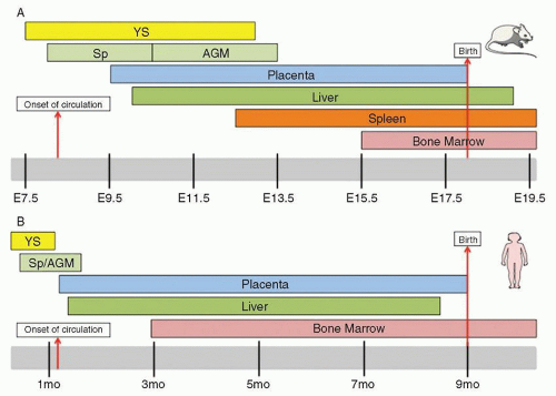
During prenatal development, the sites of hematopoiesis change several times in the mouse1,2 and 3 and human3,4 (Fig. 5.2). Since better characterized in mice (Fig. 5.2A) than humans (Fig. 5.2B), the discussion below will focus on murine developmental hematopoiesis. In humans and other vertebrates, the first hematopoietic cells arise during late gastrulation in the extraembryonic yolk sac (YS) in structures known as blood islands. This initial hematopoiesis is termed primitive hematopoiesis and serves a supportive role to rapidly produce erythroid cells, platelets, and macrophages prior to the formation of the circulatory system. Primitive hematopoiesis is transient, occurring on embryonic
days 7.25 (E7.25) through 13 (E13) in mice and day 19 through week 8 in humans. Primitive erythrocytes, which are the first embryonic hematopoietic cells, are large nucleated cells morphologically resembling erythrocytes of phylogenetically lower primitive vertebrate groups, such as birds, amphibians, and fish. These primitive erythrocytes have reduced erythropoietin (EPO) requirements during their development compared to definitive erythroid cells5 that develop later. Also unlike their definitive counterparts, primitive erythrocytes typically circulate as nucleated cells before enucleating, and additionally express ζ, βH1, and εy globin genes.6,7 These cells and primitive platelets8 derive from a primitive bipotent megakaryocyte erythroid progenitor found in the yolk sac in mice (E7.25) and humans.9,10 Along with maternally derived macrophages (MΦ) that exist, but are declining in numbers, in the yolk sac at E8, two other MΦ progenitors exist in the yolk sac: one with strictly MΦ potential
and one with bipotential for MΦ and erythrocytes.11 Importantly, since circulation does not commence until E8.25, this indicates in situ MΦ development in the yolk sac. Thus, primitive hematopoiesis in the yolk sac provides the developing embryo with three crucial hematopoietic cell types prior to contribution from multipotent stem cells deriving from definitive hematopoiesis (see below).


FIGURE 5.1. Cells of the blood and lymphoid organs and their precursors in the bone marrow. CMP, common myeloid progenitors; DC, dendritic cells; EB, erythroblast; GMP, granulocyte macrophage progenitor; HSC, hematopoietic stem cell; MDP, macrophage dendritic cell progenitor; MEP, megakaryocyte erythrocyte progenitor; MK, megakaryocyte; Mø, macrophage; mono, monocyte; MPP, multipotent progenitors; RBC, red blood cell; Retic, reticulocyte.


FIGURE 5.2. Sites of hematopoiesis. A: Sites of mouse hematopoietic development. AGM, aorta-gonad-mesonephros; Sp, splanchnopleura; YS, yolk sac. B: Sites of human hematopoietic development.

Since the first hematopoietic cells arise in the extraembryonic yolk sac, it was widely believed in the 1970s that the first HSCs developed in the yolk sac. However, experiments in avian chimeras demonstrated for the first time that although the YS had early contribution, the hematopoietic cells present in the stages closer to birth were exclusively derived from the intraembryonic compartment.12 Similar avian chimeric experiments subsequently demonstrated that the intraembryonic compartment, rather than the YS, was the exclusive source of B and T cells in the adults.13 Godin and colleagues subsequently demonstrated in mice that the aortic region of E9 embryos, but not YS precursors, were capable of contributing to B cells in irradiated adult recipients.14 In the same journal issue, Medvinsky, Dzierzak, and colleagues demonstrated that the E10.5 AGM (aorta-gonad-mesonephros) region had substantially higher and earlier onset of CFU-S activity, an early coarse assay for multipotency, compared to YS cells.15 Soon afterward, Dzierzak’s group demonstrated the ability of E10.5 AGM precursors to provide long-term multilineage reconstitution activity (LTR) in lethally irradiated adult mice.16 Together, these seminal publications affirmed the intraembryonic contribution to adult mammalian hematopoiesis.

Since the AGM region above was harvested after the establishment of circulation (E8.25), migration of HSCs from a separate undescribed site of origin could not be excluded. To investigate whether HSC development occurred de novo in the AGM, the E8 splanchnopleura (Sp, the future site of the AGM) and the concomitant yolk sac, neither of which have LTR activity, were cultured. While the cultured Sp and YS both produced hematopoietic cells, confirming two independent waves of hematopoietic generation, the YS progenitors were unable to produce lymphoid progeny or have LTR activity.17,18 Further dissection of the AGM determined that most of the HSC activity is found in hematopoietic intra-aortic clusters found on the ventral wall of the dorsal aorta.2 HSC activity is also found in the proximal vitelline and umbilical arteries, although these sites have been less characterized. Two reports from the Dzierzak and Mikkola groups established that the placenta represents a previously overlooked major site of hematopoiesis in which HSC emergence parallels that of HSC appearance at E10.5 in the AGM.19,20 In fact, when LTR HSCs are enumerated, there are 25-fold more LTR HSCs in the placenta than in the AGM.19 Since the placenta is directly upstream of the fetal liver circulation and since the dramatic expansion of HSC in the FL mirrors that of the placenta, it has been proposed that the placenta is at least a major contributor of LTR HSCs. It has also been proposed that the placenta is a site of de novo HSC emergence independent from the AGM. Indeed, explant and stromal co-culture experiments of mesodermal tissue of the placenta prior to the establishment of circulation demonstrated erythroid and myeloid potential.21,22 The concept of a de novo generated HSC was bolstered by in vitro culture of E8-9 placenta from Ncx1-/- animals, which lack a heartbeat and die by E10.5. Without circulatory contribution, the midgestation site had definitive hematopoietic cells with myelo-erythroid and lymphoid potential.23 Although LTR HSC cannot be isolated from the placenta of Ncx1-/- animals because of developmental retardation and death by E10.5, these experiments show that definitive hematopoiesis emerges in this organ de novo. Whether the AGM, uterine and vitelline arteries, placenta, or a combination of the above are the genuine origin of HSCs, this LTR activity around E10.5 represents the start of definitive hematopoiesis.

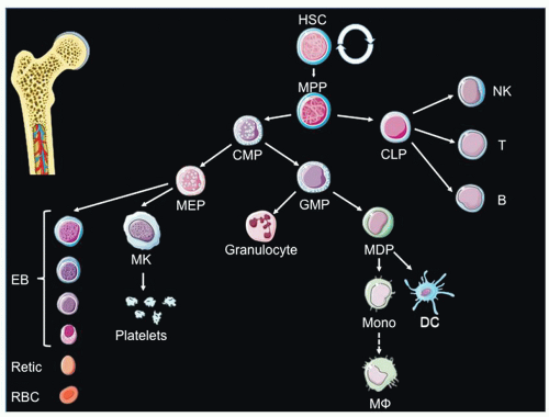
Once definitive hematopoiesis begins, lymphocytes, monocytes, granulocytes, and platelets are formed as well as definitive erythrocytes. At E10, hematopoietic cells (both primitive and definitive) colonize the fetal liver (FL). Dramatic expansion of HSCs occurs at this site (daily doubling in absolute numbers of HSC from E12.5 to E14.5).24 Eventually, LTR HSCs migrate from the fetal liver to the bone marrow via the circulation, and the bone marrow becomes the primary site of hematopoiesis, with a very small reserve of stem cells remaining in the liver. In the late stages of mammalian fetal development, the bone marrow becomes the main site of hematopoiesis. In humans, the bone marrow is the exclusive site of postnatal hematopoiesis under normal circumstances, whereas in the mouse, the spleen is also a hematopoietic organ throughout life.


Cellular Origin of Hematopoiesis

The cellular intermediates through which mesodermal tissue gives rise to hematopoietic tissue in embryonic development is an area of intense investigation. One candidate cellular ancestor is either (a) a mesoderm-derived bipotent hemangioblast capable of giving rise to either vessels and blood cells or (b) a specialized vascular cell type, called hemogenic endothelium, that serves as a precursor for blood cells. A non-mutually exclusive origin points to HSC derivation from mesenchymal tissue below the endothelial layer. Cytologic analyses of the AGM provide evidence for both endothelial-derived and sub-endothelial-derived HSCs through identification of HIACs and subaortic patches (SAPs), respectively.2 The strict temporal overlap in the appearance at E10.5 and disappearance at E12.5 of HIACs and SAPs suggests that HSCs derived in SAPs can potentially transendothelially migrate to form HIACs prior to release into the bloodstream.2 Keller and colleagues definitively showed that a bipotential hemangioblast could be found in the posterior region of the primitive streak in vivo.25 However, until recently, the existence of a bona fide endothelial intermediate had been under debate. Supportive of an endothelial origin of HSCs is the presence of numerous vessel markers on AGM HSCs, including CD31, VE-Cadherin, and Tie-2.2 Furthermore, AGM HSCs and endothelial cells in the ventral wall of the E10-E11 dorsal aorta both express Ly6A (Sca-1), c-Kit, CD34, Runx1, SCL, and GATA-2.1 Fate mapping studies elegantly showed that VE-cadherin expressing endothelium contributes to AGM and adult HSCs, while lineage tracing of subendothelial mesenchyme with Myocardin-Cre animals did not result in labeling of HSCs.26 Subsequently, novel imaging studies of embryonic stem cell-derived mesodermal cells demonstrated a hemogenic endothelial intermediate in the formation of blood cells. 27,28 Morever, when Runx1, an essential gene in definitive hematopoiesis, was specifically deleted in VE-cadherin-expressing cells (endothelial and hematopoietic cells), but not Vav1-expressing cells (only hematopoietic cells), there was a severe disruption in hematopoietic development that was associated with 65% fetal lethality.29 Finally, Nancy Speck’s group recently showed that expression of core binding factor beta (CBFβ) in Ly6a-expressing hemogenic endothelium was sufficient for HSC formation.30 Together, these observations have supported the concept of blood cell development commencing with mesodermal cells that pass through hemangioblastic and hemogenic endothelial intermediates.


Common Critical Genes in Independent Origins of Hematopoiesis

Gene knockout experiments have provided significant insight into the critical regulators of embryonic hematopoiesis. In both primitive and definitive hematopoiesis, Bmp4, Flk1, Tal1/Scl, Lmo2, Gata2, and Runx1 are all critical for HSC generation.2 Bmp4 (bone morphogenetic protein 4) is a critical signaling molecule
to specify the dorsal-ventral axis in development. Although the posterior portion of the epiblast in development is fated to give rise to hematopoietic activity, the neurally fated anterior fragment can retain the ability to produce hematopoietic cells by addition of Bmp4.31 Bmp4 is crucial for hematopoietic development as Bmp4-deficient embryos mostly die around the gastrulation stage, and those that do survive have less yolk sac mesoderm and less lateral plate mesoderm (from which the AGM will develop).32,33 In definitive hematopoiesis, Bmp-4 is expressed by endothelial cells in the ventral portion of the developing dorsal aorta and the subjacent mesoderm.2 Using murine ES cells, it was recently shown that Bmp4 is necessary for mesodermal precursor expression of the receptor tyrosine kinase Flk-1 and the bHLH transcription factor Tal-1/SCL.34

The initiation of yolk sac hematopoiesis is dependent on the mesoderm and endoderm layers acting in concert, as soluble factors from endoderm substantially bolster the production of endothelial and hematopoietic cells by murine YS mesoderm explants.35 One of the candidate soluble factor interactions is VEGF derived from endoderm and its receptor Flk1 on the mesoderm.36,37 Indeed, Flk-1-deficient embryos do not develop vessels or YS blood islands and die in utero between E8.5 and E9.5.38 To overcome this early developmental mortality, Shalaby and colleagues performed complementation studies with chimeras of Flk-1+/- and Flk1-/- ES cells and demonstrated convincingly that Flk-1 is also required for the generation of definitive endothelial and hematopoietic cells.39 It was later shown that Flk-1 signaling appears to not be required intrinsically for endothelial and hematopoietic formation, as Flk1-/- ES cells are able to give rise to endothelial and hematopoietic lineages in vitro40,41; instead, Flk-1 is likely required for the migration of mesoderm cells from the posterior primitive streak to the yolk sac.39 In concordance with the importance of the VEGF-Flk-1 signaling axis, VEGF derived from the visceral endoderm (but interestingly, not mesoderm) is sufficient for endothelial and hematopoietic differentiation.42

The transcription factor Tal-1/Scl43,44 and 45 and the transcriptional regulator Lmo246 are both expressed in the yolk sac mesoderm prior to the onset of primitive hematopoiesis and then subsequently expressed in both endothelial and hematopoietic cells. Gene knockout of both Tal-1/Scl47,48 and Lmo249 results in decreased endothelial cells and abrogates YS blood cell production. These genes are also critical for definitive hematopoiesis, as demonstrated by complementation studies with ES cells chimeras.50,51 and 52 Gata-2-deficient animals have severely impaired primitive hematopoiesis and die at E10.5.53 Gata2 haploinsufficient embryos have normal yolk sac hematopoiesis,54 but have a reduction in AGM HSCs, which is consistent with its expression on aortic endothelium55 and its proposed role in the expansion of hemogenic endothelial progenitors.55 Runx1 has also been demonstrated to be crucial in definitive hematopoiesis, as Runx1 invalidation abrogates definitive myeloid, lymphoid, and HSC accumulation in the YS, AGM, and fetal liver.56,57 and 58 Runx1 is thought to be crucial cell autonomously, as complementation studies fail to demonstrate hematopoietic contribution by Runx1-null ES cells.57 While Runx1 was initially thought to be dispensable in murine primitive erythropoiesis, recent studies have recently shown that the morphology and gene expression of erythrocytes are aberrant in Runx1-deficient animals.59


HEMATOPOIETIC STEM CELLS


Age of Morphologists

Fascinating accounts of the history of experimental efforts in hematopoiesis are presented in Wintrobe’s Blood, Pure and Eloquent.60,61 One milestone in understanding the origins and development of blood cells was the recognition by Neumann and Bizzozero in the mid-nineteenth century that the bone marrow is a site of red blood cell production throughout postnatal life. Another major advance made in the late nineteenth century by Paul Ehrlich, Artur Pappenheim, and others was the application of synthetic dyes and various staining/fixing techniques that led to precise morphologic characterization and classification of blood and marrow cells. A third milestone was the development of the idea of a multipotent stem (ancestral) cell that gives rise to all of the mature blood cell types through extensive proliferation and differentiation. By the use of refined staining methods, Pappenheim observed various transitional cells and organized them into a relational scheme—a tree whose various branches when traced backward converged to a mononuclear cell that had none of the distinct features of the end-stage blood cells or the transitional stages. He proposed the notion that this cell was so morphologically primitive that it could be the common ancestor of all blood cells. Although most morphologists between 1900 and 1940 accepted the idea of ancestral cells in a hematopoietic series leading to progressively more mature types, there was much debate about how many ancestral cell types existed. Many workers believed that lymphocytes had a separate origin from myeloblasts and thus that there were dual or perhaps plural ancestral cells. Reviews of the conflicting concepts of the origin of hematopoietic cells as of the late 1930s are presented in detail in Handbook of Hematology.62


Advent of Hematopoietic Progenitor Transplantation

In the late 1940s and the 1950s, several new approaches were developed to study hematopoiesis. Among them were radiation exposure followed by grafting of hematopoietic tissue, development of chromosome cytogenetics, and use of radioactive materials. Lorenz et al.63 showed that mice and guinea pigs can be protected against otherwise lethal whole-body irradiation by injections of bone marrow from other animals of their respective species. Ford et al.64 used bone marrow from donor mice that had a morphologically identifiable chromosomal marker to show that hematopoiesis in the irradiated recipient mice was reconstituted by cells from the donor marrow—that is, the protected animals were chimeras with respect to their hematopoietic tissues. These experiments did not settle the question about how many types of ancestral cells there were, but experiments generating radiation chimeras have since been used with great power to study the nature of stem cells and their progeny.

Till and McCulloch65 used radiation/grafting experiments to prove directly the existence of an ancestral cell with multilineage potential. In spleens of mice at 1 week after transplantation, they found growth of macroscopic colonies containing cells of multiple hematopoietic lineages. These colonies were the progeny of individual transplanted cells that were called colony-forming units spleen (CFU-S). Because the cells in these spleen colonies could, in turn, be injected into secondary, irradiated mice and give rise to spleen colonies, the CFU-S apparently replicated themselves within the colonies. When the observation time for CFU-S assays was extended from 1 to 2 weeks after transplantation, a series of evanescent colonies was found, and those appearing on later days had greater self-replication and multilineage differentiation capacities.66,67 Early studies could not demonstrate lymphoid cells in spleen colonies,68,69 but more recent studies indicate that CFU-S colonies contain lymphoid progenitors as well as myeloid progenitors.70 Several studies showed that cells with the capacity for long-term hematopoietic reconstitution of irradiated mice can be separated from most CFU-S by size and density.71 Thus, many CFU-S, although multipotent, do not have long-term repopulating capacity.


Definitive Evidence for a Multipotent Stem Cell

Animal reconstitution experiments with hematopoietic cells that were individually genetically marked have verified the existence of HSCs and demonstrated their capacity for extensive selfrenewal.72,73 In these marking experiments, hematopoietic cells were infected in vitro with a recombinant retrovirus that was able to integrate its DNA (provirus) into a cell but could not replicate and spread to other cells. The one-time, random integration of the provirus into the DNA of an individual cell provides a specific marker for the progeny of that cell that develop in an animal after transplantation. Random integration assures that each provirus has unique flanking sequences of DNA and thus has a high probability of yielding a DNA fragment of a distinguishable size after cutting with a restriction enzyme that does not cut the provirus. Several months after transplantation of the genetically marked cells and establishment of hematopoiesis, it is typically observed that all types of cells in the blood and lymphoid organs contain progeny of an individually marked cell, proving that it was multipotent. Often, these clones of marked cells continue to contribute to all of the hematopoietic lineages in the animal for an extended period. Also, when these primary recipient animals are subsequently used as donors for secondary recipient animals, frequently the same clones of HSCs are apparent in these secondary recipients. This persistence can even be demonstrated in tertiary recipients.74,75 Thus, clearly many HSCs reproduce themselves (self-renew) over a long period. Long-term reconstitution of the myeloid and lymphoid compartments can be achieved by transplantation of a single murine HSC,76,77,78 indicating that a single HSC is the smallest repopulating unit. Dick and colleagues recently demonstrated that a single cell transplant of human CD34+ CD38 CD45RA Thy1+ Rholo CD49f+ cells into immunocompromised mice was able to provide multilineage reconstitution,79 indicating that the HSC is also the smallest repopulating unit in humans.

It has been noted that not all HSC clones are long lived; some produce progeny for varying periods and then apparently become extinct. Finally, marked clones have been observed to begin contributing to hematopoiesis after some period of post-transplantation latency, indicating that dormancy is possible. Thus, these studies have demonstrated that, after transplantation, some HSCs contribute continuously to hematopoiesis for a long time—in mice, apparently for the whole lifetime of the animal. Other HSCs contribute and then become extinct, and finally, some may remain dormant for some period and then contribute. Additional transplantation studies of marked HSCs in mice80 have suggested that polyclonal hematopoiesis is more common and that long-term contribution by individual stem cells is more rare than the earlier studies indicated.74,75 Recent novel technologies combining viral barcoding and high throughput sequencing of HSC confirmed this polyclonal contribution of HSCs.81 Studies using retroviral insertion site analyses for larger animals, particularly non-human primates, have provided some evidence of polyclonal hematopoiesis.82,83,84 To what extent these possible behaviors are manifest in normal, nontransplanted mice or larger animals is not clear.


Enrichment of Hematopoietic Stem Cells

The identification of relatively immature HSCs from more committed progenitor cells on the basis of various physical properties, immunophenotypic markers, and functional attributes has greatly advanced the field of hematopoiesis.85 HSC markers that are expressed from fetal stages through adult life include CD34, CD31 (PECAM1), and Kit, but these markers can also be identified in endothelial cells.86 In humans, CD34+, CD38, CD90 (Thy-1)+, CD45RA cells that are negative for lineage markers (CD2, CD3, CD4, CD7, CD8, CD10, CD11b, CD14, CD19, CD20, CD56, CD235a) are considered highly enriched for in vivo repopulating HSCs.87,88 In mice, there is no single consensus panel of HSC markers to identify, enumerate, and sort HSCs. Many markers derive from work by Weissman’s lab, which proposes the combination of Sca-1 (Ly-6A/E)+, Kit+, Flk2, CD90 (Thy-1)+ and negative for lineage markers (CD3, B220, Mac-1, Gr-1 and Ter119) as a highly purified population enriched for in vivo repopulating HSCs89,90,91,92 (Table 5.1). Nakauchi’s group subsequently showed the enrichment of long-term repopulating activity in the CD34lo fraction.76 In 2005, Morrison’s group developed a novel marker set to identify highly enriched HSCs. They demonstrated that signaling lymphocyte and activation molecule (SLAM) markers were found to be differentially expressed on BM Lineage Sca-1+ c-kit+ populations such that CD150+ CD244 CD48 CD41 was the population enriched for murine HSC in vivo repopulation capacity.93 Since the aforementioned phenotypic descriptions involve the use of multiple markers, which are moreover not exclusively expressed on HSC, Mulligan’s group proposed the use of endothelial protein C receptor (EPCR, CD201) as a novel “explicit” marker of HSC, since HSCs express high levels of EPCR, while downstream progenitors express only intermediate levels.94 Importantly, prospective isolation with only EPCR enriched for hematopoietic reconstitution activity. The most immature HSCs with in vivo hematopoietic repopulation potential are detectable within the CD150+CD244CD48 population. Other studies have pointed to CD105 (endoglin) as an enriching marker for HSCs.95,96 In addition to immunostaining, HSCs can also be identified by the ability of HSCs to efficiently efflux dyes. The most common methods utilize the dye Hoescht 33342, which when excited at two wavelengths yields a characteristic “side population” on flow cytometry97 due to dye efflux.


TABLE 5.1 FLOW CYTOMETRIC DEFINITIONS OF HSCs, MPPs, AND SINGLE LINEAGE PROGENITORS


Population


Phenotype


Reference


Hematopoietic stem Cell (HSC)


Lin Sca-1+ Kit+ Flk2 CD34


CD90 (Thy-1)+


76, 89


91, 92


CD150+ CD244 CD48


93


EPCR+


94


Hoescht 33342 Side Population+


97


Hoescht 33342 Side Population+ CD105+


95


Lin Sca-1+, Kit+ CD105+ CD150+


96


Multipotent progenitors (MPP)


Lin Sca-1+ kit+ Thy1- Flk2+


Lin Sca-1+ kit+ CD150 CD105


90


96


Common lymphoid progenitor (CLP)


Lin Sca-1lo Kitlo Thy1lo IL-7R+


183


Common myeloid progenitor (CMP)


Lin Sca-1 Kit+ FcYRint CD34+


184


Granulocyte macrophage progenitor (GMP)


Lin Sca-1 Kit+ FcYRhi CD34+


184


Megakaryocyte erythrocyte progenitor (MEP)


Lin Sca-1 Kit+ FcYR CD34


184


Macrophage dendritic cell progenitor (MDP)


Lin Sca-1 Kithi Flk2+ CX3CR1+ CD115+


185


Lin, Lineage negative (Gr1 CD11b CD3 B220 Ter119); Sca-1, stem cell antigen 1; Flk2, fms-like kinase 2; CD, cluster of designation.


With the advances in technology, procedures have been developed to enrich greatly the proportion of HSCs in isolated cell populations from mouse, human, and other sources. In mice, with immunophenotyping alone, 50% to 96% of prospectively isolated HSCs have long-term repopulating activity.98 Isolation of candidate HSCs based on phenotypic markers expressed on the cell surface was first tested in congenic mouse transplantation models and subsequently purified human HSCs were successfully transplanted in a xenogeneic immunodeficient mouse model.85 The successes in mouse models have led to three human phase I clinical trials that successfully demonstrated sustained hematopoiesis when HSCs purified by immunophenotyping were transplanted into irradiated patients.99,100 and 101


Hematopoietic Stem Cell Assays

While immunophenotyping is the only feasible option for human HSC therapeutics because of a narrow window for successful transplantation, reliance on surface markers has the major limitation that different clinical scenarios may modulate the expression of utilized markers. For example, the content of long-term repopulating HSCs within the same immunophenotyped Lineage Sca-1+ c-kit+ (LSK) Thy1lo fraction can change in aged mice, previously transplanted mice, and mobilized mice.102 Reliability of CD34 in mice of various age has also been questioned.103 Use of the CD150+ CD48 gating scheme, rather than Thy1, with LSK gating retains the fidelity in frequency of long-term repopulating HSCs in aged, transplanted, and mobilized mice.102 Nonetheless, it still is unknown if the SLAM markers retain fidelity in mutant mice. Moreover, other surface markers can also be modulated by environmental cues. This is illustrated by the Sca-1 upregulation that occurs in inflammatory settings likely secondary to type I interferon exposure,104,105 which can erroneously lead to conclusions about HSCs based on a population of committed progenitors that artifactually acquired the Sca-1 antigen. Furthermore, not all mouse strains express the prospective HSC antigens, such as Thy1.1 or Sca-1.106,107 Thus, conclusions drawn about HSCs in mouse or man need to be verified by functional assays in order to demonstrate (a) multipotency and (b) long-term repopulation. There are numerous assays with differing levels of stringency, limitations, and appropriateness to the question being addressed, as recently reviewed.98


Long-term In Vitro Assays

In humans and mice, two types of progenitor cells called long-term culture initiating cells (LTC-ICs) and cobblestone area-forming cells (CAFC) can be detected using long-term cell culture assays. Since most committed progenitors have differentiated by 3 weeks in culture, they can be quantified by counting colonies at this time. The various progenitors quantified by in vitro assays are shown in Table 5.2. By 5 weeks or more of long-term culture, more immature progenitors that are dormant during the initial weeks but which possess extensive proliferating capacity continue to proliferate. Counting colonies at these later time points allows quantification of the number of more immature progenitors at the time of culture initiation. One type of long-term culture assay detects early-stage hematopoietic progenitors that are capable of initiating long-term hematopoiesis in culture after seeding them onto irradiated stromal cell monolayers (human108,109; mouse110,111). These LTC-ICs108 sustain production of multilineage progenitors for 4 to 6 weeks. In some instances, these cultures have been extended for more than 10 to 12 weeks.112,113 This continued production of hematopoietic progenitors of multiple lineages in individual cultures is measured after several weeks by harvesting the cultured cells and doing secondary assays for various types of lineage-committed progenitors. Long-term cultures require a supporting stromal monolayer that is commonly generated from bone marrow-derived mesenchymal or fibroblast cells. The stromal layer supports the proliferation and differentiation of seeded hematopoietic progenitor cells, but at later times, it sloughs from the culture dish and fails to sustain continuation of the culture. In CAFC assays, islands or colonies of hematopoietic cells can be recognized morphologically in situ.110 These cobblestone colonies integrate within the supporting stromal layer, forming clusters of flattened, optically dense, morphologically homogenous-appearing cells tightly adherent with the stromal layer.114,115 CAFC assays are one-step cultures in contrast to LTC-IC assays, which require plating of fresh hematopoietic cells on established stromal layers. Using limiting dilution and Poisson statistics, the frequency of CAFC or LTC-IC in a test population or following culture can be determined.109,110 and 111,113,116


TABLE 5.2 COLONY-FORMING CAPACITY OF HEMATOPOIETIC PROGENITORS ASSAYED IN VITRO


In Vitro Progenitor Name


Progenitor Stage/Potential


Factors


CAFC (#)—“cobblestone area-forming cell”


Mouse CAFC (28-40), possible stem cells


Mouse CAFC (<28), multilineage


Irradiated BM stromal layer with horse serum and hydrocortisone


LTC-IC—“long-term culture-initiating cell”


Multilineage, possibly stem cells


Irradiated BM stromal layer with horse serum and hydrocortisone


CFU-GEMM—“CFU-granulocyte, erythrocyte, macrophage, megakaryocyte”


Multi-lineage


Kit ligand, IL-11, GM-CSF, EPO


CFU-GM—“CFU-granulocyte macrophage”


Granulocytes and macrophages


Kit ligand, GM-CSF


CFU-E—“CFU-erythrocyte”


Late stage erythrocyte progenitor


EPO and IGF-1


BFU-E—“burst forming unit-erythrocyte”


Early stage erythrocyte progenitor


EPO, Kit ligand, IGF-1


CFU-G—“CFU-granulocyte”


Granulocytes


G-CSF


CFU-M—“CFU-macrophage”


Macrophages


CSF-1


CFU-Mk—“CFU-megakaryocyte”


Megakaryocytes


TPO, IL-3, Kit ligand


CFU-preB—“CFU-pre-B lymphocytes”


B cells


Kit ligand, IL-7


CFU-DL—“CFU dendritic/Langerhans cells”


Dendritic cells/Langerhans cells


GM-CSF, TNF-α


CFU, colony-forming unit; EPO, erythropoietin; G-CSF, granulocyte colony-stimulating factor; GM-CSF, granulocyte-macrophage colony-stimulating factor; IGF-1, insulin-like growth factor-1; IL, interleukin; #, number of days of culture.


Assays of murine bone marrow cells for LTC-ICs and for day 28 CAFCs yield estimates of 1 to 4 LTC-ICs or CAFCs per 105 marrow mononuclear cells—a value comparable to that obtained for HSCs in repopulation assays.117,118 A modification of the mouse LTC-IC assay 119,120 has led to a demonstration that some LTC-ICs form lymphoid as well as myeloid progenitors in vitro. However, LTCICs do not necessarily correspond in a 1:1 ratio to hematopoietic repopulating units. For example, several studies have shown that
ex vivo expansion of hematopoietic cell populations with growth factors in culture leads to a loss of in vivo repopulating cells,121,122 although measured LTC-ICs do not decrease in parallel. For these shortcomings, the LTC-IC assays is advantageous when an estimate of HSC frequency is required in scenarios in which the test population of HSCs have a defect in homing or engraftment capability, which would result in underestimation of the reduction in HSC activity when used in transplant assays (see below).


In Vivo Hematopoietic Assays

In vivo assays can measure various features of HSCs including homing, survival, proliferation, and differentiation into hematopoietic lineages. Homing and subsequent development of donorderived blood cells is termed hematopoietic engraftment. To sustain life-long hematopoiesis in the host, transplanted HSCs must self-renew and re-establish an HSC pool. Because in vivo assays can be monitored for a prolonged period for survival, proliferation, and differentiation of transplanted HSCs and, ultimately, the establishment of donor-derived hematopoiesis, they remain the gold standard for measuring the true functional potential of HSC grafts. It is worth noting here again that these assays confirm HSC activity and these methods cannot prospectively isolate HSCs. There are broadly three ways to assess long-term repopulating HSC activity in vivo: competitive repopulation assay,123 limiting dilution assay,117 and serial transplantation.124 The nomenclature of these assays is unfortunate since the former two are both actually competitive repopulation assays. Further confusing the matter, limiting dilution assays are frequently called competitive repopulating unit (CRU) assays, while competitive repopulation assays use the unit RU. All three assays rely on availability of a method to discriminate between the test and standard cells (see below). Whereas previous studies utilized retroviral marks to label and track cell lineages, the availability of congenic mice has made the process more convenient. Congenic mice are two strains of mice that are genetically identical with the exception of one gene, which allows discrimination between the two populations. The most commonly utilized congenic mice are CD45.1 and CD45.2 mice, which are both on a C57BL/6 background, and antibodies that specifically recognize the two CD45 markers are widely available.

In the competitive repopulation assay and limiting dilution assay, lethally irradiated animals are typically supplied with a standard, quantified competitor cell population to provide shortterm hematopoietic reconstitution. These competitor “support” marrow cells eliminate the chance of potential replicative stress on the small number of HSCs in the test sample following transplantation.118 Competitive repopulation assays involve transplanting a test population with unknown HSC content (i.e., total BM or a population of cells sorted based on immunophenotypic markers) along with a population of standard, although not definitively known, HSC content (typically 1-2 × 10⁵ total BM cells). The clonal contribution of HSCs is not a linear process and can display stochastic fluctuations in the short term after transplantation. Jordan and colleagues determined that individual oncoretroviralmarked HSCs gave stable contribution to hematopoiesis starting at 6 months post-transplant.74 The laboratory of Eaves et al. has demonstrated in a congenic system that this stability arises 16 weeks after transplantation, while more committed stem cells gradually lose reconstituting capacity by 16 weeks post-transplant.125 Thus, Purton and Scadden have suggested 16 weeks and an optimal time-frame of 6 months after transplant to assess donor contribution in transplantation-based HSC assays.98 The major drawback of the competitive repopulation assay is that it makes a statement regarding the relative number of HSCs, precluding a definitive statement on the actual HSC content of a test sample.

In order to enumerate the actual number of HSCs in a test population, limiting dilution analyses with Poisson statistics is used instead.117 In this assay, serial dilutions of a test cell population are transplanted into a group of animals. From the known dilutions of test cells given in the transplants and the percentage of mice without donor chimerism (defined as <0.1% or <1%, see below) yielded by each test cell dose, one can calculate the number of HSCs in a test sample by using limiting dilution analysis and Poisson statistics.80,117 A variation of this assay uses limiting dilutions of genotypically distinct donor cells to transplant into stem cell-deficient W/Wv mice that can be used as hosts rather than lethally irradiated mice.126 A second variation uses, as hosts, mice that have been transplanted previously and thus have a reduced or weakened endogenous stem cell competition capacity. While the limiting dilution assay is the gold standard to enumerate HSCs, it is resource-intensive. In addition to being time- and resource-intensive, other vagaries and considerations must be undertaken when designing limiting dilution experiments. When chimerism studies relied on southern blot detection, a threshold of <5% test-derived cells was considered as a mouse negative for engraftment. However, as flow cytometry and congenic markers have allowed for much enhanced sensitivity in detecting fine changes in engraftment, most studies utilize <1% as the threshold. While some investigators propose a <0.1% threshold, it is controversial whether detecting such low levels of chimerism is accurate.98 Caution should also be taken when enumerating HSC numbers in animals with mutations that affect the proliferation kinetics of progenitors. If progenitors specifically have increased proliferative capacity, they may erroneously indicate enhanced HSC repopulating capacity; and likewise decreased proliferative potential of progenitors might artifactually suggest reduced HSC repopulating activity.

The most stringent functional test (although not necessarily as sensitive or quantitative) is the serial transplantation assay, which involves successive rounds of transplantation, a 16-week engraftment period, and re-transplantation of recipient BM into new recipients. This is the preferred method to demonstrate changes in HSC numbers when there is a perturbation in homing, engraftment, differentiation, or altered progenitor proliferative capacity.98 Using serially diluted amounts of BM in the primary transplant, the serial transplantation assay can be combined with the limiting dilution assay to add further stringency to this assay. However, this is rarely done because of the enormous resource requirements.


Hematopoietic Stem Cell Studies in Xenograft Models

Humans vary from mice in many aspects, including their body size, life span, and daily demand for hematopoietic cell production.84,121,127 These differences result in species-specific selective pressures regarding genotoxic stress, tumorigenesis, telomerase activity, and genetic fidelity during proliferation. For example, because of larger body size, proliferative demand on human HSCs is greater than that for mice. The substantially greater life span also places unique selective pressure on human HSCs to not develop deleterious oncogenic mutations.128 With this said, there are numerous evolutionarily conserved facets of hematopoiesis, and both mouse and human studies are essential and complementary. The need to study human hematopoiesis generated a demand to create xenogenic transplant models into mice. The first humanized mouse models were developed in 1988, as recently reviewed.128 Murine models that are commonly used are derivatives of the NOD/SCID strain,129,130 strains deficient in the RAG1 or RAG2 genes necessary for T- and B-cell receptor rearrangements,131,132 and a fetal ovine system.133,134 NOD/SCID/β2-microglobulinnull mice support proliferation and differentiation of immature human hematopoietic progenitors.129,135,136,137 Residual NK cell activity of NOD/SCID mice has been inhibited by administration of monoclonal antibodies against IL-2Rβ138 or by genetic manipulation to create γc null strains (NSG mice)139.
NSG mice have 50-fold higher CD34+ cell engraftment compared to NOD/SCID mice. The hematopoietic repopulation ability of transplanted human cells in a sublethally irradiated mouse is quantified as SCID mouse repopulating cells (SRC), the frequency of which can also be determined by limiting dilution analyses.140 A technical advance in the study of human hematopoiesis in these xenogenic models was the injection of human cells directly into the femurs of NSG mice, leading to more rapid early engraftment of CD34+ cells.141 Another recent technical advance was the observation that female NSG mice have as much as 11-fold higher chimerism compared to male syngenic NSG mice.142 One of the most significant recent reports has been the identification of Thy1+ Rholo CD49f+ as being a marker set to purify human HSCs such that 14% to 28% of single Thy1+ Rholo CD49f+ cells could give rise to multilineage reconstitution in NSG mice.79 With the ability to assess the long-term repopulating contribution of single human HSCs for over 8 months and with subsequent serial transplantation studies, the field is moving toward the capability to stringently test human HSC activity.


Hematopoietic Stem Cell Studies in Large Animal Models

Abkowitz et al. have identified important differences between the kinetics and behavior of HSCs in large animals and rodents.143 The production of blood cells for the whole life span of a mouse is equivalent to the blood cell production of a human in a single day. This limited replication demand due to the relatively short murine life span poses a significant challenge to determine the long-term repopulation activity of human hematopoietic cell populations transplanted into immunodeficient mice. Human cells have been found to persist for several years after transplantation in a pre-immune in utero fetal sheep model.144 Several large animal models are available for HSC studies, including feline, canine, ovine, and non-human primates,145 but the genetic and biologic similarities between humans and non-human primates suggest that the non-human primate model is probably the best available model with which to study human hematopoiesis.84,127 Another advantage of using non-human primates is that their relatively long life span (up to 30 years) compared to rodents (up to 3 years) allows long-term monitoring after transplantation, irradiation, cytokine therapy, chemotherapy, etc. Simultaneous transplantation of genetically marked autologous cells in lethally irradiated non-human primates and immune-deficient mice demonstrated that the reconstituting cells in primates and in mice are distinct, suggesting a lack of overlap between these two cell populations.146


Quiescence of Hematopoietic Stem Cells

Compared to downstream CD34+ LSK multipotent progenitors (Fig. 5.2), 90% of which are in cell cycle, <10% of CD34 CD48 CD150hi LSK HSCs are cycling.165 Even within this phenotypically identified HSC pool, the laboratories of Trumpp and Hock have used label-retaining tracking approaches to functionally distinguish the existence of two types of murine HSC: homeostatic (or hematopoietic stress-activated) and dormant HSCs, which represent ˜70% and ˜30% of the HSC pool, respectively.105,165 Whereas homeostatic HSCs divide every 28 to 36 days, dormant HSCs divide only every 145 to 193 days, or about 5 times per lifetime.165,166 This differential cycling has functional consequences for transplantation, as although both homeostatic and dormant HSCs provide long-term repopulation in lethally irradiated recipients, only dormant HSCs provide complete long-term repopulation in secondary transplants.159,165 Notably, activated HSCs can return to the dormant state.


HEMATOPOIETIC PROGENITOR CELLS


Committed Hematopoietic Progenitor Cells

Committed hematopoietic progenitor cells are progeny of HSCs that have begun to differentiate and can no longer convey longterm reconstitution of all hematopoietic lineages in ablated animals. Figure 5.1 depicts the HSCs and downstream committed progenitors, and notably, only HSCs have the capacity to selfrenew, as indicated by the reflexive arrows. This schematic is a working model that is constantly under revision, with numerous nuances that preclude neat boundaries in differentiation potentials of progenitor cells.167,168 Nonetheless, whichever branching scheme is utilized, each successive stage has a more restricted differentiation potential, and there is a succession of commitment steps. Just as molecular processes determining self-renewal versus commitment decisions for stem cells are not completely understood, neither are the molecular events that lead to subsequent commitment steps. Although phenotypic markers can largely, although not definitively, differentiate cells with stem cell, as opposed to just progenitor cell, potential, the unique contribution of progenitor cells, as opposed to progenitor cells that derive from transferred HSCs, is not well understood. It is known that although a single HSC can yield long-term, multilineage donor contribution, supporting total BM or progenitor cells must be coinfused to allow short-term hematopoiesis; otherwise, survival is not possible. This demonstrates that in clinical transplantation or hematopoietic recovery from myeloablative regimens, progenitor cells are just as critical, if not more critical, than HSCs in the short-term after conditioning.


Multilineage Progenitors

The first committed progenitor without capacity to self-renew is the multipotent progenitor (MPP). Although initially regarded as an HSC assay, the majority of CFU-S colonies cannot provide long-term reconstitution of ablated animals, and what are being quantified are mostly MPPs. Under culture conditions in semisolid medium with adequate supportive growth factors, these progenitor cells can form colonies of multiple cell lineages in vitro.169,170 Similar multilineage colonies can also be demonstrated in vitro in human hematopoietic cell populations.171 When the hematopoietic cells reach maturity, the lineage composition of the colonies can be determined by picking out the colonies and spreading the cells on microscope slides, followed by conventional staining or by immunostaining using lineage marker antibodies. Not all multilineage colonies that appear in in vitro or in vivo assays contain all cell lineages. For example, some colonies contain granulocytes, erythrocytes, macrophages, and megakaryocytes (mixed colonies); other colonies contain granulocytes and macrophage (GM colonies); and so forth. Table 5.2 describes a variety of hematopoietic progenitor stages that are defined by in vitro assays.

Only gold members can continue reading. Log In or Register to continue

Stay updated, free articles. Join our Telegram channel

Oct 21, 2016 | Posted by in HEMATOLOGY | Comments Off on Origin and Development of Blood Cells

Full access? Get Clinical Tree

Get Clinical Tree app for offline access