Sideroblastic Anemias


Sideroblastic Anemias


Sylvia S. Bottomley


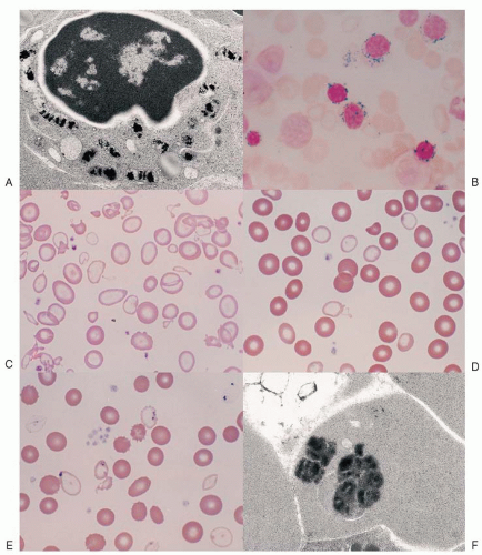
The sideroblastic anemias are a heterogeneous group of disorders1,2 and 3 uniquely characterized by pathologic iron deposits in erythroblast mitochondria4,5 (Fig. 24.1A) that are housed within a distinct, mitochondrial ferritin.6,7 The iron-glutted mitochondria account for the so-called ring sideroblast, an erythroblast in which numerous Prussian blue-positive granules often appear in a perinuclear distribution, particularly in the later stages of its maturation (Fig. 24.1B).

The basis for the mitochondrial iron accumulation in the various sideroblastic anemias can be regarded as either insufficient generation of heme as a result of primary defects in the heme biosynthetic pathway or from faults in mitochondrial functions that involve iron pathways, creating an imbalance between mitochondrial iron import and its utilization. Iron delivery to the erythroid cell does not appear to be downregulated in the face of these alterations, and iron continues to be transported normally to mitochondria, where it accumulates.8,9 Globin synthesis is also reduced, but this effect is secondary, as it can be corrected in vitro by the addition of heme.10,11


FIGURE 24.1. Morphologic features of sideroblastic anemia. A: Electron micrograph of an erythroblast with iron-laden mitochondria. B: Bone marrow smear (Prussian blue stain) with ring sideroblasts. C, D: Blood smears (Wright stain) of severe and mild sideroblastic anemia. E: Siderocytes (Wright stain). F: Electron micrograph of a Pappenheimer body in a peripheral red blood cell.

Kinetically, the sideroblastic anemias are characterized by ineffective erythropoiesis, like other erythroid disorders with defective cytoplasmic or nuclear maturation.12,13 Erythroid hyperplasia of the bone marrow is accompanied by a normal or only slightly increased reticulocyte count. The plasma iron turnover rate is increased, but
iron incorporation into circulating red cells is reduced. Red cell survival tends to be normal or slightly reduced. Slight hyperbilirubinemia may be noted, as well as an increase in urobilinogen excretion, as a result of a raised erythropoietic component of the “early-label” bilirubin peak.14 Thus, it can be inferred that a substantial proportion of the developing ring sideroblasts are nonviable, and their expiration through enhanced apoptotic mechanisms within the marrow15,16,17 accounts for the kinetic abnormalities.

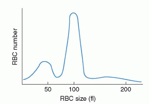
The progeny of surviving ring sideroblasts are often but not always hypochromic and microcytic erythrocytes, a finding that provides morphologic evidence of impaired hemoglobin production as well as an initial clue to the diagnosis. The degree of hypochromia and microcytosis varies considerably from one form of sideroblastic anemia to another (Fig. 24.1C,D). Often, dimorphism is pronounced, with a hypochromic/microcytic population of cells existing side by side with a normal or even a macrocytic one. The siderotic mitochondria of the developing cell may be retained in some circulating erythrocytes (Pappenheimer bodies) and are regularly found with concurrent hypofunction or absence of the spleen; these cells are the nearly pathognomonic siderocytes in the Wright-stained blood smear18 (Fig. 24.1E,F).

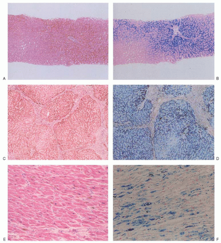
A common feature of many sideroblastic anemias that are not reversible is an excess of total body iron. The serum iron concentration is increased, often to the point of complete saturation of transferrin, and the level of serum ferritin roughly reflects the degree of iron overload. The ineffective erythropoiesis mediates increased intestinal absorption of iron by suppressing hepcidin production.19,20 The consequent iron overload state is called erythropoietic hemochromatosis, and its clinical and pathologic features and course can rival those of hereditary hemochromatosis21,22 (see Chapter 25). The occasional concomitant presence of alleles for genetic hemochromatosis accentuates the iron overload,23,24 and 25 but their prevalence in patients with sideroblastic anemia does not appear to be greater than in the general population.22,26,27 and 28

Diverse defects affecting the utilization of iron by the developing red cell are reflected in the various forms of sideroblastic anemia (Table 24.1). Within the congenital group, the majority appear as isolated anemia, the X-linked and a recently identified autosomal recessive type being the most frequent; however, in a large proportion of cases the cause remains unexplained. Very uncommon are several genetically defined syndromic forms involving multiple systems. Acquired sideroblastic anemia is considerably more common than the congenital forms and occurs as a clonal disorder manifesting only anemia or multilineage dysplasia or even myeloproliferative features. Several diverse factors, such as ethanol, certain drugs, copper deficiency, and hypothermia, produce the ring sideroblast abnormality that is fully reversible.


TABLE 24.1 CLASSIFICATION OF THE SIDEROBLASTIC ANEMIAS (SAs)


Congenital SA


X-linked (XLSA)


Mitochondrial carrier protein SLC25A38 deficiency


Glutaredoxin 5 deficiency


Associated with erythropoietic protoporphyria


Cause unknown


Associated with genetic syndromes


X-linked with ataxia (XLSA/A)


Myopathy, lactic acidosis, and sideroblastic anemia (MLASA)


Congenital sideroblastic anemia and B cell immunodeficiency (SIFD)


Pearson marrow-pancreas syndrome


Thiamine-responsive megaloblastic anemia (TRMA)


Acquired Clonal SA


Refractory anemia with ring sideroblasts (RARS)


Refractory anemia with ring sideroblasts and thrombocytosis (RARS-T)


Refractory cytopenia with multilineage dysplasia and ring sideroblasts (RCMD-RS)


Reversible SA: Associated with


Alcoholism


Certain drugs (isoniazid, chloramphenicol, linezolid)


Copper deficiency (nutritional, malabsorption, zinc ingestion, copper chelation)


Hypothermia


HISTORICAL ASPECTS

The history of sideroblastic anemias can be said to have begun in 1945 with Cooley’s report of a family with X-linked microcytic hypochromic anemia29 (Table 24.2). Near that time ironcontaining granules in erythroblasts, including their perinuclear distribution, were described separately,31,32 and 10 years later were demonstrated to represent iron-laden mitochondria.4 Over the next two decades these ring sideroblasts were recognized in pyridoxine-responsive anemia and in numerous patients with “refractory” anemia of unknown cause, hereditary or acquired, forming the basis for a 1965 symposium and adoption of the term sideroblastic anemia.1 By then, the iron overload of these disorders had also become fully appreciated.35,36

The spectrum of the sideroblastic anemias was widened further by finding various reversible causes of the phenotype as well as by descriptions of certain congenital syndromic forms (Table 24.2). With the advent of molecular biology tools, the genetic causes of several clinically distinct types of congenital sideroblastic anemia and in part their pathogenesis have been elucidated during the last two decades.


HEME SYNTHESIS IN ERYTHROID CELLS

Developing erythroid cells have the greatest requirement of any cell type for heme for hemoglobin, and they produce more than 80% of the heme in the body. The expression and regulation of erythroid heme synthesis are unique in that they are linked (a) to the differentiation events after the signaling action of erythropoietin on erythroid precursors when they acquire the machinery for hemoglobin synthesis, (b) to the availability of iron, and (c) to the production of globin during development of the red cell. As in hepatocytes, 5-aminolevulinate (ALA) synthase and porphobilinogen (PBG) deaminase have considerably lower relative activities than the remaining enzymes in the heme biosynthetic pathway (see Chapter 6) and are sites of pathway regulation.62 In contrast to the liver, the relative activity of ferrochelatase, the terminal enzyme in the pathway, appears also to be low in erythroid cells.62 Furthermore, developing red cells express erythroid-specific isozymes or messenger RNA (mRNA) transcripts of the first four enzymes of heme synthesis, namely ALA synthase, ALA dehydratase, PBG deaminase, and uroporphyrinogen III synthase.

ALA synthase, the first and rate-controlling enzyme of heme synthesis, is generated in the cytosol as a precursor protein with an N-terminal signal sequence that is proteolytically cleaved and processed on transport of the protein into the mitochondrial matrix.63,64 The mature mitochondrial protein catalyzes the formation of ALA from glycine and succinyl coenzyme A (CoA) and requires pyridoxal 5′-phosphate (PLP) as an obligate cofactor. Recently it was discovered that the erythroid-specific mitochondrial carrier family protein SLC25A38 importing glycine across the mitochondrial inner membrane to likely meet the high molar requirement of this substrate by the ALAS enzyme is
necessary for normal erythroid heme synthesis.60 Two separate genes encode the ALA synthase isoenzymes.65,66 The housekeeping gene (ALAS1), located on chromosome 3,66,67 is expressed ubiquitously,68 whereas the erythroid-specific gene (
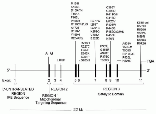
ALAS2) is on the X chromosome.65,66,69,70 Expression of the housekeeping gene, at least in hepatocytes, is controlled by glucose levels and is increased by certain steroids, various drugs, and chemicals (see Chapter 26). This gene is repressed by administration of heme, the end product of the pathway, so that heme levels tightly regulate transcription of its mRNA in a feedback manner.68 Expression of the erythroid gene is essential for hemoglobin production, and ALAS1 cannot compensate if ALAS2 is lacking.
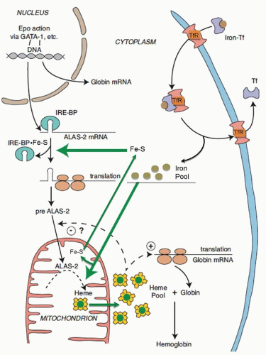
71 ALAS2 is activated and transcribed in concert with other erythroid genes through the action of erythropoietin63,68 (Fig. 24.2); it is not repressed by heme but is upregulated by hypoxia.72 Cellular iron supply may also control ALAS2 mRNA levels,73 whereas exogenous heme inhibits translation of ALAS2 mRNA,74 although the mechanisms by which these occur are not known. Translational regulation of erythroid ALA synthase mRNA is mediated through interaction of its cis-acting iron-responsive element (IRE)75,76 with iron regulatory proteins (IRP1 and IRP2; also called IRE binding proteins 1 and 2)77,78,79 that are modulated through iron-sulfur (Fe-S) clusters generated in mitochondria and by cellular iron status80,81 (Fig. 24.2) (see Chapter 23). In this manner, regulation of protoporphyrin production is linked to iron availability and to mitochondrial function for the formation of heme. The low ALA synthase activity observed in erythroid cells in iron deficiency is consistent with such a control mechanism.82 While the signal sequences of the ALAS1 and ALAS2 precursor proteins contain two heme-binding motifs implicated in regulating translocation of the enzyme into mitochondria by interaction of heme with these motifs83,84 (Fig. 24.2), mitochondrial import of ALAS2 does not appear to be affected by heme.85 Within the mitochondrion, the ALAS2 isoform uniquely associates with the succinyl-CoA synthetase βA subunit, seemingly to stabilize the ALAS2, to control the generation of its substrate succinyl-CoA, or both.86 The role of a major splice isoform of ALAS2 that is functional in vivo87 in the regulation of the enzyme or erythroid heme synthesis remains to be determined.


TABLE 24.2 MILESTONES IN THE HISTORY OF SIDEROBLASTIC ANEMIAS


First Description or Discovery


Year


Reference(s)


X-linked microcytic hypochromic anemia by T. Cooley


1945


29


Three brothers with pyridoxine-responsive anemia mentioned


1940s


30


Perinuclear iron-positive granules in erythroblasts


1947


31, 32


Case report of pyridoxine-responsive anemia


1956


33


Acquired refractory anemia with sideroblastic bone marrow


1956


34


Iron deposits in erythroblast mitochondria


1957


4


Sideroblastic anemia term adopted at international symposium


1965


1


Associated iron overload


1965


35, 36


Reversible sideroblastic anemia from antituberculosis agents


1965


37


Experimental sideroblastic anemia with antituberculosis drugs


1965, 1974


38, 39


Reversible sideroblastic anemia from chloramphenicol


1967


40


Reversible sideroblastic anemia from alcohol


1969


41


Thiamine-responsive megaloblastic anemia (Rogers syndrome)


1969


42


Sideroblastic anemia in erythropoietic protoporphyria


1973, 1993


43, 44


Sideroblastic anemia from copper deficiency


1974


45, 46


Pearson marrow-pancreas syndrome


1979


47


Sideroblastic anemia associated with hypothermia


1982


48


X-linked sideroblastic anemia with ataxia (XLSA/A)


1985


49


RARS inclusion in classification of myelodysplastic syndromes


1986


50


Mitochondrial DNA deletion as cause of Pearson syndrome


1989


51


Mutation in ALAS2 as cause of XLSA


1992


52


Mutation in ABCB7 as cause of XLSA/A


1999


53


Mutations in SLC19A2 as cause of TRMA


1999


54,55 and 56


MLASA and causative mutation in PUS1


2004


57,58


Mutation in GLRX5 as a cause of AR sideroblastic anemia


2007


59


Mutations in SLC25A38 as cause of AR sideroblastic anemia


2009


60


Mutation in YARS2 as another cause of MLASA


2010


61


AR, autosomal recessive; MLASA, myopathy, lactic acidosis, and sideroblastic anemia; RARS, refractory anemia with ring sideroblasts; TRMA, thiamine-responsive megaloblastic anemia; XLSA, X-linked sideroblastic anemia; XLSA/A, X-linked sideroblastic anemia with ataxia.


ALA dehydratase, a cytosolic enzyme, catalyzes the formation of the pyrrole PBG from two molecules of ALA. For this enzyme, two tissue-specific isozymes are produced by a single gene, which contains two promoter regions, generating housekeeping and erythroid-specific transcripts with alternative first noncoding exons (exons 1A and 1B).88,89 Although both transcripts encode identical polypeptides, the erythroid-regulated form would provide for the production of the large amounts of heme for hemoglobin. Not being a rate-limiting enzyme for heme biosynthesis, its expression in erythroid cells in manyfold excess amounts62 may also serve as the proteasome inhibitor CF-2 to inhibit protein degradations.90


FIGURE 24.2. Pathways of regulation of erythroid 5-aminolevulinate synthase (ALAS2). Epo, erythropoietin; Fe-S, iron-sulfur cluster; Tf, transferrin; TfR, transferrin receptor; IRE-BP, iron-responsive element binding protein; – denotes inhibition, + denotes stimulation. (Modified from May BK, Dogra SC, Sadlon TJ, et al. Molecular regulation of heme biosynthesis in higher vertebrates. Progr Nucleic Acid Res Mol Biol 1995;51:1-51.)

In the case of PBG deaminase, which acts in the cytosol to form the linear tetrapyrrole hydroxymethylbilane from four molecules of PBG (see Chapter 6), two tissue-specific isoenzymes also are produced by a single gene.91 The gene has two overlapping transcription units, each with its own promoter: an upstream ubiquitous promoter and another downstream promoter active only in erythroid cells. Two mRNAs are generated by alternative splicing, one encoding the ubiquitous PBG deaminase isoenzyme and the other encoding the erythroid isoenzyme. To what extent the erythroid-specific enzyme has a regulatory role in the overall production of heme in erythroid cells is not known. In response to erythropoietin or hypoxia, bone marrow PBG deaminase activity increased 3.5-fold, apparently by de novo synthesis.92

The gene for uroporphyrinogen III synthase likewise has two promoters generating housekeeping and erythroid-specific transcripts with unique 5′-untranslated sequences (exons 1 and 2A).93 As for ALA dehydratase and PBG deaminase, the erythroidpromoter activity is increased during erythroid differentiation. Uroporphyrinogen decarboxylase, the fifth enzyme of the pathway with its site of action in the cytosol, is not known to have erythroid-regulatory features; however, its mRNA is markedly increased in erythroid tissue94 and the enzyme activity is higher in erythroid cells than in the liver.62

After translation, all three terminal enzymes of heme biosynthesis (coproporphyrinogen oxidase, protoporphyrinogen oxidase, ferrochelatase), like ALA synthase, are transported to their mitochondrial sites of action. Single genes encode these enzymes, and erythroid-specific transcription products are not known for them. However, erythroid-specific regulation of their expression is accommodated by the presence of promoter sequences in their genes for binding of erythroid transcription factors (e.g., GATA-1, NFE-2)95,96 and 97 to enhance production of these enzymes during erythropoiesis.98 Ferrochelatase, the last enzyme of the heme synthetic pathway, catalyzes the insertion of iron into protoporphyrin to form heme. A repressor sequence in the promoter region of its gene is believed to be involved in the regulation of the binding of the erythroid transcription factors GATA-1 and NFE-2 to their recognition sites.97 The activity of ferrochelatase relative to the activity of ALA synthase is in considerable excess,62,99 but the enzyme becomes rate-limiting as a defective protein in erythropoietic protoporphyria (see Chapter 26). As it contains an Fe-S cluster, its expression and stability are also dependent on cellular iron levels and intact Fe-S cluster assembly machinery,100,101 and its activity is regulated by the essential mitochondrial ATPase inhibitory factior 1 (Atpif1) through modulation of mitochondrial pH and redox potential.102


The large amounts of iron required for erythroid heme synthesis are delivered through transferrin receptor-mediated endocytosis of iron transferrin (see Chapter 23), and iron availability ultimately limits the normal rate of heme synthesis in erythroid cells.103 High expression of transferrin receptors is also linked to erythropoietin-induced differentiation68,103; an erythroid-specific isoform of human transferrin receptor has been described104; and erythroid-active elements have been identified in the promoter of the murine gene.105,106 During the height of hemoglobinization, the erythroid cell may not depend upon the “standard” mode for regulation of intracellular iron homeostasis via the IRE/IRP system to ensure a maximal supply of iron to mitochondria.107 With erythroid maturation and the accumulation of cellular hemoglobin, the transferrin receptor number progressively decreases.103 While surface transferrin receptors and iron uptake are increased in iron-deficient erythroblasts,108,109 they are not altered in states of impaired heme synthesis, such as in the presence of succinylacetone110 or in erythroid cells from patients with sideroblastic anemia.8 Transport of iron out of the endosome by the divalent metal transporter DMT1 requires an endosomal ferrireductase (Steap3) in erythroid cells.111 How the further transfer of iron to mitochondria and to apoferritin is accomplished is not understood. It may be facilitated by a cytoplasmic chaperone,112 or through direct interaction of endosomes with mitochondria.113 Iron is imported across the mitochondrial inner membrane by mitoferrin 1 that is highly expressed in erythroid cells and stabilized by interacting with the ATP-binding cassette protein ABCB10.114,115 An oligomeric complex of mitoferrin 1, ABCB10 and ferrochelatase appears to facilitate the incorporation of iron into protoporphyrin to form heme.116 Iron imported into the mitochondrion is also used for the generation of Fe-S clusters, which in part are exported to the cytosol for their addition to IRP1 in the regulation of cellular iron uptake81 and translation of ALAS2,117 and for expression of ferrochelatase.100,101


TABLE 24.3 GENETIC AND HEMATOLOGIC FEATURES OF THE CONGENITAL SIDEROBLASTIC ANEMIAS (CSAs)


Erythrocyte


Mode of Inheritance


Defective Enzyme/Protein


Gene/Chromosomal Location of Gene


Mean Corpuscular Volume


Protoporphyrin


Severity of Anemia


X-linked sideroblastic anemia


X-linked ALAS2


ALAS2/Xp11.21


Decreaseda


Decreased


Mild to severe


Mitochondrial carrier protein deficiency


Autosomal


SLC25A38


SLC25A38/3p22.1


Decreased


Decreased


Severe


Glutaredoxin 5 deficiency


Autosomal


Glutaredoxin 5


GLRX5/14q32.13


Decreased


Not reported


Mild to severe


Protoporphyria


Autosomal


Ferrochelatase


FECH/18q21.3


Decreased


Markedly increased


Mild


Unknown cause Varied


?


?


Variable


Variable


Mild to severe


X-linked sideroblastic anemia with ataxia


X-linked


ABCB7 mitochondrial transporter


ABCB7/Xq13.1-q13.3


Decreased


Increased Mild


Myopathy, lactic acidosis, and sideroblastic anemia


Autosomal


Pseudouridine synthase 1


PUS1/12q24.33


Normal/increased


Not reported


Severe in time


Mitochondrial tyrosyl tRNA synthetase


YARS2/12p11.21


Not reported


Not reported


Severe


CSA and B cell immunodeficiency


Autosomal


?


?


Decreased


Not reported


Severe


Pearson syndrome


Sporadic/maternal


Respiratory chain components


Multiple/mitochondrial DNA


Increased


Increased


Severe


Thiamine-responsive megaloblastic anemia


Autosomal


Thiamine transporter


SLC19A2/1q23.3


Increased


Normal/increased


Mild to severe


a Often normal or increased in females expressing the disorder.


The export of heme out of the mitochondrion into the cytosol for its pairing with globins of hemoglobin and other heme proteins is mediated at least in the mouse by an isoform of the feline leukemia virus C receptor (FLVCR1), named FLVCR1b118. This heme exporter is essential for erythropoiesis as its loss leads to heme accumulation in the mitochondrion and termination of erythroid differentiation.

The events coordinating the production of globin chains with the rate of heme synthesis are well understood, and appear to occur at more than one level. Heme is required for initiation of globin mRNA translation and it acts by inhibiting a protein kinase called heme-regulated inhibitor (HRI), which inactivates the translational initiation factor 2 (eIF-2α) in the absence of heme119 (Fig. 24.2). Moreover, absence of this kinase adversely modifies the phenotype of disorders of heme synthesis (iron deficiency, protoporphyria) and of globin production (β-thalassemia).119 In addition, heme controls globin gene expression at the transcriptional level.120,121


CONGENITAL SIDEROBLASTIC ANEMIAS

Congenital sideroblastic anemias have emerged as not uncommon disorders. They are clinically and genetically heterogeneous, with diverse underlying causes, inheritance patterns, phenotypes, and associated features. Genetic analysis and molecular approaches have revealed a spectrum of specific defects, some appearing as isolated anemia and others involving multiple systems3,122,123 (Table 24.3). Defined underlying defects affect heme biosynthesis directly or indirectly by disrupting Fe-S cluster biogenesis, or they involve pathways related to mitochondrial protein synthesis. However, in at least one third of cases the root cause remains undiscovered. Severity of anemia is highly variable within some types, and morphologic features overlap among them.


Genetic Defects Expressed Only in the Erythron


Inheritance Patterns and Pathogenesis


X-Lined Sideroblastic Anemia

The inheritance follows an X-linked pattern, the anemia occurring most commonly in males and their maternal uncles and cousins.29,124,125,126,127 and 128,129,130 Minimal expression of the erythroid abnormality may be seen in carrier females that is consistent with variable X inactivation affecting the mutant locus of the disorder. However, in many kindreds, the anemia has occurred only in females and may have been lethal in hemizygous male conceptions.131,132,133,134,135,136

Early observations already implicated defects in ALA synthase as underlying the impaired heme biosynthesis in this form of sideroblastic anemia. In patients who responded to pyridoxine supplementation, the incorporation of glycine, but not of ALA, into heme was reduced in reticulocytes.30 ALA synthase activity in bone marrow was low before pyridoxine administration and returned to normal or supranormal levels after an erythropoietic response.129,137,138 and 139 It was presumed that the residual activity or stability of a defective erythroid ALA synthase was enhanced by additional supply of its coenzyme PLP through a mass action effect (e.g., if the enzyme had a reduced affinity for the coenzyme138 or was abnormally sensitive to proteolysis137). Usually, pharmacologic amounts of pyridoxine are required when an erythropoietic response occurs, but the response is variable and rarely complete. Individuals may present with profound anemia only in adulthood or even late in life,129,140,141,142 suggesting that the disorder can progress with time. In some cases, prior additional dietary or medicinal intake of pyridoxine,143 possible changes in pyridoxine metabolism with advancing age,144 or initiation of hemodialysis145,146 can be factors in unmasking mild phenotypes that were not symptomatic at a younger age. In female patients expressing the disease, skewed X inactivation in hematopoietic tissue that occurs with advancing age147 and involves progressive inactivation of the X chromosome bearing the normal allele has been the explanation as the anemia usually evolved in adulthood135,136 or late in life.148 Constitutive skewed X inactivation to account for disease expression in females in childhood is very uncommon.136


FIGURE 24.3. Diagram of the structure of the human erythroid 5-aminolevulinate synthase gene (ALAS2) and the location of mutations identified to date in X-linked sideroblastic anemia. Two mutations in the promoter region (c.-206C>G; c.-91_-44del) and two mutations disrupting the intron 1 enhancer of ALAS2 are not indicated. IRE, iron-responsive element; kb, kilobases. *Codon K391, the pyridoxal 5′-phosphate binding lysine. (Data from HGMD® Professional 2012.2 and unpublished material.)

After the cloning and characterization of the erythroid ALA synthase gene75,149 and its localization to the X chromosome, linkage of the disorder to the ALAS2 locus was established,129,150 and many heterogeneous missense mutations involving invariant or highly conserved amino acid residues in the catalytic domain of the enzyme have been found to cause the disorder151 (Fig. 24.3). A nonsense mutation in one case,135 nucleotide deletions, and regulatory mutations in the promoter region and in the intron 1 enhancer sequence151a of the ALAS2 gene have been the exceptions. Hence, in at least four families described up to 68 years ago,29,125,128,152 the underlying molecular defect in ALAS2 could also be defined.25,153,154 and 155 A majority of the mutations identified to date reside in exons 5 and 9 (Fig. 24.3); the latter contains the PLP-binding lysine (K391) of the enzyme.156 Among the 66 distinct mutations so far encountered, only 22% occurred in more than one unrelated family or proband, and nearly one third of the probands are female. An apparent somatic mutation in the ALAS2 gene was found in an older male patient with acquired sideroblastic anemia.157

Sites of ALAS2 mutations and severity of anemia or extent of its responsiveness to pyridoxine supplements can be correlated to a considerable extent. The activity of the recombinant enzyme is reduced for many, but not all, ALAS2 mutants so far examined and is variably enhanced by PLP.52,129,130,142,153,158,159 Some mutants have altered substrate kinetics159,160 or fail to bind to the β-subunit of succinyl-CoA synthetase.86,160 A three-dimensional structure model of the human enzyme161 and the resolved crystal structure of the significantly homologous ALAS of Rhodobacter capsulatus162 made it possible to explain how altered structure by many of the naturally occurring mutations that give rise to X-linked sideroblastic anemia (XLSA) affects the function of the enzyme. For example, mutations changing an amino acid located in the vicinity of the PLP-binding site exhibit a response to pyridoxine, whereas mutations involving sites of substrate binding, enzyme stability, or folding are refractory to pyridoxine.162 However, while clinical severity can thus be related at least in part to the effect of a specific mutation on enzyme function, marked variation in severity of anemia has been observed between some kindreds bearing the same mutation as well as within a few
kinships,129,130,163,164 implicating undefined genetic differences or environmental factors for the apparent variable penetrance.


Mitochondrial Carrier Protein SLC25A38 Deficiency

A significant segment of congenital sideroblastic anemias (˜15%) is associated with biallelic mutations in the gene encoding the erythroid-specific mitochondrial inner membrane carrier protein SLC25A38.60,165 The SLC25A38 gene has an amino terminal targeting signal and three mitochondrial carrier family protein domains that encode six transmembrane helices. From its structural features, the protein is predicated to function as an amino acid transporter. Data so far obtained in the yeast Saccharomyces cerevisiae deficient in the protein indicate a heme biosynthetic defect and suggest that it serves as a glycine importer across the mitochondrial inner membrane.60

Heterogeneous mutations spread throughout the SLC25A38 protein domains have been reported in 26 families, including three sibling pairs.60,166 The mutations are missense type occurring in conserved amino acids at substrate contact points as well as nonsense and splicing errors. Two thirds of patients are homozygous and one third are compound heterozygotes for defects.


Glutaredoxin 5 Deficiency

The human counterpart of a zebrafish mutant (shiraz) deficient in glutaredoxin 5,80 which is essential for the synthesis of Fe-S clusters such as for IRP1 and thus ALAS2 translation, was identified in a single patient.59 A homozygous mutation in the GLRX5 gene that affects intron 1 splicing and markedly reduces GLRX5 RNA production was associated with microcytic sideroblastic anemia. The anemia was detected in the fifth decade, became severe by age 60, and improved with iron chelation therapy for the associated iron overload. Studies in cell lines derived from the patient indicated severe impairment of Fe-S cluster biogenesis and also revealed markedly reduced levels of ferrochelatase.167


Sideroblastic Anemia in Protoporphyria

Marked deficiency of ferrochelatase underlies erythropoietic protoporphyria as a result of a large variety of mutations in the gene encoding the enzyme (see Chapter 26). The defect is manifested mainly, if not exclusively, in erythroid cells and leads to marked accumulation of free protoporphyrin, the substrate of the enzyme, during the final stages of erythroid maturation when the defective ferrochelatase becomes rate-limiting for heme production.168 Erythroid heme synthesis appears to be compromised in most patients as reflected in a mild microcytic hypochromic anemia.169 In ten patients marrow ring sideroblasts with typical mitochondrial iron deposits were observed43,44 but not in one sibling pair with mild microcytosis without anemia.168 Bone marrow examination has generally not been performed in this disorder so that the incidence of the ring sideroblast feature is not known. The genetic heterogeneity in protoporphyria may account for the phenotypic variation of hematologic features.


Undefined Congenital Sideroblastic Anemia(s)

It is estimated that at least one third of nonsyndromic cases of congenital sideroblastic anemia are currently molecularly unexplained.165 These may have autosomal or X-linked defects not yet detectable in the known causative genes, or defects in novel genes to be discovered. Novel genetic defects are also likely in some previously described kindreds. For example, in two families sideroblastic anemia occurred in a vertical distribution including father-to-son transmission and consistent with a dominant trait.170,171 In another family manifesting the anemia in both genders, a defect in mitochondrial DNA was postulated172; the mild anemia was characterized by erythrocyte dimorphism and macrocytosis.


Clinical and Laboratory Features of XLSA and SLC25A38 Deficiency

If severe, XLSA is recognized in infancy or early childhood. However, not infrequently the disorder is milder or asymptomatic and may be discovered only in young adulthood or even in later life. Because severity of anemia can also vary within kindreds,129,130,164 diagnosis in family members may be delayed or overlooked unless complete pedigree studies or DNA analyses for an ALAS2 mutation identified in the proband are performed. In contrast, patients with SLC25A38 genetic defects typically present at birth or in early childhood with severe anemia.

All patients develop manifestations of iron overload. Linkage to human leukocyte antigen (HLA) is not evident22 as HFE mutations were underrepresented in a series of XLSA patients examined.26 Mild to moderate enlargement of the liver and spleen is common, but liver function usually is normal or only mildly disturbed at presentation. Liver biopsy reveals iron deposition that is indistinguishable from hereditary hemochromatosis36 (Fig. 24.4). In the X-linked form the iron burden does not correlate with the severity of anemia, and, not infrequently, well-established but asymptomatic micronodular cirrhosis is discovered in the third or fourth decade.22,36,134,141,173 Hepatocellular carcinoma developed in two reported cases.163,174 Clinical diabetes or abnormal glucose tolerance may or may not be related to the iron overload process. Skin hyperpigmentation is uncommon. The most dangerous manifestations of the iron overload are cardiac arrhythmias and congestive heart failure, which usually occur late in the disease course. In severely affected infants or young children, growth and development tend to be impaired.127

The hallmark is a microcytic anemia. In severe cases, microcytosis and hypochromia are pronounced (mean corpuscular volume [MCV], 50 to 60 fl), and striking anisocytosis, poikilocytosis, target cells, and occasional siderocytes are prominent findings on blood smear (Fig. 24.1C). The red cell volume distribution is usually abnormally wide and, notably, dimorphism is seen in males with the X-linked form124 as well as in autosomal forms of the disease.60,166,170 Some female carriers of the X-linked trait have a biphasic Coulter counter red cell histogram25,128 (Fig. 24.5), or only a very small microcytic erythrocyte population. However, most women who express XLSA exhibit macrocytosis, although dimorphism may be evident (Fig. 24.1D). Presumably few viable erythrocytes with a markedly defective or nonfunctional mutant ALAS2 enzyme as often encountered in women reach the circulation, so that most of the erythrocytes are progeny of the residual normal clone that are released from the marrow at an accelerated rate in response to the anemic hypoxia. Leukocyte and platelet values usually are normal; they may be reduced in the presence of splenomegaly (hypersplenism). Erythroid hyperplasia is found on marrow examination, and maturation is usually normoblastic with poorly developed cytoplasm. Megaloblastic changes may be observed if complicating folate deficiency is present. Marrow reticuloendothelial iron is increased, and the telltale ring sideroblast emblem is prominent in late, nondividing erythroblasts.175,176 Transferrin saturation is increased, as is the serum ferritin level, and transferrin levels tend to be decreased. Ferrokinetic studies reflect ineffective erythropoiesis. A reduced serum haptoglobin level is consistent with the ineffective erythropoiesis.

The erythrocyte protoporphyrin level usually is low or normal.12,131 In one female patient, the low protoporphyrin level was shown to be restricted to the microcytic red cells.132 Kindreds in which the erythrocyte protoporphyrin is increased131,170 can be considered to represent disorders other than XLSA or SLC25A38 deficiency.


Treatment and Prognosis

Approximately two thirds of patients with sideroblastic anemia due to identified ALAS2 defects can be expected to respond to
pyridoxine administration. Doses of 50 to 100 mg/day are large compared with the estimated adult daily requirement for vitamin B6 of 1.5 to 2.0 mg and are sufficient for a maximal response, although in some cases a supplement of only 2 to 4 mg/day was found to be effective.129 Higher doses may be toxic. No convincing evidence is available that the parenteral route or PLP, the active coenzyme form, is more effective than oral administration. However, the response to pyridoxine is quite variable. With an optimal response, reticulocytosis is observed, blood hemoglobin concentration returns to normal or near-normal levels in 1 to 2 months, and low erythrocyte protoporphyrin levels increase to normal.12,177 The morphologic red cell abnormalities then
diminish but very rarely completely resolve, even when ALA synthase activity and the hemoglobin level are restored with pyridoxine supplementation. Approximately two thirds of responding patients experience a distinct but suboptimal improvement with pyridoxine administration, and the hemoglobin concentration stabilizes at less than normal levels. When an effect of pyridoxine is achieved, continued maintenance treatment is necessary because relapses follow within several months after discontinuance of the vitamin. In a few instances, subsequent remissions with resumed treatment were less complete.12 In the occasional case with accompanying megaloblastic changes, folic acid should be given, which usually leads to normoblastic maturation, suboptimal reticulocytosis, and some increase in the hemoglobin level. In severely anemic individuals who do not respond to pyridoxine, periodic red cell transfusions are necessary to relieve symptoms and to allow normal growth and development of children. Transfusion of red cells should be kept to a minimum to lessen or delay the development of iron overload.


FIGURE 24.4. Histopathology of the iron overload in congenital sideroblastic anemia. A, B: Liver section of a 26-year-old man with SLC25A38 deficiency and moderate hemochromatosis. C, D: Autopsy liver section of a 45-year-old man with X-linked sideroblastic anemia, micronodular cirrhosis, and hemochromatosis. E, F: Section from the heart of the latter patient with marked hemosiderosis. (A, C, and E: Hematoxylin stain; B, D, and F: Prussian blue stain.)


FIGURE 24.5. Dimorphic red cell distribution in a female patient with mild X-linked sideroblastic anemia. Redrawn from the output of a Coulter analyzer. RBC, red blood cell.

Based on the assessed extent of iron overload, including liver biopsy, an iron depletion program should be instituted to prevent or stabilize already established organ damage.134,178,179,180 Therapeutic phlebotomies are well tolerated and preferred in patients with mild or moderate anemia in the absence of contraindications such as heart disease.134,173,181,182 and 183 After the initial removal of all storage iron, maintenance phlebotomies should be continued indefinitely. For patients with severe anemia, or for those who depend on regular transfusions and thus become massively iron loaded,184,185 an iron-chelating agent is administered. As recommended for thalassemia,185 deferoxamine is infused over 12 hours subcutaneously or intravenously, at 40 mg/kg/day, and for at least 5 days each week. Although iron removal with deferoxamine is enhanced by ascorbate, large supplements can cause acute cardiac toxicity by facilitating excessive mobilization of ferritin iron, and any intake of the vitamin should be limited to 200 mg daily.186 The risks of deferoxamine treatment are minimal.179,187 Occasional local reactions can be controlled with inclusion of small amounts of hydrocortisone in the infusate. Rare hypersensitivity is amenable to desensitization.188 Reported visual and auditory neurotoxicity is unlikely without excessive doses of the drug. The new, orally active tridentate iron chelator deferasirox (Exjade) has an efficacy similar to deferoxamine or better180,189; the recommended initial daily dose is 20 mg/kg and can be increased to 30 mg/kg. Although the long-term safety profile of this agent is not known, it has emerged as a preferred iron chelator. The goal of iron chelation therapy is to maintain the serum ferritin concentration below 500 µg/L. The increased risk of infection with Yersinia (and perhaps other organisms) in iron overload, although uncommon, increases further with deferoxamine treatment.179,190 Removal of the iron excess has occasionally reduced severity of the anemia8,25,182,183 by improving erythroblast mitochondrial function, such as restoration of secondary ferrochelatase deficiency183; by enhancing pyridoxine responsiveness25; and by diminishing the ineffective erythropoiesis.182

Only gold members can continue reading. Log In or Register to continue

Stay updated, free articles. Join our Telegram channel

Oct 21, 2016 | Posted by in HEMATOLOGY | Comments Off on Sideroblastic Anemias

Full access? Get Clinical Tree

Get Clinical Tree app for offline access