Red Cell, Platelet, and White Cell Antigens


Red Cell, Platelet, and White Cell Antigens


Kathryn E. Webert

James W. Smith

Donald M. Arnold

Howard H. W. Chan

Nancy M. Heddle

John G. Kelton


INTRODUCTION

This chapter reviews the biochemistry and importance of various blood group antigens with a focus on red cells, platelets, and white blood cells. Because the circulating blood cells originate from a common progenitor cell, it is not surprising that there are a number of common blood group antigens. But what is surprising is the fact that the various cell lineages have so many unique and different antigens.

Studies characterizing the red cell antigens were first performed approximately 100 years ago, whereas studies investigating the platelet and white cell antigens were performed much more recently. There are a number of explanations for this, including the earlier recognition of red cells and the early attempts to transfuse these cells. Additionally, red cells have proved to be not only more plentiful, but also easier for investigators to work with. Indeed, the early techniques used to study red cell antigens, such as primary and secondary agglutination reactions using direct and indirect antiglobulin tests, proved so robust that they are still used today.

The nomenclature used for red cells is complex, yet historically interesting. Until recently, there was no attempt to be systematic. In the past, some blood groups were named after the individual (Kell is derived from Mrs. Kellner) or animal (Rh is derived from the rhesus monkey) lacking the antigen. Others were named after the discoverers (e.g., LW was named for Landsteiner and Weiner). Finally, some names are best described as quaint (e.g., the Lutheran blood group system was named according to a mislabeled blood sample). As is discussed, a more systematic approach for naming antigenic systems of red cells, platelets, and white cells is now used. However, the traditional names are still frequently used and, to add to the confusion, a number of laboratories continue to use other traditional nomenclatures (e.g., the Fisher-Race terminology continues to be used for the Rh system).

For biologists, a compelling question presents itself: What are the purposes of blood group antigens? For the majority of antigens, the answer is unknown, but there is increasing evidence that suggests certain antigens participate in host defense. Blood group antigens of red cells, platelets, and white cells can be made of proteins, carbohydrates attached to proteins, or lipids. Within each category, the antigen can be intrinsically produced during the formation of that cell, or it can be adsorbed from the plasma. Similarly, the antigens can be attached to the surface of the cell, can be partially embedded within the membrane (phosphatidylinositol glycan), or can be transmembrane. Recognizing this, investigators have grouped red cell antigens according to the functional activities of their associated carbohydrate, lipid, or protein.1,2 These include functions such as membrane stabilization, transport across the membrane, receptor function, enzymatic activity, and others. However, it is important to emphasize that although these blood group antigens are one component of this particular function, there is little evidence that they exist solely for this function. Consequently, although it is interesting to understand how a protein or carbohydrate associated with a blood group antigen contributes to a cell’s integrity, it may not be the sole explanation for the presence of the antigen.

Any discussion of blood group antigens must touch on disease associations. In this review, we also comment on generally agreed on associations. Readers should be cautious about the reported associations between certain blood groups and diseases, as these associations could be collateral to other factors. However, there is general agreement that infectious agents, especially parasites and some bacteria, have led to blood group antigenic selection. Perhaps this is best exemplified by the geographic distribution of the Duffy blood group system, which is encoded by two alleles. The Fy(a-b-) phenotype is rare in most populations, with the exception being blacks originating from West Africa. Studies have demonstrated that the Duffy glycoprotein can serve as a receptor for Plasmodium vivax, an etiologic agent of malaria; hence, there is a strong selection advantage for those individuals not expressing this glycoprotein (i.e., Fy [a-b-]).

These dramatic observations and other studies provide indirect evidence that perhaps the major biologic advantage of antigens on cells is to enhance the ability of the species to distinguish self from non-self. Hence, one can anticipate in the ongoing “evolutionary warfare” between species and their invaders that when a microorganism uses a component of a blood group to invade that cell, spontaneous mutations producing another blood group would have a selective advantage. Additionally, nonhazardous mutations, which do not provide advantage or disadvantage, might not necessarily be deleted. It is likely that this helps to explain the enormous number of blood groups found on red cells, platelets, and white cells. Some of these blood groups are common (typically termed public systems within the platelet nomenclature), whereas others are rare (termed private systems).

This chapter summarizes red cell and platelet blood groups by first summarizing the approach to nomenclature and then reviewing the common blood groups.


RED BLOOD CELL ANTIGENS

In humans, 33 blood group systems with 298 antigens have been identified.3,4,5 Additional antigens have been identified but have not been assigned to established systems. Red blood cell antigens may be proteins, glycoproteins, or glycolipids. Most red cell antigens are synthesized by the red cells; however, some antigens, such as those of the Lewis and Chido/Rodgers systems, are adsorbed onto the red cell membrane from the plasma. Some red cell antigens are specific to red cells; however, others are found on other cells throughout the body.

The importance of red cell antigens is multifold. Since the work of Landsteiner in the early 1900s, it has been recognized that knowledge and understanding of blood groups are essential for transfusion therapy. In routine practice, it is necessary to determine the compatibility of certain red blood cell antigens between the blood donor and the blood recipient. This is because individuals who lack antigens on their red blood cells can form alloantibodies, or be alloimmunized, if they are exposed to blood expressing the antigen. This might occur with transfusion of blood products or during pregnancy. Antibodies that react with red blood cell antigens can cause problems such as delayed and immediate hemolytic transfusion reactions (HTRs) and hemolytic disease of the newborn (HDN). Furthermore, the study of red cell antigens and the associated membrane structures allows for a greater understanding of the physiology of red blood cells. For example, abnormalities of some blood group systems, such as Rh and Kell, may be associated with both functional and morphologic changes in the red cells. Finally, study of the inheritance of blood group antigens provides a greater understanding of the mechanisms of gene expression.

In this chapter, the red cell antigen groups are presented in order of relative clinical importance. The summary tables, given in each antigen section, list important clinical information and the most important antigens of each blood group system. As noted, many red cell antigens (as well as the platelet antigens) have an interesting but inconsistent approach to nomenclature. However, because these names are commonly used, they are used along with the nomenclature proposed by the International Society of Blood Transfusion (ISBT).


International Society of Blood Transfusion Terminology

The ISBT Working Party on Terminology for Red Cell Surface Antigens was established in 1980 with the goal of creating a uniform nomenclature. Blood antigens were categorized into systems, collections, and series. The most recent monograph produced by the Working Party was produced in 2004 and included blood group antigens categorized into 29 systems, six collections, and two series.6 Updates to this monograph have been published online (http://ibgrl.blood.co.uk/ibgrl/ISBT%20Pages/ISBT%20ter-minology%20pages/terminology%20Home%20page.htm#system) and there are now 30 systems recognized.


Systems

A blood group system is genetically discrete from other blood group systems and consists of one or more antigens governed by either a single gene locus or a complex of two or more closely linked genes with virtually no recombination events occurring among them. Furthermore, it must be defined by a human alloantibody, and the gene encoding it must have been identified and sequenced. There are currently 30 recognized systems (Table 20.1).


Collections

The concept of collections was introduced into the ISBT terminology in 1988 to bring together related sets of antigens (genetically, biochemically, or serologically), which could not correctly be classified as systems because they have not been shown to be genetically distinct from all existing systems. Seven collections are currently recognized (Table 20.2).


Series

An antigen may be assigned a number if it is a low-frequency antigen (the 700 series) or if it is a high-frequency antigen (the 901 series). A low-frequency antigen is an antigen that has an incidence of <1% in most populations tested, similar to the platelet designation of “private.” The 700 series currently consists of 18 antigens (Table 20.3). High-frequency antigens are antigens with an incidence of >90% in most populations, similar to the platelet “public” system. Originally, high-frequency antigens were assigned to the 900 series. However, because so many of the original antigens assigned to the 900 series have been reassigned to collections, the 901 series was created. There are currently six antigens in the 901 series (Table 20.4).

Each blood group antigen is given an identification number consisting of six digits. The first three numbers represent the system to which the antigen has been assigned. The second three digits identify the antigen. Each system also has an alphabetic symbol. For example, the ABO system is number 001, and the A antigen is the first antigen of that system; thus, it has the ISBT number 001001 or ABO001. By convention, the zeros may be omitted, and numbers are separated by a dot (i.e., the A antigen would be 1.1 or ABO1). This terminology is useful for databases and as a classification system; however, most clinical laboratories still use traditional terminology.


Red Cell Blood Group Systems


ABO (ISBT 001) and Hh (ISBT 018) Blood Group Systems


SUMMARY OF IMPORTANT CHARACTERISTICS OF ABO ANTIBODIES


Antibody Specificity


Clinically Significant


Antibody Class


HDN


HTR


Frequency of Antigen-Negative Blood (White Population)


Anti-A


Yes


IgM; some IgG


Yes


Yes


Common (53%)


Anti-B


Yes


IgM; some IgG


Yes


Yes


Common (87%)


HDN, hemolytic disease of the newborn; HTR, hemolytic transfusion reaction; Ig, immunoglobulin.


The ABO blood group system was discovered by Landsteiner in 1900 when he noticed that the red cells of some individuals could be agglutinated by the serum of others. It remains the most important of all blood group systems for several reasons: (a) when the ABO antigen is not expressed on the red cell, individuals always have ABO antibodies in their plasma, with the stimulus for antibody production being a variety of environmental agents; and (b) the ABO antibodies formed are frequently mixtures of both immunoglobulin M (IgM) and IgG antibodies, both having thermal reactivity at 37°C and both capable of activating complement.

These unique characteristics of the antigens and antibodies of the ABO blood group system provide optimal conditions for rapid

red cell destruction if ABO-incompatible blood is transfused: A clinical scenario termed an acute HTR.


TABLE 20.1 BLOOD GROUP SYSTEMS


Gene Name(s)


ISBT No.


System Name (symbol)


No. of Antigens


Antigen(s)


ISBT


HGNC


Gene Product(s)


Chromosomal Location


001


ABO (ABO)


4


A, B, AB, A1


ABO


ABO


A = α-3-N-acetyl-D-galactosaminyltransferase


9q34.2


B = α-3-D-galactosyltransferase


002


MNS (MNS)


46


M, N, S, s, U, He, Mia, Mc, Vw, Mur, Mg, Vr, Me, Mta, Sta, Ria, Cla, Nya, Hut, Hil, Mv, Far, sD, Mit, Dantu, Hop, Nob, Ena, EnaKT, N′, Or, DANE, TSEN, MINY, MUT, SAT, ERIK, Osa, ENEP, ENEH, HAG, ENAV, MARS, ENDA, ENEV, MNTD


MNS


GYPA


GYPB


GYPE


Glycophorin A (GYPA)


Glycophorin B (GYPB)


Glycophorin E (GYPE)


4q31.21


003


P (P1)


3


P1, PK, NOR


P1


22q11.2-qter


004


Rh (RH)


54


D, C, E, c, e, f, Ce, Cw, Cx, V, Ew, G, Hro, Hr, hrs, VS, CG, CE, Dw, c-like, cE, hrH, Rh29, Goa, hrB, Rh32, Rh33, HrB, Rh35, Bea, Evans, Rh39, Tar, Rh41, Rh42, Crawford, Nou, Riv, Sec, Dav, JAL, STEM, FPTT, MAR, BARC, JAHK, DAK, LOCR, CENR, CEST, CELO, CEAG, PARG, CEVF


RH


RHD


RHCE


Acetylated RhD protein


Acetylated RhCE protein


1p36.11


005


Lutheran (LU)


20


Lua, Lub, Lu3, Lu4, Lu5, Lu6, Lu7, Lu8, Lu9, Lu11, Lu12, Lu13, Lu14, Lu16, Lu17, Aua, Aub, Lu20, Lu21, LURC


LU


B-CAM


B-cell adhesion molecule


19q12-q13


006


Kell (KEL)


35


K, k, Kpa, Kpb, Ku, Jsa, Jsb, Ula, K11, K12, K13, K14, K16, K17, K18, K19, Km, Kpc, K22, K23, K24, VLAN, TOU, RAZ, VONG, KALT, KTIM, KYO, KUCI, KANT, KASH, KELP, KETI, KHUL, KYOR


KEL


KEL


Zinc endopeptidase


7q33


007


Lewis (LE)


6


Lea, Leb, Leab, LebH, ALeb, BLeb


LE


FUT3


α-1,3/1,4-L-Fucosyltransferase


19p13.3


008


Duffy (FY)


6


Fya, Fyb, Fy3, Fy4, Fy5, Fy6


FY


DARC


Duffy antigen receptor for chemokines


1q21-q22


009


Kidd (JK)


3


Jka, Jkb, JK3


JK


SLC14A1


Urea transporter


18q11-q12


010


Diego (DI)


22


Dia, Dib, Wra, Wrb, Wda, Rba, WARR, ELO, Wu, Bpa, Moa, Hga, Vga, Swa, BOW, NFLD, Jna, KREP, Tra, Fra, SW1, DISK


DI


SLC4A1


Anion exchanger 1, solute carrier family 4/band 3


17q12-q21


011


Yt (YT)


2


Yta, Ytb


YT


ACHE


Acetylcholinesterase


7q22


012


Xg (XG)


2


Xga, CD99


XG


XG


Xga glycoprotein


Xp22.32


013


Scianna (SC)


7


Sc1, Sc2, Sc3, Rd, STAR, SCER, SCAN


SC


ERMAP


Erythrocyte membrane-associated protein (ERMAP)


1p34


014


Dombrock (DO)


8


Doa, Dob, Gya, Hy, Joa, DOYA, DOMR, DOLG


DO


ART4


ADP-ribosyltransferase 4


12p13.2-q13.3


015


Colton (CO)


4


Coa, Cob, Co3, Co4


CO


AQP1


Aquaporin-1 (AQP1)


7p14


016


Landsteiner-Wiener (LW)


3


LWa, LWab, LWb


LW


ICAM4


Intracellular adhesion molecule 4 (ICAM4)


19p13.2-cen


017


Chido/Rodgers (CH/RG)


9


Ch1, Ch2, Ch3, Ch4, Ch5, Ch6, WH, Rg1, Rg2


CH/RG


C4B/C4A


Complement component 4A protein[en]Complement component 4B protein


6p21.3


018


H (H)


1


H


H


FUT1


Galactoside 2-α-L-fucosyltransferase 1


19q13.1-qter


019


Kx (XK)


1


Kx


KX


XK


Membrane transport protein XK


Xp21.1


020


Gerbich (GE)


11


Ge2, Ge3, Ge4, Wb, Lsa, Ana, Dha, GEIS, GEPL, GEAT, GET1


GE


GYPC


Glycophorin C (GPC) and GPD (glycophorin C precursor)


2q14-q21


021


Cromer (CROM)


18


Cra, Tca, Tcb, Tcc, Dra, Esa, IFC, WESa, WESb, UMC, GUTI, SERF, ZENA, CROV, CRAM, CROZ, CRUE, CRAG


CROM


CD55


CD55/decay accelerating factor (DAF)


1q32


022


Knops (KN)


9


Kna, Knb, McCa, S11, Yka, McCb, S12, S13, KCAM


KN


CR1


CD35/CR1


1q32


023


Indian (IN)


4


Ina, Inb, INFI, INJA


IN


CD44


CD44


11p13


024


Ok (OK)


3


Oka, OKGV, OKVM


OK


BSG


Basigin


19p13.3


025


Raph (RAPH)


1


MER2


RAPH


CD151


CD151


11p15.5


026


John Milton Hagen (JMH)


6


JMH, JMHK, JMHL, JMHG, JMHM, JMHQ


JMH


SEMA7A


Semaphorin 7A


15q22.3-q23


027


I (I)


1


I


I


GCNT2


I- β-1, 6-N-acetylglucosaminyltransferase A


6p24.2


028


Globoside (GLOB)


1


P


GLOB


B3GALNT1


UDP-N-acetyl-galactosamine globo-triaosylceramide 3-βN acetylgalactosaminyl-transferase


3q25


029


Gill (GIL)


1


GIL


GIL


AQP3


Aquaporin-3 (AQP3)


9p13


030


Rh-associated glycoprotein (RHAG)


4


Duclos, Ola, DSLKa, RHAG 4


RHAG


RHAG


6p12.3


031


Forssman (FOR)


1


FORS1


GBGT1


GBGT1


globoside alpha-1,3-N-acetylgalactosaminyltransferase 1


9q34.13-q34.3


032


JR


1


Jra


ABCG2


ABCG2


ATP-binding cassette, sub-family G (WHITE), member 2


4q22.1


033


LAN


1


LAN


ABCB6


ABCB6


ATP-binding cassette, sub-family B (MDR/TAP), member 6


2q36


HCNC, HUGO gene nomenclature committee (www.genenames.org); ISBT, International Society of Blood Transfusion; No, number.


Data from Daniels GL, Fletcher A, Garratty G, et al. International Society of Blood Transfusion Working Party on terminology for red cell surface antigens. Vox Sang 2004;87:304-316; Denomme GA, Rios M, Reid ME. Molecular protocols in transfusion medicine. San Diego, CA: Academic Press, 2000; Logdberg L, Reid MA, Lamont RE, et al. Human Blood Group Genes 2004: chromosomal locations and cloning strategies. Transfus Med Rev 2005;19:45-57; Costa FP, Hue-Roye K, Sausais L, et al. Absence of DOMR, a new antigen in the Dombrock blood group system that weakens expression of Do(b), Gy(a), Hy, Jo(a), and DOYA antigens, Transfusion 2010; 50:2026-2031; Smart EA and Storry JR. The OK blood group system: a review. Immunohematology 2010; 26:124-126; Walker PS, Reid ME. The Gerbich blood group system: a review. Immunohematology 2010; 26:124-126; and International Society of Blood Transfusion Working Party on terminology for red cell antigens web site: http://www.isbtweb.org/fileadmin/user_upload/WP_on_Red_Cell_Immunogenetics_and/Updates/Table_of_blood_group_antigens_within_systems_v3_2_130331.pdf Accessed April 3, 2013


There are three alleles of the ABO gene in the ABO blood group system (A, B, and O) that are inherited in Mendelian fashion (Table 20.1). Both A and B are codominant alleles, whereas O is a recessive allele. Hence, these three result in four different phenotypes: A, B, AB, and O. An individual with the A phenotype can be homozygous for the A gene (AA) or heterozygous (AO). Similarly, the B phenotype can be the result of homozygous (BB) or heterozygous (BO) gene inheritance. The genotype of the AB phenotype is AB, and the group O phenotype is always genetically OO. Thus, there are four ABO group phenotypes (A, B, AB, and O) that arise from six possible genotypes (AA, AO, BB, BO, AB, and OO). The alleles normally occupy the same position of a paired chromosome. Rarely, individuals may express a cisAB phenotype in which the A and B alleles appear to be carried on the same chromosome. This phenomenon is caused by a collection of mutant ABO alleles that encode a glycosyltransferase capable of synthesizing both A and B antigens.7

The frequencies of ABO phenotypes are variable among different ethnic populations. In whites, the O and A phenotypes are the most common, occurring in >40% of the population. The B phenotype is found in approximately 10% of whites, and the AB phenotype is encountered in only 3% of individuals. In contrast, around the world, the O phenotype is the most common ABO phenotype, particularly in South and Central America. The A phenotype is found in 10 to 35% of individuals throughout the world with the highest frequency among the aborigines of northern Scandinavia and northern America.


ABO Gene

The ABO gene is located at 9q34.2. The gene consists of at least seven exons spanning over 18 kb in the DNA genome. The A and B alleles result from differences in seven nucleotides, resulting in different substrate specificity of the encoded enzyme. The difference in substrate specificity is mainly determined by the amino acids 266 and 268 in exon 7.8 The O phenotype is due to either a frameshift mutation leading to a stop codon or, rarely, a mutation producing a nonfunctional enzyme. Numerous mutations are found in ABO, but the most common mutation results in the A2 phenotype. In A2, the A allele of the ABO gene has two nucleotides different from the A1 phenotype, which results in diminished enzymatic activity and, consequently, weakened antigen expression. For example, a red cell with A1 phenotype carries more than 800,000 A antigens, but only 250,000 A antigens are present in a red cell with the phenotype A2.9 Similarly, weak subgroups of group B have been described due to mutations of the B allele of the ABO gene.


ABO Antigens

The antigens of the ABO system are located on carbohydrate oligosaccharide chains, which are parts of glycosphingolipids or gp molecules. There are four different types of oligosaccharide chains: Type 2 and type 4 oligosaccharide chains are predominantly on the red cell membrane; type 1 chains are found in plasma, saliva, and body fluids; and type 3 chains are found in the mucins secreted by gastric mucosa or ovarian cysts.

The ABO gene does not encode directly for the antigens but encodes for enzymes that add specific sugars to the red cell membrane. These sugars are the ABO red cell antigens that are detectable with serologic testing. The A allele encodes for the α(1,3) N-acetyl-galactosaminyl-transferase which adds an N-acetylgalactosamine to the red cell membrane. The B allele encodes for the α(1,3) galactosyltransferase which adds a galactose to the red cell membrane. In an individual with the AB phenotype, the A and B transferases coexist and compete for the same substrate. The O
allele encodes for a nonfunctional transferase; hence, a specific sugar is not attached to the red cell membrane. Mutations of the A and B alleles result in amino acid substitutions within the transferases, and this translates into weakened expression of the A and B antigens (frequently classified as subgroups). The most common subgroups associated with the A allele are A1 and A2. The A1 subgroup occurs in approximately 80% of group A individuals, and the A2 subgroup is present in approximately 20%. The other subgroups of A are less frequently encountered, with the A3 subgroup being the next most frequent, occurring in 1 in 1,000 individuals. The clinical relevance of A and B subgroups is of greater significance in blood donors than recipients. Because of the weakened antigen expression on the red cells of an individual who has inherited a subgroup allele, it is possible that serologic phenotyping of red cells results in misclassification of the red cell phenotype as group O. For a blood recipient, this would not be a problem, as group O blood is compatible with all other groups (universal donor); however, if a donor unit of blood from an individual with an A or B subgroup is misclassified as group O and transfused to an O individual, intravascular hemolysis could result.


TABLE 20.2 COLLECTIONS OF ANTIGENS


ISBT Number


Name


Symbol


Antigen Number


Antigen Symbol


Antigen Incidence (%)


205


Cost


COST


205001


Csa


95


205002


Csb


34


207


Ii


I


207002


I


a


208


Er


ER


208001


Era


>99


208002


Erb


<1


208003


Er3


>99


209


GLOB


209002


Pk


>99a


209003


LKE


98


210


210001


Lec


1


210002


Led


6


212


Vel


VEL


212001


Vel


>99


212002


ABTI


>99


213


MN


213001


Hu


CHO


213002


M1


213003


Tm


213004


Can


213005


Sext


213006


Sj


ISBT, International Society of Blood Transfusion.


a By standard serologic tests, may appear to be low incidence. Reference: International Society of Blood Transfusion Working Party on Terminology for Red Cell Antigens web site: http://www.isbtweb.org/fileadmin/user_upload/WP_on_Red_Cell_Immunogenetics_and/Table_of_collections_v3.0_121028.pdf. Accessed April 3, 2013.


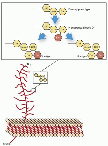
On the red cell membrane, both the A and B transferases add sugar moieties to a substrate that is encoded by the H (FUT1) gene. FUT1 is located at chromosome 19q13.1-qter, and the genes inherited at this locus are inherited in a Mendelian manner. Two alleles have been identified at this locus: H and h. The allele H, most frequently inherited, encodes for an enzyme termed H transferase type II [α(1,2) fucosyl-transferases; FUT1], which adds an L-fucose to the terminal galactose molecule of oligosaccharide chains in an α(1-2) linkage. This structure is called H substance, and it is to this structure that the A and B transferases add specific sugars resulting in A and B antigens. The rare allele sometimes inherited at the H locus is h. This h allele encodes for a nonfunctional transferase. If the h allele is inherited in the homozygous state (hh), L-fucose molecules (H substance) are not present on the red cell membrane. Without the presence of H substance on the red cell membrane, the A and B transferases, even when present, are not able to add the specific sugars that give A and B antigen specificity. This hh genotype is known as the Bombay phenotype: Serologically, the red cells group as O; however, unlike the true O phenotype, which has large amounts of H antigen on the red cells, red cells from the Bombay phenotype lack H antigen (Fig. 20.1). Children of a parent with the Bombay phenotype (hh) may have normal A or B antigen expression, or both, if they inherit
the dominant H allele from the other parent. The clinical relevance of the Bombay phenotype relates to the ability of these individuals to form not only anti-A and anti-B but also anti-H. The presence of all three of these antibodies makes it difficult to find compatible blood if transfusion is required. The only compatible blood for an individual with the Bombay (hh) phenotype is blood from another Bombay individual, and this phenotype is extremely rare.


TABLE 20.3 THE 700 SERIES (LOW-INCIDENCE ANTIGENS)


ISBT Number


Name


Symbol


700002


Batty


By


700003


Christiansen


Chra


700005


Biles


Bi


700006


Box


Bxa


700017


Torkildsen


Toa


700018


Peters


Pta


700019


Reid


Rea


700021


Jensen


Jea


700028


Livesay


Lia


700039


Milne


700040


Rasmussen


RASM


700044


JFV


700045


Katagiri


Kg


700047


Jones


JONES


700049


HJK


700050


HOFM


700052


SARA


700054


REIT


ISBT, International Society of Blood Transfusion.


Reference: International Society of Blood Transfusion Working Party on Terminology for Red Cell Antigens web site: http://www.isbtweb.org/fileadmin/user_upload/WP_on_Red_Cell_Immunogenetics_and/Table_of_low_incident_antigens_700_series_v2.0_110914.pdf. Accessed April 3, 2013.


TABLE 20.4 THE 901 SERIES (HIGH-INCIDENCE ANTIGENS)


ISBT Number


Name


Symbol


Incidence (%)


Implicated in Hemolytic Disease of the Newborn and/or Hemolytic Transfusion Reaction


901003


August


Ata


>99


Yes


901008


Emm


>99


No


901009


Anton


AnWj


>99


Yes


901011


Sid


Sda


91


No


901014


PEL


>99


Yes


901016


MAM


>99


Yes


ISBT, International Society of Blood Transfusion.


Adapted from International Society of Blood Transfusion Working Party on Terminology for Red Cell Antigens web site: http://www.isbtweb.org/fileadmin/user_upload/WP_on_Red_Cell_Immunogenetics_and/Table_of_high_incidence_antigens_901_series_v3.0_121028.pdf. Accessed April 3, 2013.


FIGURE 20.1 Biosynthesis of ABO blood group antigens. The antigens of the ABO system are located on the carbohydrate of type II oligosaccharides. H transferase is required to add fucose to the oligosaccharide chain and form H substance. Without the presence of H substance, A transferase and B transferase are not able to add terminal sugar moieties to the oligosaccharide chain. Fuc, l-fucose; Gal, d-galactose; Glc-NAc, d-N-acetyl-glucosamine.

The ABO(H) antigens are found not only on red cells, but also on other blood cells; in most body fluids (except cerebrospinal fluid); and on the cell membranes of tissues such as intestine, urothelium, and vascular endothelium. The expression of ABO(H) antigens on the red cell membrane and tissue membranes is controlled by the FUT1 (H) gene. The expression of ABO(H) antigens into body fluids is controlled by the FUT2 (Se) gene. The FUT2 gene, similar to the FUT1 (H) gene, is located at chromosome 19 (19cen-qter); however, they are not part of the ABO system. The dominate Se allele codes for H transferase type 1 [α(1,2) fucosyltransferase; FUT2]. Without the prior addition of a fucose to the oligosaccharide chains, A and B antigens would not be expressed in the body secretions, irrespective of the presence of A and B transferases (Fig. 20.1).

Despite the wide distribution of ABO(H) antigens in various cell membranes and body fluids, the normal physiologic function of these glycoproteins and glycolipids is largely unclear. The carbohydrate moieties of the ABO(H) antigens might contribute to the formation of glycocalyx. However, based on the observation that individuals who lack all ABH antigens (Bombay phenotype) have normal red cell survival and function, the role of ABO antigens in maintaining a state of health is unknown.10 There is some evidence that ABO blood groups may be associated with certain diseases.11 For example, the normal range of von Willebrand factor (vWF) antigen level varies among individuals with different ABO blood groups. Individuals with blood group O have the lowest vWF antigen level, followed by group A, then group B, and the highest levels in those with group AB.12 However,
the normal range of vWF is generally defined as the level in the normal general population irrespective of the ABO blood group. It has been postulated that ABO antigen expression can affect the glycosylation, thus altering the proteolysis and clearance of vWF.13,14 Studies have shown that individuals with blood group O are less susceptible to arterial and venous thromboembolism possibly associated with a lower level of vWF.14,15 and 16 In addition, blood group ABO may be associated with resistance to certain infections and, therefore, may offer a survival advantage. For example, individuals with blood group O may be less susceptible to severe Plasmodium falciparum malaria,17,18 but may be more likely to have severe infection with Vibria cholerae O13919,20 or Escherichia coli O139.21 Gastric cancer and peptic ulcer disease have been reported to be more prevalent in individuals with blood group O22,23 because of the association between blood group O and a gastric pathogen, Helicobacter pylori. It has been shown that H. pylori binds more readily to gastric epithelium in group O individuals because of lack of expression of ABO and Lewis antigens in non-hematopoietic tissue (see also section on Lewis antigens).24 In contrast, secretors of ABO and Lewis antigens in the gastrointestinal tract are more susceptible to norovirus,25 rhinoviruses, echoviruses, influenza viruses, and respiratory syncytial virus.26


Antibodies and Clinical Significance

All immunocompetent individuals produce antibodies against the missing ABO(H) blood group antigens (Table 20.5). Anti-A and anti-B production does not require red cell stimulation through transfusion or pregnancy but occurs predominately through environmental exposure, such as bacteria.27 Anti-A and anti-B are usually detectable within 3 to 6 months after birth.28 By the age of 5 years, the titers of anti-A and anti-B antibodies reach maximum and persist throughout adulthood. The titers of IgM anti-A and anti-B antibodies may gradually decline with advanced age.29 Newborn infants do not usually have a significant amount of anti-A or anti-B in their plasma; therefore, pretransfusion testing is not usually required for transfusions within the first 4 months of life. Infants born to alloimmunized mothers are an exception to this rule, as other specific blood group antibodies may have crossed the placenta and may be present in the infant’s circulation.

The “naturally occurring” anti-A and anti-B antibodies are predominantly IgM, although variable amounts of IgG may be present. Like most IgM immunoglobulins, ABO antibodies are especially effective at complement activation for two reasons: the antibodies have thermal activity most reactive at body temperature, and the high density of antigen sites on the red cell membrane allows for large numbers of antibodies to bind to the cell membrane. Therefore, the transfusion of ABO-incompatible blood typically presents as acute intravascular hemolysis. Anti-ABO antibodies, anti-human leukocyte antigen (HLA) antibodies, and anti-human platelet antigen (HPA) antibodies can cause alloimmunization resulting in platelet transfusion refractoriness.30 The ABH expression on the platelet is linked to the red cell phenotype; yet the expression on the platelet may be determined mainly by the enzyme H transferase type II (FUT1).31


TABLE 20.5 SUMMARY OF ABO GENES AND ANTIGENS


Phenotype


Antibody


Antigen


Gene Product


Gene


A


Anti-B


N-acetylgalactosamine


A transferase


9q34.1-q34.2


B


Anti-A


D-galactose


B transferase


9q34.1-q34.2


AB


None


N-acetylgalactosamine and D-galactose


A transferase and B transferase


9q34.1-q34.2


O


Anti-A and anti-B


L-fucose


Absent or nonfunctional A or B transferase


Absent or nonfunctional gene


A2


Anti-B and variable amount of anti-A1


N-acetylgalactosamine


A transferase


9q34.1-q34.2


In allogeneic stem cell transplant, ABO incompatibility may cause hemolysis, pure red cell aplasia, and delayed engraftment of donor cells.32 However, ABO incompatibility may not affect the long-term survival.32,33 After ABO unmatched hematopoietic stem cell transplantation, the patient’s red cells will switch to the donor’s blood group. Due to the fact that ABO antigens are not limited to the red cells, peripheral tissue may continue to express the recipient’s original antigens for life.34 These isohemagglutinin will be adsorbed onto the red cells and cause ABO discrepancy during crossmatch.35,36 Similarly, in kidney transplantation, ABO-incompatible recipients have higher graft loss in the immediate posttransplant period, but the long-term survival is similar to those with ABO-mismatched recipients.37 Moreover, delay in the production of ABO antibodies in infants may provide an “immunologic gap” for ABO-incompatible organ transplantation.38

The antibodies against ABO antigens are not a major cause of HDN for several reasons. First, soluble A or B substance in the fetal plasma can neutralize the alloantibodies. Second, antigens A or B expressed on other body tissues may also bind alloantibodies. Third, alloantibodies against ABO antigens are specific for sugar molecules; these antibodies generally have a weaker binding affinity than anti-bodies reacting with protein antigens such as the D antigen.

In routine blood group typing, the transfusion recipient’s red cells are typed using commercial sources of anti-A and anti-B antibodies (forward or cell typing). The presence of anti-A and anti-B in the serum/plasma of the recipient is detected by testing the serum/plasma against group A and group B red cells (reverse or serum typing). The interpretation of these two tests must agree for the patient’s blood group to be assigned. Sometimes, the serum and cell grouping do not agree; this is termed ABO discrepancy. In these circumstances, the laboratory should proceed with additional testing to correctly identify the patient’s ABO blood group. New technologies in blood group genotyping may identify and resolve these discrepancies.

Discrepancies in ABO grouping can be found in various conditions. For example, the ABO(H) antigens may be weakened in the donors with mutations of the ABO alleles. In patients with some types of leukemia or cancer, or having diseases associated with chromosome 9 translocations, the ABO(H) antigens may also be weakened and serologically typed as negative for that particular antigen. Alternatively, an individual may acquire an ABO antigen on the red cells; for example, one can acquire a B antigen after bacterial infections, or acquire an A antigen associated with Tn activation of the red cells.39


Rh Blood Group System (ISBT 004)


SUMMARY OF IMPORTANT CHARACTERISTICS OF RH ANTIBODIES


Antibody Specificity


Clinically Significant


Antibody Class


HDN


HTR


Frequency of Antigen-Negative Blood (White Population)


Anti-D


Yes


IgG; some IgM


Yes


Yes


Common (15%)


HDN, hemolytic disease of the newborn; HTR, hemolytic transfusion reaction; Ig, immunoglobulin.


The Rh blood group system was discovered by Landsteiner and Weiner in 1940.40 They injected rabbits with red cells from the rhesus monkey, and the antibody produced was initially termed Rh and is now known as anti-D. Unlike the ABO blood group system, Rh antibodies are not environmentally stimulated; however, the D antigen is highly immunogenic, causing anti-D formation in up to 70% of D-negative individuals who are exposed to D-positive blood. Anti-D is IgG and is known to cause HTRs and HDN.


Rh Terminology

Three different systems of nomenclature have been developed to describe the genes and antigens of the Rh blood group system antigens: The Wiener system, the Fisher-Race system, and the Rosenfield numeric terminology. Wiener proposed that the Rh antigens were the products of a single gene.41 The Fisher-Race nomenclature was based on the theory that reactions observed with various Rh antisera could be explained by three pairs of allelic genes: Cc, Dd, and Ee.42 Genetic analysis does not support either of these models. However, both the Wiener notation and the Fisher-Race nomenclature remain widely used today because of familiarity. In 1962, Rosenfield et al. proposed a system of nomenclature that was based on serologic findings.43 The symbols were used to convey phenotypic information rather than genetic information.44 In this system, the antigens are numerically named in order of their discovery or assignment to the Rh blood group system. The various nomenclatures for common Rh system antigens are listed in Table 20.6.


TABLE 20.6 RH BLOOD GROUP SYSTEM: ANTIGENS


Rosenfield Numeric Terminology


Fisher-Race Terminology


Wiener Terminology


Rh1


D


Rho


Rh2


C


rh′


Rh3


E


rh″


Rh4


c


hr′


Rh5


e


hr″


Rh6


ce (f)


hr


Rh7


Ce


rhi


Rh8


Cw


rhw1


Rh9


Cx


rhx


Rh10


V (Ces)


hrv


Rh11


Ew


rhw2


Rh12


G


rhG


Genes

The Rh antigens are encoded by two genes: RHD and RHCE. The genes are located at chromosome 1p36.11 (Table 20.1). RHD encodes for the D antigen, whereas RHCE encodes for the Cc and Ee antigens. The d antigen does not exist; however, by convention, d is used to connote the absence of the D antigen. RHD and RHCE each contain ten exons and are distributed over 69 kilobase pairs (kbp).45 Both the RHD and the RHCE genes encode for similar polypeptides of 417 amino acids with 12 membrane-spanning domains.45 In the red cell membrane, these two polypeptides form a complex with a glycoprotein termed the Rh-associated glycoprotein (RhAG), which is encoded by the RHAG gene (RH50) on chromosome 6. The RhAg and associated antigens have recently been designated the thirtieth blood group system. The structure of the Rh antigens suggests that they are transport proteins and the RhAG protein may play a role in the transport of ammonium. The Rh-associated proteins are transport proteins involved in ammonia/ammonium transport.46,47 The physiologic role of the Rh protein is poorly defined, but it has been suggested that these proteins may act to keep total blood ammonia low by trapping ammonium within the red blood cell.46,48


Antigens

There are 54 antigens that have been assigned to the Rh blood group system: D, C, E, c, e, f, Ce, Cw, Cx, V, Ew, G, Hro, Hr, hrs, VS, CG, CE, Dw, c-like, cE, hrH, Rh29, Goa, hrB, Rh32, Rh33, HrB, Rh35, Bea, Evans, Rh39, Tar, Rh41, Rh42, Crawford, Nou, Riv, Sec, Dav, JAL, STEM, FPTT, MAR, BARC, JAHK, DAK, LOCR, CENR, CEST, CELO, CEAG, PARG, and CEVF (Table 20.1). Of the 54 antigens in the Rh blood group system, the most common and important are D, C, E, c, and e. Although individuals can become alloimmunized to the C, c, E, and e antigens after red cell exposure through transfusion or pregnancy, these antigens are much less immunogenic than D. Less than 3% of individuals exposed to the C, c, E, and e antigens become alloimmunized; hence, pretransfusion testing is not routinely performed to match for these antigens. The principal phenotypes of the Rh blood group system and their frequencies are outlined in Table 20.7.

An individual is considered to be Rh positive if his or her red cells express the D antigen. The term Rh negative refers to the absence of the D antigen. The absence of the D antigen occurs in approximately 15% to 17% of individuals in white populations and is less frequent in other populations.49 In white populations, the absence of the D antigen is usually due to the deletion of the RHD gene.49 In Asian and black populations, the absence of the D antigen is usually due to an inactive RHD rather than a gene deletion.50,51


TABLE 20.7 RH BLOOD GROUP SYSTEM: PRINCIPAL PHENOTYPES


Haplotype Based on Antigens Present


Frequency (%)


Fisher-Race


Wiener


Whites


Blacks


Asians


DCe


R1


42


17


70


DcE


R2


14


11


21


Dce


R0


4


44


3


DCE


Rz


<0.01


<0.01


1


ce


r


37


26


3


Ce


r′


2


2


2


cE


r″


1


<0.01


<0.01


CE


ry


<0.01


<0.01


<0.01


Cis Product Antigens. Ce(rh1) is an antigen that almost always accompanies C and e when they are encoded by the same haplotype.44

Cc and Ee Variant Antigens. Various Rh antigens appear to be determined by alleles of the Cc and Ee antigens. Variants of C include Cw(Rh8), Cx(Rh9), and MAR. Variants of the E antigen include Ew(Rh11) and ET(Rh24). Variants of the e antigen include hrB (Rh31), hrs (Rh19), and es(Rh20).

G Antigen (Rh12). The G antigen is found on any red cell that also has the C or D antigen.


Weak D Phenotype

Some red cells that express the D antigen require prolonged incubation with the anti-D reagent or application of the antiglobulin test for agglutination to occur. These red cells are considered to be D antigen-positive and are described as weak D, formerly termed Du. The weak D phenotype is thought to occur by one of three mechanisms: (a) inheritance of an RHD gene encoding for a weakened expression of D, (b) interaction of the D allele with other genes, and (c) inheritance of an RHD gene missing some epitopes. In the first mechanism, the weak D phenotype is due to an RHD gene encoding for a D protein with reduced D antigen expression.44 This is more common in the black population and generally occurs in association with the Dce haplotype. In the white population, weak D is less common but may occur in association with the haplotype DCe or DcE.44 In the second mechanism, the weak D phenotype occurs as a result of the position of the D allele. This is most easily conceptualized using the Fisher-Race terminology. Red cells from individuals with a C allele in a trans position to the D allele (i.e., Dce/Ce) have weakened expression of D due to C being on the opposite chromosome. Individuals with the weak D phenotype by either of these two mechanisms do not form alloantibodies after exposure to D-positive red cells. Finally, the third mechanism, sometimes termed partial D, occurs when individuals lack part of the D antigen complex. The D antigen is thought to be a mosaic consisting of several individual parts or epitopes. Most D-positive individuals inherit the gene encoding the entire D antigen. However, the partial D phenotype describes red cells that are deficient in components of D, resulting in a decreased expression of the D antigen (weak D). Individuals with partial D may produce anti-D if transfused with D-positive red cells.

The weak D and partial D phenotypes have implications for the practice of transfusion medicine. Donors with weak D red cells should be considered Rh(D) positive. It is important that donor blood with weak D not be mislabeled as Rh(D) negative because the weak D antigen could induce an immune response if this blood were transfused to an Rh(D)-negative individual.33 It is generally recommended that patients who phenotype as weak D can be transfused with Rh(D)-positive blood. The most common types of weak D do not appear to be at risk of alloimmunization to the D antigen, therefore, these individuals can safely receive D-positive blood and would not require Rh immune globulin prophylaxis during pregnancy. However, it should be noted that some weak D types, identified by molecular analysis or the RHD gene are susceptible to alloimmunization.52


Rhnull Phenotype

The Rhnull phenotype occurs when red cells do not express Rh antigens. This phenotype occurs because of at least two mechanisms. First, the inheritance of an abnormal RHAG gene appears to result in the absence of the Rh antigen expression despite the presence of normal RHD and RHCE genes.30 This is termed regulator type Rhnull. The second mechanism is termed amorph type of Rhnull and involves the inheritance of a mutation in the RHCE genes in association with a D-negative background.53 The Rhnull phenotype is associated with abnormalities in the red cell membrane causing stomatocytosis and hemolysis. The presence of the Rh proteins in the red cell membrane appears to be necessary for the expression of other membrane proteins such as the LW, Duffy, and U antigens. Rhnull cells have been demonstrated to lack the LW and Fy5 antigens and have weakened expression of the S, s, and U antigens.54 Fortunately, the Rhnull phenotype is rare, as these individuals form an alloantibody (anti-Rh29) that reacts with all other red cells except Rhnull when they are transfused. Thus, obtaining compatible blood can be challenging.


Antibodies and Clinical Significance

Most Rh antibodies are IgG, although some may be IgM. They are usually not capable of activating complement. Anti-D is one of the most common Rh antibodies because of the high immunogenicity of the D antigen. Anti-D can cause severe HDN and HTR. The frequency of anti-D has greatly decreased with the use of prophylactic Rh immune globulin administration to Rh(D)-negative mothers during pregnancy and at delivery if the infant is Rh(D) positive. Antibodies against antigens of the Rh system including C, c, E, and e can be associated with mild HTR or HDN.49


Kell Blood Group System (ISBT 006)


SUMMARY OF IMPORTANT CHARACTERISTICS OF KELL ANTIBODIES


Antibody Specificity


Clinically Significant


Antibody Class


HDN


HTR


Frequency of Antigen-Negative Blood (White Population)


Anti-K


Yes


IgG; rarely IgM


Yes


Yes


Very common (98%)


Anti-k


Yes


IgG; rarely IgM


Yes


Yes


Rare (<2%)


HDN, hemolytic disease of the newborn; HTR, hemolytic transfusion reaction; Ig, immunoglobulin.


The Kell blood group system was discovered in 1946 and was named after Mrs. Kellner, the mother of the first child discovered to be affected with HDN because of Kell antibodies.55,56


Genes and Antigens

The Kell blood group system consists of 35 antigens: K, k, Kpa, Kpb, Ku, Jsa, Jsb, Ula, K11, K12, K13, K14, K16, K17, K18, K19, Km, Kpc, K22, K23, K24, VLAN, TOU, RAZ, VONG, KALT, KTIM, KYO, KUCI, KANT, KASH, KELP, KETI, KHUL, and KYOR. The antigens are coded by a complex of genetic loci, known as the Kell locus, which is located at 7q33 (Table 20.1). The KEL gene contains 19 exons distributed over 21.5 kbp.45 There are at least four subloci in the complex, each of which has an allele for a high-frequency antigen (k, Kpb, Jsb, and KEL11) and for one or more alleles for a lower-frequency antigen (K, Kpa and Kpc, Jsa, and KEL17).57 The most common haplotype is k/Kpb/Jsb/Kel11. The phenotype frequency of the common Kell antigens is shown in Table 20.8. The k antigen is a high-frequency antigen that is present in more than 98% of whites and blacks. The K antigen is much less common.

The KEL gene encodes a type 2 integral membrane protein, called zinc endopeptidase, containing 732 amino acids, which is present at 3,500 to 17,000 copies per red blood cell.45,56 The protein has enzymatic activity, and it has been demonstrated in vitro to cleave big endothelin-3, which is a peptide with vasoconstrictor activity.58 The Kell protein is associated with the Kx protein in the red cell membrane (see section Kx Blood Group System [ISBT 019]).59 Individuals have been identified who do not express the Kell protein (Kellnull phenotype) and who are healthy with no structural or functional abnormalities of their red cells identified.


TABLE 20.8 PRINCIPAL PHENOTYPES OF BLOOD SYSTEMS


Frequency (%)


Frequency (%)


System


Phenotype


Whites


Other


System


Phenotype


Whites


Other


ABO


O


45


49a


Xg


Xg(a+)


Males, 65.6;


A


40


27


females, 88.7


B


11


20


Xg(a-)


Males, 34.4;


AB


4


4


females, 11.3


MNS


M+N-


28


26a


Scianna


Sc:1,-2


99.7c


M+N+


50


44


Sc:1,2


0.3


M-N+


22


30


Sc:-1,2


Very rare


S+s-U+


11


3


Sc:-1,-2


Very rare


S+s+U+


44


28


Dombrock


Do(a+b-+)


17.2


11a


S-s+U+


45


69


Do(a+b+)


49.5


44


S-s-U-


0


<1


Do(a-b+)


33.3


45


S-s-U+w


0


Rare


Colton


Co(a+b-)


89.3


P


P1


79


94a


Co(a+b+)


10.4


Lutheran


Lu(a+b-)


0.15


Co(a-b+)


0.3


Lu(a+b+)


7.5


Co(a-b-)


Very rare


Lu(a-b+)


92.35


Landsteiner-Wiener


LW(a+b-)


>99


93.9d


Lu(a-b-)


Very rare


LW(a+b+)


<1


6.0


Kell


K+k-


0.2


Rarea


LW(a-b+)


Very rare


0.1


K+k+


8.8


2


LW(a-b-)


Very rare


Very rare


K-k+


91.0


98


Chido/Rodgers


Ch+, Rg+


95.0


Kp(a+b-)


Rare


0


Ch-, Rg+


2.0


Kp(a+b+)


2.3


Rare


Ch+, Rg-


3.0


Kp(a-b+)


97.7


100


Ch-, Rg-


Very rare


Js(a+b-)


0


1


Hh


H+


>99.9


Js(a+b+)


Rare


19


Kx


Kx+


˜100


Js(a-b+)


100


80


Kx-


Rare


Ko


Very rare


Very rare


Gerbich


Ge2, Ge3, Ge4


100


Lewis


Le(a+b-)


22


23a


Wb, Lsa, Ana, Dha


Rare


Le(a+b+)


72


55


Cromer


Cra, Tca, Dra, Esa, IFC, WESb, UMC


100


Le(a-b+)


6


22


Tcb, Tcc, WESa


Rare


Le(a-b-)


Rare


Rare


Duffy


Fy (a+b-)


17


9a


Knops


Kn(a+b-)


94.5


99.9a


Fy(a+b+)


49


1


Kn(a-b+)


1


0


Fy(a-b+)


34


22


Kn(a+b+)


4.5


0.1


Fy(a-b-)


Rare


68


McC(a+)


98


94


Kidd


Jk(a+b-)


28


57a


Sla(a+)


98


60


Jk(a+b+)


49


34


Yka(a+)


92


98


Jk(a-b+)


23


9


Indian


In(a+b-)


Very rare


Very raree


Jk(a-b-)


Very rare


Very rare


In(a+b+)


<1


7


Diego


Di(a+b-)


Rare


Rarea


In(a-b+)


>99


93


Di(a+b+)


Rare


Rare


Ok


Ok(a+)


100


Di(a-b+)


>99.9


>99.9


Raph


MER2+


92


Yt


Yt(a+b-)


91.9


97b


MER2-


8


Yt(a+b+)


7.9


23


Yt(a-b+)


0.2


0


a Blacks;

b Israelis;

c Most populations;

d Finns;

e Iranians/Arabs.


Data from AABB technical manual, 14th ed. Bethesda, MD: American Association of Blood Banks, 2002; and Denomme GA, Rios M, Reid ME. Molecular protocols in transfusion medicine. San Diego, CA: Academic Press, 2000.


Antibodies and Clinical Significance

The K antigen is very immunogenic. Only the D antigen has greater potential to induce alloimmunization. Because of this, anti-K is often encountered. Anti-K typically is induced by blood transfusion. The antibody tends to be IgG. Anti-K is clinically significant and has been implicated in both HTR and HDN. There is poor correlation with maternal anti-K titer and disease severity.60 Furthermore, HDN associated with anti-K tends to be more severe than HDN caused by anti-D. This is thought to occur because the Kell antigens are well expressed on fetal cells and appear on erythroid progenitor cells. It is postulated that anti-K, in addition to causing hemolysis, also causes a suppression of erythropoiesis.61,62

The k antigen is also highly immunogenic. However, because only individuals not expressing the k antigen (i.e., KK phenotype) produce anti-k, and because the k antigen is present in most individuals, anti-k is much less common. Anti-k has been associated with both HDN and HTR. The other Kell blood group system antibodies are much less common but are also clinically significant.


Duffy Blood Group System (ISBT 008)


SUMMARY OF IMPORTANT CHARACTERISTICS OF DUFFY ANTIBODIES


Antibody Specificity


Clinically Significant


Antibody Class


HDN


HTR


Frequency of Antigen-Negative Blood (White Population)


Fya


Yes


IgG


Yes


Yes


Common (34%)


Fyb


Yes


IgG


Yes


Yes


Common (17%)


HDN, hemolytic disease of the newborn; HTR, hemolytic transfusion reaction; Ig, immunoglobulin.


The Duffy blood group system was discovered in 1950 in the serum of a multiply transfused male patient with hemophilia, Mr. Duffy.63


Gene and Antigens

The Duffy system consists of six antigens, Fya, Fyb, Fy3, Fy4, Fy5, and Fy6, which are encoded at the Duffy locus on chromosome 1q21-q22 (Table 20.1). The gene, DARC (or FY), contains two exons distributed over 1.521 kbp.45 The antigens, Fya and Fyb, are encoded by a pair of codominant alleles. The phenotypes of the Duffy system and their frequencies are presented in Table 20.8. The most common phenotype in the white population is Fy(a+b+), and the most common phenotype in the black population is Fy(a-b-). The Fyx antigen represents a form of weak Fyb. The Fy5 antigen is defined by an interaction of the Duffy and Rh gene products.44 The antigens Fya, Fyb, and Fy6 are sensitive to denaturation by enzymes such as papain, ficin, or α-chymotrypsin. Fy3 and Fy5 are not sensitive to enzyme denaturation.

The Duffy DARC gene encodes for a glycoprotein (DARC) that is found on red cells as well as other tissues including brain, heart, lung, kidney, and spleen. On red cells, the glycoprotein has been identified as a receptor for various chemokines and may contribute to chemokine-induced leukocyte migration to sites of inflammation.64,65 DARC may also play a role in renal disease as increased DARC expression has been noted in different causes of renal injury.66 It is thought that DARC expression is increased in an attempt to control inflammation.66 The glycoprotein is also the receptor for P. vivax and P. knowlesi. Therefore, individuals who do not express Fya or Fyb on their red cells are not susceptible to these forms of malaria. In parts of Africa where malarial infection is common, most individuals are Fy(a-b-), likely because of natural selection.44


Antibodies and Clinical Significance

The Duffy antibodies are usually IgG. Anti-Fya is a common alloantigen. Fya is considered clinically significant, as it has been associated with HDN and HTR. Anti-Fyb is uncommon. It has been associated with mild HTR and only rarely with HDN.44,67 The other Duffy antibodies are much less common.


Kidd Blood Group System (ISBT 009)


SUMMARY OF IMPORTANT CHARACTERISTICS OF KIDD ANTIBODIES


Antibody Specificity


Clinically Significant


Antibody Class


HDN


HTR


Frequency of Antigen-Negative Blood (White Population)


Jka


Yes


IgG; rarely IgM


Yes


Yes


Common (23%)


Jkb


Yes


IgG; rarely IgM


Yes


Yes


Common (28%)


HDN, hemolytic disease of the newborn; HTR, hemolytic transfusion reaction; Ig, immunoglobulin.


The Kidd blood group system consists of three antigens: Jka, Jkb, and JK3 (Table 20.1). The system was named for the woman (Mrs. Kidd) whose serum was found to contain the antibody, and the antigen was named Jk for the initials of the woman’s child (John Kidd) affected by HDN.68


Gene and Antigens

The Kidd blood group system gene is located at chromosome 18q11-q12. The gene, SLC14A1, also known as JK or HUT11, is distributed over 30 kbp and contains 11 exons.45 It encodes for the urea transporter hUT-B1.69 The principal phenotypes of the Kidd blood group system and their frequencies are outlined in Table 20.8. The antigens Jka and Jkb are found at relatively the same frequencies in the white populations but differ in other ethnic groups such as blacks and Asians.45 The Jk(a-b-) phenotype is rare and is found primarily in the Polynesian population.70 These null red cells have been demonstrated to be resistant to lysis by 2M urea; however, this phenotype is not associated with shortened red cell survival or clinical symptoms.71


Antibodies and Clinical Significance

The Kidd antibodies are usually IgG but occasionally are IgM. These antibodies tend to be short-lived; therefore, they are frequently not detected before transfusion. However, they are capable of a rapid amnestic response and have been associated with severe delayed HTRs. The antibodies have rarely been associated with HDN, usually of mild severity.56


MNS Blood Group System (ISBT 002)


SUMMARY OF IMPORTANT CHARACTERISTICS OF MNS ANTIBODIES


Antibody Specificity


Clinically Significant


Antibody Class


HDN


HTR


Frequency of Antigen-Negative Blood (White Population)


Anti-M


Seldom


IgG; some IgM


Few


Few


Common (22%)


Anti-N


Rarely


IgM; rarely IgG


Rare


Rare


Very rare (<1%)


Anti-S


Occasionally


IgG; some IgM


Yes


Yes


Common (45%)


Anti-s


Yes


IgG; rarely IgM


Yes


Yes


Very rare (<1%)


HDN, hemolytic disease of the newborn; HTR, hemolytic transfusion reaction; Ig, immunoglobulin.


The MNS blood group system was discovered in 1927. The M and N antigens get their names from the letters in the word immune, because anti-M and anti-N antibodies were discovered in the sera of rabbits immunized with human red cells. The letter I was not used because it was felt this would be confused with the number 1.72,73 The S antigen was named after the city in which it was discovered—Sydney, Australia.73


Genes and Antigens

The MNS blood group system consists of 46 antigens: M, N, S, s, U, He, Mia, Mc, Vw, Mur, Mg, Vr, Me, Mta, Sta, Ria, Cla, Nya, Hut, Hil, Mv, Far, sD, Mit, Dantu, Hop, Nob, Ena, ENKT, N′, Or, DANE, TSEN, MINY, MUT, SAT, ERIK, Osa, ENEP, ENEH, HAG, ENAV, MARS, ENDA, ENEV, and MNTD (Table 20.1). The M and N antigens are carried on glycophorin A, whereas the antithetical antigens, S and s, are carried on glycophorin B. They are encoded by the GYPA, GYPB, and GYPE genes located at chromosome 4q31.21. GYPA has seven exons distributed over 60 kbp.45 GYPB has five exons and is distributed over 58 kbp.45 GPA and GPB contribute to most of the carbohydrate on the red cell membrane. The sialic acid of the O-glycans on these sialoglycoproteins results in the Tred cell membrane being negatively charged. This negative charge prevents red cells from sticking together, prevents red cell adherence to endothelial cells of the blood vessel walls, and protects from invasion by pathogens.74,74,75,76 The presence of glycophorin A has been demonstrated to be necessary for the adhesion of certain malarial parasites (P. falciparum) as well as for bacteria and viruses (e.g., influenza virus and encephalomyocarditis virus).76,77,78,79 and 80 Other proposed functions of glycophorin A include regulation of transport of band 3 to the red cell membrane and complement regulation.70,78,81,82 and 83 The main phenotypes and frequencies of the MNS blood group system are listed in Table 20.8.


Antibodies and Clinical Significance

Anti-M and anti-N antibodies are typically IgM and are reactive at cold temperatures. Antibodies against M and N are naturally occurring (environmentally stimulated). They are not generally considered to be clinically significant. Rarely, anti-M has been implicated in cases of HDN and HTR.61,62,63 and 64 Anti-N has only rarely been associated with HDN or HTR. In contrast, antibodies against S, s, and U are capable of causing HTR and HDN.70 Anti-S and anti-s tend to be IgG and occasionally are IgM. Antibodies to many low-prevalence antigens in the MNS system have also been associated with HDN.76


P Blood Group System and Related Antigens (ISBT 003)


SUMMARY OF IMPORTANT CHARACTERISTICS OF P ANTIBODIES


Antibody Specificity


Clinically Significant


Antibody Class


HDN


HTR


Frequency of Antigen-Negative Blood (White Population)


P1


Rare


IgM; rarely


No


Rare


Common (21%)


IgG


HDN, hemolytic disease of the newborn; HTR, hemolytic transfusion reaction; Ig, immunoglobulin.


The first P system antigen was discovered by Landsteiner and Levine in 1927.44 Initially, the system was thought to include the P, Pk, and LKE antigens, but these antigens have subsequently been assigned to the globoside antigens (see section GLOB Collection [ISBT 209]).44


Genes and Antigens

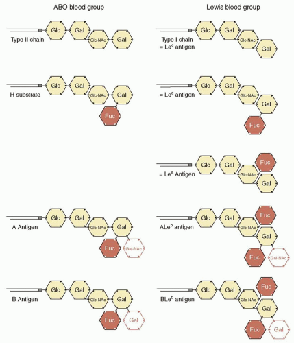
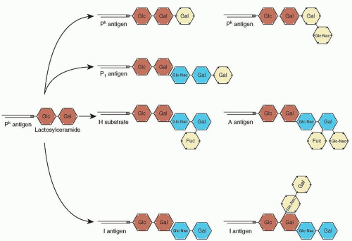
The P blood group system consists of a single antigen: P1 (Table 20.1). P1 is the product of a galactosyltransferase encoded by the gene P1, which is located at chromosome 22q11.2-qter.45 It has been suggested that this transferase is encoded by the gene A4GALT, but this has been contradicted by recent studies.52 The P1 antigen is similar to the ABO antigens because it is composed of a chain of sugars linked to glycolipids on red cells (Fig. 20.2). P1 is formed when β-D-galactose (Gal) is added in an α(1-4) linkage to paragloboside.44 In the white population, 79% of individuals express the P1 antigen.44


Antibodies and Clinical Significance

Anti-P1 is naturally occurring and is commonly found in individuals lacking the P1 antigen. Anti-P1 is usually IgM. It has not been reported to cause HDN and has been associated with HTR only in rare instances.84 Identification of anti-P1 is aided by the fact that the activity of the antibodies is inhibited by hydatid cyst fluid or pigeon egg white.44

Individuals who lack the P1 antigen who also do not express the P and Pk antigens may produce anti-P,P1,Pk, also known as anti-Tja. This antibody can be either IgM or IgG and has been associated with severe hemolytic reactions.85,86


Lutheran Blood Group System (ISBT 005)


SUMMARY OF IMPORTANT CHARACTERISTICS OF LUTHERAN ANTIBODIES


Antibody Specificity


Clinically Significant


Antibody Class


HDN


HTR


Frequency of Antigen-Negative Blood (White Population)


Lua


Seldom


IgM; some IgG


Mild


No


Very common (92%)


Lub


Seldom


IgG; some IgM


Mild


No


Very rare (0.1%)


HDN, hemolytic disease of the newborn; HTR, hemolytic transfusion reaction; Ig, immunoglobulin.


FIGURE 20.2 Differences between ABO (A) and Lewis (B) blood groups. ABO blood group antigens are synthesized in the red cells on type II oligosaccharides, but Lewis blood group antigens are produced in the plasma on type I oligosaccharides and then adsorbed onto the red cell surface. Type II oligosaccharide chains differ from type I chains in the linking position of the terminal galactose moiety. The Le (FUT3) gene encodes type III H transferase, which adds a fucose group (redcolored fucose group) to the second-last sugar moiety of the type I oligosaccharide chain. Synthesis of the Lec and Led antigens does not depend on the activity of the Le gene. Fuc, l-fucose; Gal, d-galactose; Gal-NAc, d-N-acetyl-galactosamine; Glc, d-glucosamine; Glc-NAc, d-N-acetyl-glucosamine.

The first Lutheran blood group antibody (anti-Lua) was found in 1946 in the serum of a previously transfused patient named Lutteran. The label on the sample was misread, and the blood group system was named Lutheran.70


Genes and Antigens

The Lutheran blood group system consists of 20 antigens: Four pairs of antigens (Lua/Lub, Lu6/Lu9, Lu8/Lu14, and Aua/Aub) and 11 independent antigens (Lu3, Lu4, Lu5, Lu7, Lu11, Lu12, Lu13, Lu16, Lu17, Lu20, Lu21, and LURC) (Table 20.1).6 The antigens are encoded by the B-CAM (LU) gene located at chromosome 19q12-q13. The gene contains 15 exons and is distributed over approximately 12 kbp.39 The gene products include the Lutheran glycoprotein, which is 597 amino acids long, and a spliced version of the B-cell adhesion molecule (B-CAM), which is 557 amino acids in length.45,70 The Lutheran glycoproteins are members of the Ig superfamily and have been demonstrated to act as a receptor for laminin.87,88 The Lutheran blood group may play a role in the pathophysiology of sickle cell disease and polycythemia vera. In sickle cell disease, B-CAM and Lu are overexpressed on red cells, which may mediate increased red cell adhesion to laminin.89 In polycythemia, there is increased phosphorylation of the Lu glycoprotein, which increases red cell adhesion.90 The principal phenotypes of the Lutheran blood group system and their frequencies are outlined in Table 20.8.


Antibodies and Clinical Significance

The Lutheran antigens are not very immunogenic; therefore, antibodies in this system are rare.70 Anti-Lua and anti-Lub are usually IgG and are reactive on the indirect antiglobulin test. Anti-Lua has not been implicated in HTR but has been implicated in mild HDN.70 Anti-Lub has been associated with mild, subclinical HDN.91,92


Lewis Blood Group System (ISBT 007)


SUMMARY OF IMPORTANT CHARACTERISTICS OF LEWIS ANTIBODIES


Antibody Specificity


Clinically Significant


Antibody Class


HDN


HTR


Frequency of Antigen-Negative Blood (White Population)


Lea


Seldom


IgM


No


Rare


Rare (6%)


Leb


Seldom


IgM


No


Rare


Common (22%)


HDN, hemolytic disease of the newborn; HTR, hemolytic transfusion reaction; Ig, immunoglobulin.


The Lewis blood group system is different from other blood group systems, as the antigens (Lea and Leb) are formed in the plasma and absorbed onto the red cell membrane. This unique feature has implications for transfusion practices for several reasons: (a) transfused red cells always absorb Lewis antigens from the plasma of the transfusion recipient; hence, within several days of the transfusion, the phenotype of the circulating transfused red cells is the same as the patient’s red cell phenotype; and (b) soluble antigen in transfused plasma has the potential to inhibit Lewis system antibodies that may be in the plasma of some individuals.


Genes and Antigens

The two alleles (Le and le) are inherited in Mendelian fashion. Le is the dominant allele, and le is the recessive allele. The FUT3 (Le) gene is located on chromosome 19 (19p13.3)93,94 and is closely linked to the FUT1 (Hh) and FUT2 (Sese) genes. There are 11 different mutations of the FUT3 gene.95,96 The silent le allele is due to mutations that result in defective gene products. The FUT3 gene encodes for an enzyme, α(1,3/1,4) fucosyltransferase (FUT3, H transferase type 3), which adds fucose molecules to carbohydrate precursor chains (Fig. 20.3). The enzyme itself is a type II membrane-bound protein of 361 amino acids.97


FIGURE 20.3 The relationship among ABO, P, and Ii blood group systems. These antigens are located on terminal oligosaccharides. Among these blood groups, the structure in common is lactosylceramide (red sugar moieties). Lewis blood group antigens also share similar structures, but they are not synthesized in red cells. Fuc, l-fucose; Gal, d-galactose; Gal-NAc, d-N-acetyl-galactosamine; Glc, d-glucosamine; Glc-NAc, d-N-acetyl-glucosamine.


Antigens

As mentioned previously, Lewis antigens (Lea and Leb) on the red cells are not intrinsic to the red cell membrane but are absorbed from the plasma onto the cells.98 The antigenic epitopes are located at the fucose moieties of glycosphingolipids. The biosynthesis of the antigens Lea and Leb involves two different pathways. The formation of antigen Lea is catalyzed by the enzyme FUT3 that adds a fucose group to the type 1 oligosaccharide precursor chain (Fig. 20.3). In individuals with FUT2 (SeSe or Sese genotypes), most type 1 oligosaccharide precursor chains are converted to an intermediate product similar to the H antigen. The intermediate product is subsequently catalyzed by the enzyme FUT3 and forms the Leb antigen. Therefore, the phenotypes Le(a-b+) and Le(a+b-) are not due entirely to the FUT3 gene but depend on the presence or absence of the FUT2 (Se) gene. This relationship between the FUT2 and FUT1 genes has practical implications for the laboratory. For example, the easiest way to determine an individual’s secretor status is to type his or her red cells to determine their Lewis phenotype. The Le(a+b-) phenotype indicates that the individual is a nonsecretor; the Le(a-b+) phenotype indicates that the individual is a secretor; and the Le(a-b-) phenotype does not allow secretor status to be assigned. If the FUT2
product (FUT2) is partially active, such as in individuals with Sew, some type 1 precursor chain is converted to Lea antigen, and the remaining is converted to Leb. The resultant phenotype, Le(a+b+), is rare in whites but common in Asians. The fucose residual may also be added to the type 2 oligosaccharide precursor chain and forms the Lex and Ley antigens that are similar to ABO(H) antigens in biochemical structure.

In the Lewis blood group system, the phenotype distribution varies among different ethnic groups. The Le(a-b+) phenotype is found in 70% of the white population and approximately 50% of the black population. The Le(a-b-) phenotype is less common in the white population but is found in approximately 30% of the black population. The Le(a+b+) phenotype is rare in both the white and black populations (Table 20.8).

In addition to red blood cells, Lewis antigens are found on other cell surfaces, such as gastric mucosa. They are also found in the lipopolysaccharide envelope of Helicobacter pylori.99 Although the gastric mucosa predominately expresses Lea and Leb, the cell envelope of H. pylori mainly expresses Lex and Ley.99 The relationship between the Lewis antigens and the pathogenesis of H. pylori infection is uncertain. The similarity of the antigens may deceive the host immune system and facilitate the colonization of H. pylori.100 In a murine model, H. pylori infection has been shown to induce antibodies cross-reactive to gastric mucosa and to contribute to the development of chronic gastritis and peptic ulcers.101 However, this has not been firmly established in humans because no concordance in the expression of the Lewis antigen has been found between the bacteria and the host.102,103 Some studies showed that Leb and H antigens on the gastric mucosa mediated the binding of H. pylori via a binding protein (blood group antigen-binding adhesin).104,105 However, other studies demonstrated that H. pylori adherence is not dependent on the Lewis antigen.106 Similarly, it has been disputed whether blood group O is a risk factor for peptic ulcer disease.107,108 and 109 Also, the isoform antigens Lex and Ley may be found as neoantigens on malignant tissue.110


Antibodies and Clinical Significance

Like the ABO system, antibodies specific for Lewis antigens are naturally occurring, being formed through exposure to environmental antigens. The antibodies are usually IgM, complement activating, and reactive at or below room temperature. Although Lewis antibodies have been reported to cause HTRs, this does not usually occur for several reasons. First, although Lewis antibodies can activate complement, the process is relatively slow, thus allowing the inhibitors within the complement cascade to stop the process before the membrane attack complex is activated. Second, soluble antigen present in the plasma of the transfused blood product can neutralize the antibody, preventing subsequent binding of antibody to transfused red cells. Finally, within 24 to 48 hours of transfusion, the transfused red cells absorb Lewis antigens from the patient’s plasma, taking on the Lewis phenotype of the patient’s own red cells. This latter mechanism also prevents delayed transfusion reactions from occurring. Lewis antibodies may be clinically relevant if the antibody causes in vitro hemolysis during serologic laboratory testing. When these “in vitro hemolytic antibodies” are detected, they should be considered clinically relevant, and antigen-negative blood should be selected for transfusion. Lewis antibodies do not cause HDN because they are IgM and do not cross the placenta. Furthermore, Lewis antigens are poorly developed on fetal red cells.

Anti-Lea antibody is more common than anti-Leb antibody. Both anti-Lea and anti-Leb antibody are found in individuals with the Le(a-b-) phenotype. In contrast, individuals with the Le(a-b-) phenotype do not develop anti-Lea because of the presence of residual Lea antigen in the secretions.


Diego Blood Group System (ISBT 010)


SUMMARY OF IMPORTANT CHARACTERISTICS OF DIEGO ANTIBODIES


Antibody Specificity


Clinically Significant


Antibody Class


HDN


HTR


Frequency of Antigen-Negative Blood (White Population)


Dia


Yes


IgG; some IgM


Yes


Rare


Very common (>99.9%)


Dib


Yes


IgG; some IgM


Yes


Rare


Very rare (<0.1%)


HDN, hemolytic disease of the newborn; HTR, hemolytic transfusion reaction; Ig, immunoglobulin.


The Diego blood group system consists of 22 antigens: Dia, Dib, Wra, Wrb, Wda, Rba, WARR, ELO, Wu, Bpa, Moa, Hga, Vga, Swa, BOW, NFLD, Jna, KREP, Tra, Fra, SW1, and DISK (Table 20.1).6


Gene and Antigens

The Diego system antigens are carried on the band 3 protein. The antigens are encoded by the gene SLC4A1, which is found at chromosome 17q12-q21 (Table 20.1). SLC4A1 contains 20 exons and is distributed over 228 kbp.45 Band 3 is a multipass transmembrane protein that serves as an anion transporter. The Dia antibody was initially found in the serum of a Venezuelan woman and was implicated in HDN.111 The principal phenotypes of the Diego blood group system and their frequencies are outlined in Table 20.8. The expression of the antigen Dia is almost exclusively restricted to populations of Mongolian descent, such as American Indians, Chinese, and Japanese.100 An altered version of the band 3 protein is found in a condition known as Southeast Asian ovalocytosis. This condition may confer a degree of resistance to Plasmodium falciparum.112


Antibodies and Clinical Significance

Anti-Dia and anti-Dib are usually IgG and are detected by the indirect antiglobulin test. HDN has been reported with both anti-Dia and anti-Dib.113,114,115,116 and 117 HTR is rare but has been reported.118


Yt Blood Group System (Cartwright) (ISBT 011)


SUMMARY OF IMPORTANT CHARACTERISTICS OF YT ANTIBODIES


Antibody Specificity


Clinically Significant


Antibody Class


HDN


HTR


Frequency of Antigen-Negative Blood (White Population)


Yta


Rare


IgG


No


Yes


Very rare (0.2%)


Ytb


Rare


IgG


No


Yes


Very common (91.9%)


HDN, hemolytic disease of the newborn; HTR, hemolytic transfusion reaction; Ig, immunoglobulin.


The Yt system consists of two antigens: Yta and Ytb (Table 20.1). The system was named after Cartwright, the individual discovered producing the antibody. Because all of the other letters in the individual’s name were already being used, the last letter, T, was selected. The letter Y was placed first to denote “why not T?”.70


Gene and Antigens

The ACHE gene is located at chromosome 7q22. It encodes for an acetylcholinesterase, which is a dimerized glycosyl phosphatidylinositol (GPI)-linked glycoprotein in the red blood cell membrane. Its function is unknown; however, the molecule is enzymatically active. Yta and Ytb antigens result from a single amino acid substitution, which does not appear to affect the enzymatic activity of acetylcholinesterase.119 The antigens are antithetical. Yta occurs with a frequency of approximately 99%, and Ytb has a frequency of approximately 8% (Table 20.8).120 Because they are carried on a GPI-linked protein, Yt antigens may be absent or reduced in individuals with paroxysmal nocturnal hemoglobinuria (see Chapter 31).


Antibodies and Clinical Significance

Antibodies against the Yt antigens are usually IgG. They do not activate complement. Yt antibodies have been implicated in delayed HTR but not in HDN.112


Xg Blood Group System (ISBT 012)


SUMMARY OF IMPORTANT CHARACTERISTICS OF XG ANTIBODIES


Antibody Specificity


Clinically Significant


Antibody Class


HDN


HTR


Frequency of Antigen-Negative Blood (White Population)


Xga


No


IgG


No


No


Common (males, 34.4%; females, 11.3%)


HDN, hemolytic disease of the newborn; HTR, hemolytic transfusion reaction; Ig, immunoglobulin.


The Xg blood group system consists of two antigens: Xga and CD99 (Table 20.1). The Xga antigen was discovered in 1962 by Mann in the serum of a multiply transfused male. Because the antigen frequency appeared to differ between males and females, the antigen was named Xga, as it appeared to be controlled by the X chromosome. The γ in the name stands for Grand Rapids, the hometown of the patient.121


Gene and Antigens

The gene encoding Xga, XG, is found at Xp22.32. The gene is not subject to lyonization or X inactivation. The gene that encodes CD99, now also named CD99 (previously MIC2), is closely linked to XG. The Xga antigen is located on a sialoglycoprotein. The antigen is only weakly expressed on the red cells of infants. The Xga antigen is resistant to treatment by sialidase and dithiothreitol (DTT); however, it is sensitive to treatment with proteolytic enzymes. The principal phenotypes of the Xg blood group system and their frequencies are outlined in Table 20.8.


Antibodies and Clinical Significance

Anti-Xga antibodies are usually IgG and may be able to activate complement. Anti-Xga can be naturally occurring.122 Alloantibodies to CD99 have been detected in only two healthy individuals and are not thought to be clinically significant.122 There have been no documented cases of HTR or HDN with these antibodies.


Scianna Blood Group System (ISBT 013)


SUMMARY OF IMPORTANT CHARACTERISTICS OF SCIANNA ANTIBODIES


Antibody Specificity


Clinically Significant


Antibody Class


HDN


HTR


Frequency of Antigen-Negative Blood (White Population)


Sc1


No


IgG


Rare


No


Very rare (<1%)


Sc2


No


IgG


Rare


No Very common (>99%)


HDN, hemolytic disease of the newborn; HTR, hemolytic transfusion reaction; Ig, immunoglobulin.


The Scianna blood group system consists of seven antigens: Sc1, Sc2, Sc3, Rd, STAR, SCER, and SCAN (Table 20.1).


Gene and Antigens

The gene for this system, ERMAP, is located on chromosome 1p34 and is linked to Rh. ERMAP encodes a glycoprotein (erythroid membrane-associated protein), the function of which is not known. However, based on its structure, ERMAP is known to be a member of the butyrophilin-like family of the immunoglobulin superfamily.123 The Sc1 antigen is a high-frequency antigen, with the incidence in most populations being close to 99.9% (Table 20.8).45 Sc2 is antithetical to Sc1 and is present in approximately 1% of the population.45 The antigens are resistant to treatment of red cells with proteolytic enzymes such as papain, ficin, trypsin, α-chymotrypsin, and sialidase.70


Antibodies and Clinical Significance

The Scianna antibodies are usually IgG. Scianna antibodies can activate complement and have been reported to cause mild HDN.124,125,126 The antibodies have not been associated with severe HTR.125,126


SUMMARY OF IMPORTANT CHARACTERISTICS OF DOMBROCK ANTIBODIES


Antibody Specificity


Clinically Significant


Antibody Class


HDN


HTR


Frequency of Antigen-Negative Blood (White Population)


Doa


Rare


IgG


No


Yes


Common (33.3%)


Dob


Rare


IgG


No


Yes


Common (17.2%)


HDN, hemolytic disease of the newborn; HTR, hemolytic transfusion reaction; Ig, immunoglobulin.


Dombrock Blood Group System (ISBT 014)

The Dombrock blood group system consists of eight antigens: Doa, Dob, Gya, Hy, Joa, DOYA, DOMR, and DOLG (Table 20.1).4,127


Gene and Antigens

The gene for the Dombrock blood group system, ART4 (or DO), is found at chromosome 12q13.2-q13.3.128 The antigens
in the Dombrock system have been demonstrated to be carried on a GPI-linked glycoprotein.129,130,131 and 132 Homology studies suggest that the Dombrock glycoprotein is a member of the adenosine 5′-diphosphate ribosyl-transferase ectoenzyme gene family and has been identified as ADP-ribosyltransferase 4 (ART4).131,132 and 133,134,135 However, the function of this molecule is uncertain, as no enzymatic activity has been demonstrated on the red cells and no pathology has been associated with absence of this glycoprotein.135

The antigens Doa and Dob are antithetical. The frequencies of the three main phenotypes defined by these antigens are listed in Table 20.8. The antigens Gya, Hy, and Joa are high-incidence antigens, with gene frequencies of >99% in all populations studied.136 The antigens are resistant to papain or ficin treatment but sensitive to trypsin, pronase, or DTT (200 mmol/L) treatment.133


Antibodies and Clinical Significance

The Dombrock antibodies are usually IgG and do not activate complement. They have not been associated with clinical HDN, but the antibodies have been associated with severe HTR.135,137,138 and 139 A baby born to a mother with an anti-DOMR antibody required phototherapy for hyperbilirubinemia.4


Colton Blood Group System (ISBT 015)


SUMMARY OF IMPORTANT CHARACTERISTICS OF COLTON ANTIBODIES


Antibody Specificity


Clinically Significant


Antibody Class


HDN


HTR


Frequency of Antigen-Negative Blood (White Population)


Coa


Yes


IgG


Yes


Yes


Very rare (0.3%)


Cob


Yes


IgG


No


Yes


Very common (90.3%)


HDN, hemolytic disease of the newborn; HTR, hemolytic transfusion reaction; Ig, immunoglobulin.

Only gold members can continue reading. Log In or Register to continue

Stay updated, free articles. Join our Telegram channel

Oct 21, 2016 | Posted by in HEMATOLOGY | Comments Off on Red Cell, Platelet, and White Cell Antigens

Full access? Get Clinical Tree

Get Clinical Tree app for offline access