Platelet Structure and Function in Hemostasis and Thrombosis


Platelet Structure and Function in Hemostasis and Thrombosis


Susan S. Smyth


PLATELET STRUCTURE


Structural and Functional Anatomy


Light Microscopy

Light microscopy of Wright-stained smears (Fig. 16.1) reveals platelets as small, anucleate (i.e., lacking a nucleus) fragments with occasional reddish granules, measuring approximately 2 µm in diameter with a volume of approximately 8 fl1 and exhibiting considerable variation in size and shape. Platelets released from the marrow under “conditions of stress” such as thrombocytopenia and termed stress platelets are large and often beaded in shape, whereas young platelets, recently released from the marrow, are termed reticulated in reference to their RNA content and in analogy to young red cell reticulocytes.2


Electron Microscopy and Subcellular Features

Platelets exist in two distinct forms, resting and activated, with the resting state marked by baseline metabolic activity and the activated form resulting from agonist stimulation (e.g., response to thrombin). By scanning electron microscopy, circulating resting blood platelets appear as flat discs with smooth contours, rare spiny filopodia (Fig. 16.2), and random openings of a channel system, the surface-connected canalicular system (SCCS), which invaginates throughout the platelet and is the conduit by which granule contents exocytose after stimulation.3,4 Although the platelet is anucleate, transmission electron microscopy reveals a complex surface and a cytoplasm packed with a number of different subplatelet structures and organelles that are essential to the maintenance of normal hemostasis (Figs. 16.3 and 16.4). Initial descriptions of platelet anatomy stem from studies employing transmission electron microscopy; and platelet structure is classified into four general areas: the platelet surface, membranous structures, cytoskeleton, and granules.


Platelet Surface


Plasma Membrane

The platelet plasma membrane separates intra- from extracellular regions and, in thin sections, exhibits a typical 20-nm-thick trilaminar structure5 whose overall appearance does not differ from that of other blood cells.6 The platelet membrane is exceptionally complex in composition, distribution, and function, incorporating a number of glycoproteins (GPs) and lipids into its phospholipid bilayer and integrating a variety of extra- and intraplatelet events such as permeability, agonist stimulation, and platelet adhesion, activation/secretion, and aggregation.

The lipid composition of the membrane is distributed in an asymmetric manner, with neutral species located mainly in the outer layer, and anionic forms, such as phosphatidylserine (PS), concentrated on the inner side.7 This sequestration of PS, which promotes plasma coagulation by contributing to the prothrombinase complex,8 on the inner side of the membrane may account for the fact that resting platelets are essentially nonreactive in terms of thrombin generation.9 On the other hand, activated platelets make a major contribution to thrombin formation through the interactions of factors Xa and Va and prothrombin on their surface.9,10 The plasma membrane also contains sodium and calcium adenosine triphosphatase (ATPase) pumps, which are important for maintaining ionic homeostasis.11


Platelet Membranous Systems

Platelets have features of muscle-related cells in terms of their high content of actin and their contractile response during activation. Similar muscle-like qualities are found in the two membranous systems of platelets, the SCCS and the dense tubular system, which resemble transverse tubules and sarcotubules, respectively.12


Surface-connected Canalicular System

The SCCS, also called the open canalicular system, is fenestrated and contiguous with the surface plasma membrane.3,13,14 The SCCS has several prominent functional roles: first, as an internal reservoir of membrane to facilitate platelet spreading and filopodia formation after adhesion;15 and second, as a storage reservoir for membrane GPs, such as αIIbβ3 (GPIIb-IIIa), that increase on the platelet surface after activation.16 The system also provides a route for granule release during the secretory phase of platelet activation4 and serves as a route of ingress and egress for molecules as they translocate between the plasma and the platelet.


Dense Tubules

Unlike the SCCS, the dense tubular system is a closed-channel system consisting of narrow, membrane-limited tubules, approximately 40 to 60 nm in diameter.17 It contains residual smooth endoplasmic reticulum from the megakaryocyte.18,19

Peroxidase,20,21 and 22 glucose 6-phosphatase,23 acetylcholinesterase (in cat, rat, and mouse but not human platelets or megakaryocytes),24 adenylate cyclase, and Ca2+– and Mg2+-activated ATPases25 have been cytochemically demonstrated in the dense tubular system.

This channel system is involved in the regulation of intracellular calcium transport because it has been reported to selectively bind, sequester, and release divalent cations after activation.25
The dense tubular system is also the site of prostaglandin (PG) synthesis in platelets.26,27


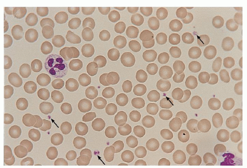
FIGURE 16.1. A human peripheral blood smear stained with Wright-Giemsa. Platelets, indicated by arrows, are interspersed between erythrocytes and a few leukocytes. The pale, grayish-blue cytoplasm contains purple-red granules. Original magnification of 35-mm slide ×100.


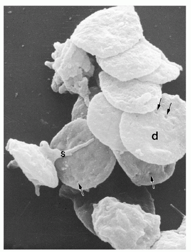
FIGURE 16.2. Scanning electron micrograph of unstimulated human platelets. Most are discoid (d) in shape. Many surface indentations, indicated by arrows, are present; these correspond to openings of the surface-connected canalicular system to the external milieu. Magnification ×15,000. (Data from Stenberg PE, Shuman MA, Levine SP, Bainton DF. Optimal techniques for the immunocytochemical demonstration of β-thromboglobulin, platelet factor 4, and fibrinogen in the alpha granules of unstimulated platelets. Histochem J 1984;16:983-1001.)


Platelet Cytoskeleton

(See additional cytoskeleton information in the section “Role of the Cytoskeleton in Platelet Function.”)

The shape of platelets and their ability to contract and spread depend on an organized cytoskeleton.28 The cytoskeleton can direct platelet shape change, send out extracellular extensions, collect and then extrude secretory granules, and affect surface reactivity (Fig. 16.5). These varied functions are performed by three distinct structures: first, the membrane skeleton, which buttresses the inner side of the plasma membrane; second, the mass of actin and intermediate filaments, which fills the cytoplasm; and third, the circumferential microtubule band, which encircles the substance of the platelet to produce the resting disclike form.15,28 Three different protein filaments/tubules contribute to the overall network: 5- to 6-nm-diameter microfilaments of actin,29,30 10- to 12-nm intermediate filaments of desmin and vimentin,31,32 and 25-nm microtubules composed mainly of tubulin.33,34 and 35 Together, these filaments, depending on the activation state of the platelet, comprise 30% to 50% of total platelet protein.


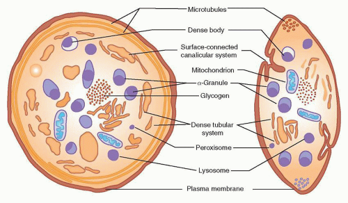
FIGURE 16.3. Diagram of a human platelet displaying components visible by electron microscopy and cytochemistry. In addition to membranous components (plasma membrane, surface-connected canalicular system, and dense tubular system), mitochondria, microtubules, and glycogen, four types of storage organelles are identified: α-granules, dense bodies, lysosomes, and microperoxisomes. Whereas the first two can be identified morphologically, microperoxisomes and lysosomes are recognizable only by cytochemical stains. (From Bentfeld-Barker ME, Bainton DF. Identification of primary lysosomes in human megakaryocytes and platelets. Blood 1982;59:472-481, with permission.)


Membrane Skeleton

The membrane skeleton was first described more than 30 years ago by electron microscopy36,37 and then analyzed biochemically by detergent lysis in Triton X-100.28,38 The membrane skeleton contains short actin filaments that connect surface receptors with the bulk of cytoplasmic actin filaments.29 Filamin-A, a component of the membrane cytoskeleton, links the cytoplasmic domain of GPIbα with actin filaments and also binds integrins. Other components of the membrane skeleton include structural and signaling molecules such as spectrin, talin, migfilin, Src, and small GTPase family members (Table 16.1).15,28 Through tight regulation, the interactions of these proteins allow platelets to sense and to respond to environmental cues.


Cytoplasmic Actin and Intermediate Filaments

The bulk of the platelet cytoskeleton consists of a large amount of actin (Mr = 42,000) that comprises approximately 25% of total platelet protein.39 Other platelet cytoskeletal proteins, such as tropomyosin and α-actinin, are present in lower amounts (2% to 5% of total platelet protein).40,41,42 and 43 The actin exists in soluble, monomeric (G-actin), and filamentous (F-actin) forms and connects to both the membrane skeleton and the microtubules.15 In resting platelets, approximately 40% of actin is in microfilaments, which are dispersed throughout the cytoplasm and obscured by their small size and the many other subplatelet structures present, such as granules.44 The intermediate filaments are more stable, resistant structures, rich in vimentin, that appear to bear tension within the cytoplasm.32

Platelet stimulation results in profound changes in cytoskeletal organization. Morphologically, platelets rapidly lose their discoid shape, become rounded, and extend filopodia.45,46 With the
rise in intracellular calcium, the proportion of F-actin increases rapidly to 60% or 70%.46 Actin monomers polymerize into filaments at platelet peripheries,45,47 and bundles of new filaments form to fill developing filopodia.48,49 and 50 Phosphorylation of myosin light chain results in binding of myosin to actin,51,52,53 providing the tension required for granule centralization and retraction of filopodia.54 Additional proteins, such as talin and surface αIIbβ3, join the developing electron-dense actin filaments, and the structure is remodeled through the action of an associated calciumdependent protease, calpain.55 The sum of these events in platelet function is critical because the combination of various additions, rearrangements, and remodeling steps underpins shape change and spreading (filopodia and lamellipodia formation), along with platelet secretion and clot retraction.


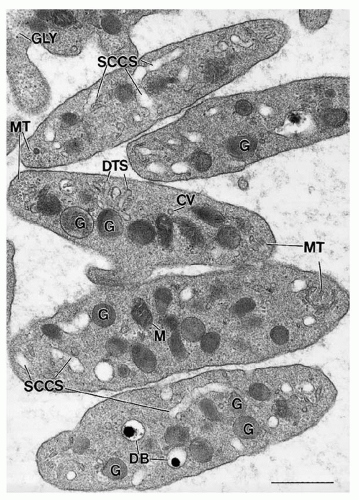
FIGURE 16.4. Ultrastructure of unstimulated human platelets. Membranous organelles, including the surface-connected canalicular system (SCCS) and dense tubular system (DTS), and cytoplasmic organelles, including mitochondria (M), α-granules (G), dense bodies (DB), coated vesicles (CV), and glycogen (GLY), are visualized at the ultrastructural level. Microtubules (MT) are present as cross-sectional and longitudinal profiles at the poles of the discoid platelets. Magnification ×46,000. Bar = 0.5 µm.


Microtubules

A circumferential microtubule band that supports the discoid form of the platelet33,34 is made of two nonidentical subunit proteins (α– and β-tubulin), associated with microtubule-associated proteins. The 25-nm-diameter microtubule coil lies adjacent to, but does not touch, the plasma membrane.56 Platelet microtubules comprise 13 protofilaments of αβ tubulin dimers and are primarily polymerized in unstimulated platelets. Platelet activation results in microtubule disassembly and then reassembly; such alterations in the marginal microtubule bundle result in platelet shape changes.56 Microtubules appear to be key determinates of platelet size57 and may be disorganized in giant platelet disorders.58


Platelet Granules and Organelles

Platelets possess secretory granules and mechanisms for cargo release to amplify responses to stimuli and influence the surrounding environment. Platelet granule structures include α– and dense granules, lysosomes, and peroxisomes. α-Granules and the dense bodies are the main secretory granules that release cargo (e.g., fibrinogen and adenosine diphosphate [ADP]) upon platelet activation.


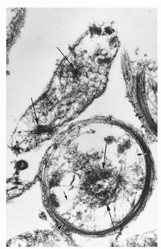
FIGURE 16.5. Human platelet cytoskeletons prepared by simultaneous fixation and lysis in Triton X-100 detergent. Single actin filaments, indicated by short arrows, course throughout the platelet cytoplasm. Clusters of filaments, indicated by long arrows, also are present. Note the microtubule coils at the platelet peripheries (arrowheads). Magnification ×30,000. (From Boyles J, Fox JEB, Phillips DR, Stenberg PE. Organization of the cytoskeleton in resting, discoid platelets: preservation of actin filaments by a modified fixation that prevents osmium damage. J Cell Biol 1985;101:1463-1472, with permission.)

Platelet granule secretion begins with a dramatic increase in platelet metabolic activity, set off by a wave of calcium release and marked by increased adenosine triphosphate (ATP) production.59 After platelet stimulation by agonists, a “contractile ring” develops around centralized granules,5,6 the granules fuse with the surface membranes, and then they extrude their contents.60 The molecular events underlying platelet granule release involve many of the same proteins and processes observed in other systems of membrane docking, fusion, and extrusion.61,62 Granule secretion in platelets is a graded process that depends on the number, concentration, and nature of the original stimulus/stimuli, either strong (e.g., thrombin and collagen) or weak (e.g., ADP and epinephrine).62


α-Granules

α-Granules, with a cross-sectional diameter of approximately 300 nm and numbering approximately 50 per platelet, are the predominant platelet granules.63 They are approximately spherical in shape, with an outer membrane enclosing two distinct intragranular zones that vary in electron density. The larger, electron-dense region is often eccentrically placed and consists of a nucleoid material that is rich in platelet-specific proteins such as β-thromboglobulin.64 The second zone, of lower electron density, lies in the periphery adjacent to the granule membrane and contains tubular structures with adhesive GPs such as von Willebrand factor (vWF) and multimerin,
along with factor V.65,66,67 Platelets take up plasma proteins and store them in their α-granules.68,69,70 Select α-granule proteins are discussed below and listed in Table 16.2.


TABLE 16.1 MAJOR PLATELET CYTOSKELETAL PROTEINS


Protein


Molecular Weight


Principal Known Function


Actin


42,000


Major protein constituent of microfilaments; 30% of platelet protein; F-actin binds myosin.


Myosin II


500,000


Binds actin; phosphorylation of light chains contracts microfilaments; 4% of platelet protein.


Talin


235,000


Interacts with α-actinin, vinculin; 2% of platelet protein.


Vinculin


130,000


Interacts with talin; links to actin.


α-Actinin


102,000


Dimer forms a gel with F-actin; promotes actin polymerization.


Actin-binding protein (filamin 1)


260,000


Cross-links actin filaments; links membrane skeleton with glycoprotein Ib-IX complex.


Gelsolin


91,000


Caps and severs actin filaments.


Thymosin β4


5,000


Binds 1:1 with G-actin monomer and inhibits its polymerization.


Profilin


15,200


Binds 1:1 with G-actin and inhibits its polymerization; adds adenosine triphosphate.


Tropomyosin


28,000


Binds groove on certain F-actins.


Caldesmon


80,000


Regulates actomyosin ATPase and actin bundling.


Myosin light chain kinase


105,000


Phosphorylates myosin, activates its ATPase, and causes contraction.


Calmodulin


17,000


Binds four Ca2+; activates myosin light chain kinase.


ATPase, adenosine triphosphatase.


Three proteins, β-thromboglobulin, PF4, and thrombospondin, are synthesized in megakaryocytes and highly concentrated in α-granules. The first two, β-thromboglobulin and PF4, show homology in amino acid sequence and share the additional features of localization in the dense nucleoid of α-granules, heparin-binding properties, and membership in the CXC family of chemokines.14,71,72,73 and 74 Together, they constitute approximately 5% of total platelet protein, and they can serve as useful markers for platelet activation in serum or plasma.75,76 Thrombospondin may comprise up to 20% of the total platelet protein released in response to thrombin, and likely participates in multiple biologic processes.77,78

vWF is also synthesized by megakaryocytes and is present in the tubular structures of the α-granule peripheral zone, similar to its localization within Weibel-Palade bodies of vascular endothelial cells.66,79 Factor V and multimerin, a factor V/Va-binding protein,67,80 co-localize with vWF in platelets but not in endothelial cells. Fibrinogen is also found in α-granules, but is incorporated actively from plasma and not synthesized by megakaryocytes.69 In fact, small amounts of virtually all plasma proteins, such as albumin, immunoglobulin G (IgG), fibronectin, and β-amyloid protein precursor, may be taken up into the platelet α-granules.79,81,82 and 83 α-Granules also contain many growth factors, including platelet-derived growth factor, transforming growth factor-β1 (TGF-β1), and vascular endothelial growth factor. These signaling molecules may contribute to the mitogenic activity of platelets.84,85 A role for platelet α-granule proteins in angiogenesis has been recently reported. Both pro- (e.g., VEGF) and antiangiogenesis proteins (e.g., endostatin) are stored within α-granules. How populations of angiogenesis regulatory proteins may be selectively compartmentalized and released from platelets is an area of active investigation.86,87,88,89 and 90 Likewise, the activation and functions of platelet-derived TGF-β1 in cardiac fibrosis and other conditions have been described.91,92

Platelet α-granules serve as an important reservoir for αIIbβ3 that contributes significantly to the surface fibrinogen receptors present on activated platelets.93,94,95 The α-granule membrane protein, P-selectin (granule membrane protein-140) is translocated to the plasma membrane after platelet activation.96,97 Finally, a number of additional proteins have been located to the surface of α-granules alone, including CD9, platelet endothelial cell adhesion molecule-1 (PECAM-1), Rap 1b, GPIb-IX-V, and osteonectin98,99 and 100 (Table 16.2).

The platelets and megakaryocytes of patients with gray platelet syndrome have decreased numbers of α-granules and reduced levels of some proteins. It is proposed that there is incorrect targeting of α-granule proteins to the α-granule in the megakaryocyte in this disease.101,102


Dense Bodies

Dense bodies, numbering approximately five per platelet, are exceptionally electron-dense and easily distinguished by electron microscopy because of their distinctive “bull’s-eye” appearance.103,104 With an approximate diameter of 250 nm, these granules contain a large reservoir of ADP, a critical agonist for platelet activation that amplifies the effect of other stimuli.132 In addition to this nonmetabolic pool of ADP, the dense bodies are rich in ATP, pyrophosphate, calcium, and serotonin (5-hydroxytryptamine), with lesser amounts of guanosine triphosphate (GTP), guanosine diphosphate (GDP), and magnesium.74 The adenine nucleotides are synthesized and segregated by megakaryocytes, whereas serotonin is incorporated into dense granules from the plasma by circulating platelets.133,134,135 There is more ADP than ATP in dense bodies, and both can lead to adenosine monophosphate (AMP). In turn, AMP can be dephosphorylated to adenosine or cyclized to produce cyclic AMP, an inhibitor of the platelet-stimulatory response. The dense granule membrane contains P-selectin and granulophysin.136


Lysosomes

Lysosomes are small, acidified vesicles, approximately 200 nm in diameter,105 that contain acid hydrolases with pH optima of 3.5 to 5.5, including β-glucuronidase, cathepsins, aryl
sulfatase, β-hexosaminidase, β-galactosidase, heparitinase, and β-glycerophosphatase. Additional proteins found in lysosomes include cathepsin D and lysosome-associated membrane proteins (LAMP-1/LAMP-2), which are expressed on the plasma membrane after activation).106,107 Lysosomal constituents are released more slowly and incompletely (maximally, 60% of the granules) than α-granules or dense-body components after platelet stimulation, and their release also requires stronger agonists such as thrombin or collagen.


TABLE 16.2 MAJOR PLATELET GRANULAR CONSTITUENTS SECRETED WITH ACTIVATION


α-Granule Protein


Comments


Amount/109 Platelets


Concentration in Platelets >Plasma


Coagulant Proteins


Fibrinogen


Critical ligand for aggregation


140 µg


3× conc. platelets >plasma


Factor V


Critical cofactor for coagulation


4 µg


30× conc. platelets >plasma


Platelet-Specific Proteins


Platelet factor 4


Marker for platelet activation


12 µg


20,000× conc. platelets >plasma


β-Thromboglobulin


Marker for activation


10-20 µg


20,000× conc. platelets >plasma


Mitogenic and Angiogenic Factors


Platelet-derived growth factor


Smooth muscle mitogen


30-100 ng


Transforming growth factor-β


Complex activation pathway; binds thrombospondin


Vascular endothelial growth factor


Relatively high concentrations in platelets


Adhesive Glycoproteins and α-Granule Membrane-Specific Proteins


Thrombospondin


Multiple complexes


40 µg


20,000× conc. platelets >plasma


von Willebrand factor (vWF)


Role in adhesion 0.3 µg


100× conc. platelets >plasma


Multimerin


Binds factor V; resembles vWF-binding factor VIII; has RDG sequence


P-selectin


Mediates platelet-leukocyte binding


20,000 copies on activated platelets


Dense Granule Constituent


Comments


Concentration in Granules (nmol/mg Dense Granule Protein)


Percent Secreted


Adenosine diphosphate


Highly concentrated; a critical mediator of aggregation


630


95% secreted with platelet activation


Adenosine triphosphate


440


40% released with activation


Calcium


2,630


70% secreted with activation


Serotonin


100


95% released with activation


conc., concentration.


Note: A number of additional elements are released or secreted from within the platelet. For example, α-granules also contain the major platelet surface glycoproteins (GPs) GPIIb-IIIa and GPIb-IX along with albumin and immunoglobulin G, adhesive glycoproteins (fibronectin, vitronectin), fibrinolytic components (α2-antiplasmin, plasminogen activator), and coagulation-related proteins (high-molecular-weight kininogen, α2-macroglobulin). In addition, dense granules contain guanosine triphosphate/guanosine diphosphate and high concentrations of pyrophosphate, phosphate, and magnesium, much of which is secreted with activation. A number of additional proteins are present, some released and some retained in the platelet cytosol, such as a subunit of factor XIII, amyloid β-protein precursor, protease nexin I, and tissue factor pathway inhibitor.


Organelles: Microperoxisomes, Coated Vesicles, Mitochondria, and Glycogen

Peroxisomes are rare, small (90 nm in diameter) granules, demonstrable with alkaline diaminobenzidine as a result of their catalase activity.108 The structure may participate in the synthesis of platelet-activating factor.109

Mitochondria in platelets are similar, with the exception of their smaller size, to those in other cell types. There are approximately seven per human platelet, and they serve as the site for the actions of the respiratory chain and the citric acid cycle.110 Glycogen is found in small particles or in masses of closely associated particles, playing an essential role in platelet metabolism.111


Platelet Biochemistry and Metabolism


Composition

The platelet is composed of approximately 60% protein, 15% lipid, and 8% carbohydrate by dry weight. Platelet minerals include magnesium, calcium, potassium, and zinc. Platelets contain substantial amounts of vitamin B12, folic acid, and ascorbic acid.112 The concentrations of sodium and potassium within the platelet are 39 and 138 mEq, respectively.113 This gradient against plasma, apparently distributed in two discrete metabolic compartments, is maintained by an active ion pump, which derives energy from a membrane ATPase of the ouabain-sensitive, Na+/K+-dependent type.114

Unstimulated platelets maintain a low cytoplasmic Ca2+ concentration (˜100 to 500 nmol/L) by limiting Ca2+ transport from plasma and promoting active efflux of this ion from the cell.115 Two pools of calcium are present in platelets: a rapidly turning over cytosolic pool regulated by a sodium-calcium antiporter in the plasma membrane and a more slowly exchanging pool regulated by Ca2+/Mg+-ATPase and sequestered in the dense tubular system.116 Platelets are therefore able to transport calcium from the cytosol by moving it against a gradient into the extracellular space or by sequestration in the dense tubular system.


Energy Metabolism and Generation of ATP

There are several similarities between the energy metabolism of the platelet (Fig. 16.6) and that of skeletal muscle. Both involve active glycolysis and the synthesis and use of large amounts of glycogen,117 and in both, the major mediator of intracellular energy use is an actomyosin-like ATPase. The platelet, like muscle, is metabolically adapted to expend large amounts of energy rapidly during aggregation, the release reaction, and clot retraction.

The major energy source for the platelet is glucose, which is rapidly taken up from the plasma (Fig. 16.6, Step 1). Under basal conditions, 40% to 50% of the absorbed glucose is used to provide energy for synthetic functions or is converted into glycogen. Electron microscopy reveals prominent masses of glycogen in some platelets. The glycolytic pathway with its regulatory enzymes (phosphorylase, pyruvate kinase, hexokinase, phosphofructokinase, and glyceraldehyde 3-phosphate dehydrogenase),117 the citric acid cycle, the pentose phosphate shunt, and the NAD-NADH (nicotinamide adenine dinucleotide-nicotinamide adenine dinucleotide [reduced form]) system are all active in the platelet. Ninety-eight percent of platelet pyruvate is converted to lactate, which leaves the platelet.118 and 119,120 In addition to glycolysis, platelets contain enzymes for oxidative phosphorylation and fatty acid oxidation (Fig. 16.6, Step 2).121,122


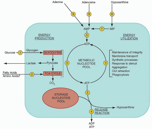
FIGURE 16.6. Simplified scheme of platelet energy metabolism. Platelet energy is derived from the metabolism of glucose and, to a lesser extent, from the metabolism of fatty acids. Energy is provided in approximately equal amounts by glycolysis and the citric acid cycle. The platelet energy reserve is provided by the metabolic pool of platelet nucleotides that is in a state of continuous turnover. This energy is used for the maintenance of the platelets’ structural integrity and in the reactions accompanying the response of platelets to stimuli. The granule-bound storage (nonmetabolic) nucleotide pool is discharged during the release reaction. ADP, adenosine diphosphate; AMP, adenosine monophosphate; ATP, adenosine triphosphate; IMP, inosine monophosphate. (Adapted from Hirsh J, Doery JCG. Platelet function in health and disease. Prog Hematol 1972;7:185-234.)

ATP production in platelets is affected greatly by the suspending medium, chelating agents, and in vitro manipulation of platelets. In plasma, oxidative ATP production by unstimulated platelets is predominant, and all ATP formed by oxidative phosphorylation may be the product of β-oxidation of fatty acids;123 glycogen turnover, the hexose monophosphate shunt, and the citric acid cycle are virtually inactive.120 Glycolysis is capable of completely compensating for reduced ATP production when oxidative phosphorylation is inhibited. Platelet stimulation by agents that induce aggregation and release is associated with a marked increase in metabolic activity involving glycogenolysis,124 as well as with glycolysis and oxidation to varying degrees.117,125

The total amounts of ATP synthesized by the two pathways are approximately equal because of the greater ATP yield per mole of glucose provided by oxidation.118,122 ATP energy is used in unstimulated platelets to maintain homeostatic levels of H+, K+, Na+, and Ca2+.126,127


Nucleotide Metabolism and the Nonmetabolic Role for ADP

Adenine nucleotides constitute 90% of free platelet nucleotides and are partitioned into at least two different pools, which undergo minimal interchange (Fig. 16.6).97 The metabolic or
cytoplasmic pool makes up 40% of total adenine nucleotides; it is used for the maintenance of various energy-consuming cell functions and is retained during platelet release. In large part made up of ATP, this pool is constantly turning over, as revealed by the rapid incorporation of 14C-adenine and 32P-phosphate into ATP. In unstimulated platelets, the relative concentrations of metabolic AMP, ADP, and ATP are maintained by the enzyme adenylate kinase (Fig. 16.6, Step 3).128

The storage pool, which is present in the dense bodies, contains approximately two-thirds of the total platelet nucleotides, mainly in the form of ADP and ATP.129 It is metabolically inactive, does not rapidly incorporate exogenous adenine or phosphate, and equilibrates slowly with the metabolic pool.130 Nucleotides in this pool are extruded from the platelet during the release reaction (Fig. 16.6, Step 4) and cannot be replenished after release. ATP hydrolysis is required for conversion of G-actin to F-actin, and the resultant ADP becomes associated with F-actin; this small percentage of platelet ADP bound to actin constitutes one-third of the nucleotide compartment.131 Perhaps as much as 40% of all ATP produced is used during the process of actin treadmilling,132 and as much as 7% is used in the turnover of the phosphoinositides PIP and PIP2.133

The ATP that is broken down to provide energy for the release reaction is not rephosphorylated; rather, it is irreversibly degraded to hypoxanthine (Fig. 16.6, Step 5), which diffuses out of the cell.128 This reaction also proceeds slowly in stored normal platelets.170 Hypoxanthine in the plasma may be reincorporated slowly into metabolic AMP by the salvage pathway (Fig. 16.6, Steps 6 and 7).129,134 Platelet stimulation results in marked activation of ATP-producing pathways.135 The steady-state level of ATP decreases and hypoxanthine accumulates. In addition, a transient but greatly increased uptake of phosphate by platelets occurs.136 Although ATP-requiring processes are activated by platelet stimulation, it is unknown whether or how these are coupled to signal processing in platelets.


Lipid Composition and Metabolism and the Generation of Arachidonic Acid

Phospholipids constitute 80% of total platelet lipid, with neutral lipids and glycolipids comprising the remainder.137 The five major phospholipids identified in human platelets are phosphatidylcholine (PC, 38% of total phospholipids), phosphatidylethanolamine (PE, 27%), sphingomyelin (17%), PS (10%), and phosphatidylinositol (PI, 5%).8 Studies of platelet subcellular fractions reveal that 57% of total human platelet phospholipids are present in the plasma membrane.138 Most of the negatively charged phospholipids (i.e., PE, PI, and PS) are contained in the inner leaflet,138,139 and 140 an asymmetric arrangement that prevents inappropriate coagulation by sequestering the phospholipids that accelerate plasma coagulation (mainly PS) away from the platelet surface. This asymmetry collapses when platelets are activated.141 These same phospholipids (i.e., PS and PE, which interact with coagulation proteins) redistribute with platelet activation and are thereby exposed on the surface to function in promoting clot formation.142 The asymmetric distribution of phospholipids on the platelet surface appears to be maintained by one or more membrane scramblase enzymes; and a “floppase” has been proposed to reverse the phospholipid asymmetry with activation.

Almost all platelet fatty acids are esterified in phospholipids, leaving only trace amounts in the free form. Platelet phospholipids are enriched in arachidonate, the precursor of prostaglandins, at their “sn-2” position.143 After agonist stimulation, phospholipase A2 activity rises, and arachidonic acid is released from membrane phospholipids, predominantly PC.144,145,146 After release, arachidonic acid is oxygenated to form the cyclic endoperoxide intermediate, PGH2, by cyclo-oxygenase-1, which leads to TxA2 formation.143 The lipoxygenase pathway accounts for a small proportion of arachidonate metabolism, producing mainly 12-HETE (12-hydroxyeicosatetraenoic acid).

Neutral lipids, mainly cholesterol, make up approximately 28% of total platelet lipids. Cholesterol is a major constituent of platelet membranes and is also present in the platelet cytoskeleton.147 It is synthesized by megakaryocytes but not by platelets. Finally, neutral glycolipids, gangliosides, and ceramides have been detected in platelets.179


Platelet Microparticles and Kinetics


Microparticles

Platelet microparticles are tiny structures rich in surface PS that are generated during platelet activation and can contribute significantly to the acceleration by platelets of plasma coagulation, specifically factor Xa and thrombin generation.148 The composition of microparticles varies with the agonist or stimulus (C5b-9, ionophore A23187, thrombin, tissue factor, shear) involved in their formation,149,150 and have been found to bind factors Xa, VIII, and Va, along with protein S.151,152 Physiologically, microparticles appear to be an important contributor to procoagulant activity as their defect in patients is associated with clinical bleeding.153,154


Platelet Heterogeneity

The normal platelet count varies between 150,000 and 400,000/µl, and normal platelet size (mean platelet volume) varies between 7.5 and 10.5 fl. Platelets are released into the blood from long proplatelet extensions of megakaryocytes.155 Young platelets, that is, those recently released by megakaryocytes, are larger and more dense, and undergo remodeling in circulation, in part by shedding some of their surface components. Macrothrombocytopenias may reflect a disturbance in the steps of platelet production,156 and the properties of large platelets may reflect unique attributes of platelets recently released from the marrow or proplatelets produced under conditions of accelerated or abnormal production.


Platelet Distribution and Survival Kinetics


Labeling

Platelets labeled with 51Cr (chromate) have been used to estimate platelet lifespan in humans at 8 to 12 days,157 and the method has been widely validated.158 Other methods reported for platelet labeling include 111In (indium) chelated with 8-hydroxyquinoline and 32P-labeled diisopropylfluorophosphate, 68Ga (gallium).159 Platelet labeling is not commonly used for clinical evaluations, but all of the published studies give the same approximate values for distribution and survival.


Distribution

Approximately one-third of the total platelet mass appears to pool in the spleen. The splenic pool exchanges freely with the platelets in the peripheral circulation. Transfusion of 51Cr-labeled platelets into normal subjects results in approximately twothirds remaining in the circulation—in contrast to nearly 100% in splenectomized patients.160,161 In addition, administration of epinephrine, which evacuates platelets from the spleen, increases the peripheral platelet count 30% to 50%.162 Platelet counts in asplenic patients are not affected by epinephrine. Some studies suggest that the splenic pool consists of the youngest, largest platelets. The mechanism of splenic sequestration has been hypothesized to result from a longer transit time through the splenic cords (which platelets enter because of their small size) or from binding to the reticular and endothelial cells of the spleen.163 Pathophysiologic states can result in 80% to 90% of platelets being sequestered in the spleen, resulting in thrombocytopenia. Release of platelets from the lungs after intracardiac administration of epinephrine has been reported.164 Also, platelet counts rise after
vigorous exercise, and this rise is not affected by splenectomy.165 This nonsplenic pool represents approximately 16% of the total platelet mass.


Life Span

Platelet life span, based on the time required to clear labeled platelets from the circulation, has been estimated to be 8 to 12 days in humans. In steady state, when platelet production equals destruction, platelet turnover has been estimated at 1.2 to 1.5 × 10¹¹ cells per day.160,166 Recommendations for estimation of platelet lifespan have been published by the Panel on Diagnostic Application of Radioisotopes in Hematology, International Committee for Standardization in Hematology,166 and multiple models for analysis of platelet lifespan have been proposed.167 As discussed above, platelets may be removed from circulation by pooling in the spleen,161 the liver, and the lung.164

Platelet life span is regulated by Bcl-2 proteins.168,169 Platelet Bcl-x(L) is required for survival by suppressing Bak. Bak activation results in classic apoptosis pathways that result in mitochondrial damage, caspase activation, and PS exposure. Platelets from Bak-deficient animals live longer than normal,169 indicating a predetermined program of platelet death.

Oct 21, 2016 | Posted by in HEMATOLOGY | Comments Off on Platelet Structure and Function in Hemostasis and Thrombosis

Full access? Get Clinical Tree

Get Clinical Tree app for offline access