Neutrophilic Leukocytes


Neutrophilic Leukocytes


Keith M. Skubitz


Three types of granulocytes are readily identified in peripheral blood smears. Neutrophils are so named because of their neutral staining with Wright stain, whereas eosinophils avidly stain with the dye eosin, and basophils have readily identified large darkstaining granules with Wright stain. Neutrophils play a critical role in host defense by phagocytizing and digesting microorganisms, and inappropriate activation of neutrophils may result in damage to normal host tissues. In the resting uninfected host, the production and elimination of neutrophils are balanced, resulting in a fairly constant concentration of neutrophils in peripheral blood. When an infection occurs, chemotactic agents are generated that result in migration of neutrophils to the site of the infection and activation of neutrophil defensive functions. This effect is often associated with an increased production and release of neutrophils from the bone marrow. This chapter reviews the structure, morphology, production, distribution and kinetics, and functions of neutrophils.


SUBCELLULAR STRUCTURE OF NEUTROPHILS

Mature neutrophils contain several types of granules and other subcellular organelles. Many studies have been performed to identify and characterize the molecular composition of the various subcellular compartments of neutrophils. Two techniques are commonly used for this purpose: immunoelectron microscopy and subcellular fractionation. Most recent subcellular fractionation studies used the technique of nitrogen cavitation to disrupt the neutrophils. In this technique, cells are equilibrated with nitrogen at high pressure and then released into a pressure of 1 atm. The rapid decrease in extracellular pressure results in disruption of the cells by the formation of nitrogen gas within the cell. The disrupted cells are then fractionated by density-gradient centrifugation.1,2 Many types of subcellular organelles and structures are present in neutrophils, as seen by electron microscopy (EM). Although the granules of neutrophils may be best viewed as a continuous spectrum of granules, possibly resulting from differential production of different granule contents during neutrophil development, studies of neutrophils have operationally defined several types of granules. Four well-defined types of granules in neutrophils are primary granules, secondary granules, tertiary granules, and secretory vesicles, although additional heterogeneity of these fractions may exist. Some of the known constituents of these granules are indicated in Table 7.1.


Azurophilic (Primary) Granules

The azurophilic, or primary, granules are formed during the promyelocytic stage and in general contain many antimicrobial compounds. These granules fuse with phagocytic vesicles, resulting in the delivery of their contents to the ingested organism. Among the azurophilic granule contents is myeloperoxidase (MPO), a protein that catalyzes the production of hypochlorite (OCl) from chloride and hydrogen peroxide produced by the oxidative burst. MPO constitutes approximately 5% of the dry weight of the neutrophil18 and imparts the greenish coloration to pus. The human neutrophil defensins (HNP-1 to -3), a group of cationic proteins that kill a variety of bacteria, fungi, and viruses,19,20,21 also constitute approximately 5% of total neutrophil protein.22 Other components of azurophilic granules include lysozyme, which degrades bacterial peptidoglycans,23 bactericidal permeability-increasing protein, which has antibacterial activity against certain gram-negative bacteria,24,25,26,27 and 28 azurocidin, which has antibacterial as well as antifungal activity against Candida albicans,29,30 and the serine proteinases elastase, cathepsin G, proteinase 3, esterase N, and others.31,32,33 and 34 The granule membrane itself contains a large amount of CD66c35,36 and CD63 antigens.37 Heterogeneity among azurophil granules is likely.3


Specific (Secondary) Granules

Although some specific (also called secondary) granules, like azurophilic granules, fuse with phagocytic vesicles, it is believed that these granules are largely for release into the extracellular space. Some of the known contents of these granules are also indicated in Table 7.1 and include apolactoferrin, the major specific granule protein, vitamin B12-binding protein, plasminogen activator, and collagenase. Lysozyme and some gelatinase are also present in specific granules. Release of specific granule contents may modify the inflammatory process. For example, collagenase may degrade collagen, thus augmenting movement through collagen and participating in tissue remodeling. Apolactoferrin, by binding iron, may have an antibacterial effect by preventing bacteria from obtaining necessary iron for growth.38 Iron binding by apolactoferrin may also modify hydroxyl radical formation and cell adhesion.39,40,41 and 42 Although the antimicrobial and proinflammatory defensins are stored in azurophil granules, proHNPs produced at more mature stages of differentiation are not cleaved to the antimicrobial form and are stored in specific granules. ProHNPs are constitutively exocytosed, though their function is unclear.15 Haptoglobin released from specific granules could also inhibit bacterial growth and inflammation.16 Specific granules also contain a number of membrane-bound molecules that are also expressed on the cell surface. This includes CD11, CD18, CD66a, CD66b, NB-1 (CD177), f-met-leu-phe (FMLP) receptors, C5a receptors, and cytochrome b558. When cells are stimulated, the surface expression of many of these membrane proteins is increased, and some of the upregulated molecules may be derived from specific granules. The importance of the specific granules in neutrophil function is shown in patients who lack specific granules; these patients are susceptible to repeated skin and respiratory infections and have defective neutrophil chemotaxis and adhesion.


Gelatinase (Tertiary) Granules

Gelatinase, or tertiary, granules, which cosediment with specific granules in some subcellular fractionation techniques, were initially identified as gelatinase-containing granules.43 Like specific granules, tertiary granules also contain many membrane proteins
that are upregulated to the cell surface with stimulation.2 The relative contribution of tertiary granules and specific granules to upregulation of membrane proteins is not clear.


TABLE 7.1 CONTENTS OF HUMAN NEUTROPHIL GRANULES


Azurophil (Primary) Granules


Specific (Secondary) Granules


Gelatinase (Tertiary) Granulesa


Secretory Vesicles


Membrane


Membrane


Membrane


Membrane


CD63


CD15 antigens


CD11b


Alkaline phosphatase


CD66c


CD66a


FMLP receptor


Cytochrome b558b


CD68


CD66b


Diacylglycerol deacylating enzyme


CD11b


Matrix


Cytochrome b558b


Cytochrome b558b


u-PA receptor


Lysozyme


FMLP receptor


Laminin receptor


FMLP receptor


Defensins


Fibronectin receptor


Matrix


CD10, CD13, CD45


Elastase


G-protein α-subunit


Gelatinasea


CD16


Cathepsin G


Laminin receptor


Acetyltransferase


DAF


Proteinase 3


CD11b


Lysozyme


CR1 (CD35)


Esterase N


NB 1 antigen


NRAMP1


Matrix


α1-Antitrypsin


α-Mannosidase


Azurocidin


Bactericidal permeability-increasing protein


Rap 1, Rap 2


Thrombospondin receptor


Tumor necrosis factor receptor


Vitronectin receptor u-PA receptor


Ficolin-1


Plasma proteins (including tetranectin and albumin) pro-u-PA/u-PA


β-Glycerophosphatase


Matrix


β-Glucuronidase


Apolactoferrin


β-Galactosidase


Lysozyme


β-Glucosaminidase β2-Microglobulin


α-Fucosidase


Collagenase


Cathepsin B


Gelatinasea


Cathepsin D


Histaminase


Acid mucopolysaccharide


Heparinase


Heparin binding protein


pro-u-PA


N-Acetyl-β-glucosaminidase


Vitamin B12-binding protein


Sialidase


Sialidase


Ubiquitin protein


Protein kinase C inhibitor


hCAP-18


SGP28


PTX3


Haptoglobin


Prodefensins (proHNPs)


DAF, decay accelerating factor; FMLP, f-met-leu-phe; u-PA, urokinase-type plasminogen activator.


a Gelatinase is present in only a subset of specific granules; most is present in gelatinase granules.

b Cytochrome b558 is also called b245.


Adapted from references (2) through (17).


Secretory Vesicles

Secretory vesicles, which largely distribute in the plasma membrane fraction using subcellular fractionation techniques, have also been described. Complement receptor (CR)1, recognized by CD35 monoclonal antibodies, has been found exclusively in the light membrane fractions containing secretory vesicles and plasma membranes using subcellular fractionation techniques.11 The observation that CR1 can be readily upregulated to the neutrophil surface with weak stimulation demonstrates that secretory vesicles provide an intracellular reservoir from which membrane proteins can be recruited to the cell surface. CR3 (HMac-1), recognized by CD11b antibodies and present in both secretory vesicles and specific granules, is also upregulated to the cell surface with weak stimulation. In contrast to CR1, a more marked upregulation of CR3 is observed with more potent stimulation, demonstrating that specific granules can also serve as an intracellular reservoir from which membrane proteins can be upregulated to the cell surface.11 By EM, the secretory vesicles appear as smooth-surfaced vesicles. A defining feature of secretory vesicles is their rapid and complete translocation to the surface membrane with weak stimulation.11 These secretory vesicles also contain alkaline phosphatase, cytochrome b558, and FMLP receptors. Secretory vesicles can be upregulated to the cell surface in the absence of extracellular calcium, in contrast to specific and gelatinase granules, which require extracellular calcium for release.11 The secretory vesicles appear to be formed by a process of endocytosis and contain albumin.


Plasma Membrane

Many constituents of the neutrophil plasma membrane have been defined. These include membrane channels, adhesive proteins, receptors for various ligands, ion pumps, and ectoenzymes. For example, CD11/CD18 and CD62L (L-selectin) are involved in neutrophil adhesion. The FMLP- and C5a-receptors sense stimuli and activate neutrophils, while aminopeptidase N (CD13) can inactivate interleukin (IL)-8, eliminating its chemotactic activity,44 and neutrophil endopeptidase (CD10) can inactivate the chemotactic peptide FMLP.45 CD66a, b, c, can activate neutrophils,46 and some CD45 antibodies, which recognize a transmembrane protein with tyrosine phosphatase activity in its cytoplasmic domain, inhibit neutrophil chemotaxis.47 Some of the various clusters of differentiation (CD) expressed on neutrophils are shown in Table 7.2. The components of the membrane are not uniformly distributed. Studies have indicated the presence of differentiated domains in the membrane called rafts, which are described in the section “Lipid Rafts.”


TABLE 7.2 SOME CD ANTIGENS EXPRESSED ON NEUTROPHILS


CD


CD Antigen


CD10


Common acute lymphoid leukemia antigen, neutral endopeptidase


CD11a


Leukocyte factor antigen-1, αLβ2


CD11b


Mac-1, αMβ2


CD11c


p150, 95, αXβ2


CD13


Aminopeptidase N


CD15


Lex (Ga1β1→4G1cNAc(Fucα1→3) β1→3Ga1β1→ 4G1cNAc→R)


CD15s


sLex


CD16


FcRIII


CDw17


LacCer


CD18


β2-Integrin


CD24


Glycosyl phosphatidylinositol-linked protein


CD31


Platelet endothelial cell adhesion molecule-1


CD32


FcRII


CD43


Leukosialin, sialophorin


CD44


Pgp-1


CD45


Leukocyte common antigen (a protein tyrosine phosphatase)


CD50


Intercellular adhesion molecule-3


CD53


Tetraspan molecule


CD55


Decay accelerating factor


CD62L


L-Selectin


CD63


Tetraspan family member


CD64


FcRI


CD65


Ga1β1→4G1cNAcβ1→3Ga1β1→G1cNAc(Fucα1→3)→R


CD65s


Sialylated CD65


CD66a


CEACAM1 (Biliary glycoprotein)


CD66b


CEACAM8 (CGM6)


CD66c


CEACAM6 (NCA)


CD66d


CEACAM3 (CGM1)


CD82


Tetraspan family member


CD88


C5a receptor


CD95


Fas, APO-1


CD114


Granulocyte colony-stimulating factor receptor


CD156


ADAM-8 (a disintegrin and metalloprotease domain)


CD157


Bifunctional ectoenzyme (adenosine diphosphate ribosylase)


CD177


NB1, HNA-2a


NOTE: When used to describe the antigen, the term CD antigen should be used.


Cytoskeletal Matrix

Like many other cells, neutrophils contain a complex cytoskeleton. Alterations in the distribution of cytoskeletal elements may be important in chemotaxis, phagocytosis, and exocytosis. Many protein components of this cytoskeleton have been identified, including actin, actin-binding protein, α-actinin, gelsolin, profilin, myosin, tubulin, and tropomyosin. Actin accounts for approximately 10% of neutrophil protein.48 The role of the cytoskeleton in neutrophil function is reviewed in detail elsewhere.49


Neutrophil Lipids

Although most studies of neutrophil structure have concentrated on proteins, lipids and carbohydrates also serve important functions. Lipids account for approximately 5% of neutrophils by weight,50,51 of which approximately 35% is phospholipid.52 Phosphatidylcholine and phosphatidylethanolamine account for approximately 75% of the phospholipid in intact neutrophils.52 Subcellular distribution studies reveal that plasma membranes and secretory vesicles contain approximately half the cellular phospholipid. Most of the phosphatidylinositol and phosphatidylcholine are present in plasma membrane and secretory granules, whereas a large part of the phosphatidylethanolamine is found in the specific granules.52 Among the phospholipids, the phosphoinositides are important as sources of inositol 1,4,5-triphosphate (IP3) (a signal-transduction molecule that results in calcium release) and diacylglycerol (which activates protein kinase C [PKC]). The occurrence of arachidonic acid in phospholipids, especially phosphatidylcholine, is important as a precursor for the production of leukotrienes, prostaglandins, thromboxanes, and lipoxins.53,54 Cholesterol and triglycerides constitute most of the nonphospholipid neutrophil lipid. Glycolipids, which include both neutral glycosphingolipids and gangliosides, constitute the remaining neutrophil lipids. The study of glycolipids is complex, and the glycolipid composition of neutrophils is not well understood. Glycolipids are important because their carbohydrate components contain many neutrophil differentiation antigens with a multitude of potential functions. The major neutrophil glycolipid is lactosylceramide (LacCer: Galβ1 → 4Glcβ1 → 1Cer) 55,56,57 and is recognized by CDw17 monoclonal antibodies. Interestingly, the surface expression of LacCer decreases after neutrophil stimulation. More than 75% of neutrophil LacCer is found in intracellular granules.56,58 It has been hypothesized that most LacCer in the granule membranes is found in the outer leaflet and may contribute to the ability of these membranes to form the highly convex surfaces necessary to form these submicrometer particles.54 Approximately 75% of the five major glycosphingolipids are located intracellularly.58 Studies of the subcellular distribution of glycosphingolipids in neutrophils have found no differences among the plasma membrane, primary granules, or secondary granules in the relative amounts of these five glycosphingolipids.58


Lipid Rafts

Studies have demonstrated the existence of large noncovalent detergent-resistant complexes in cell extracts that contain important signaling molecules, including protein kinases and many glycosyl phosphatidylinositol-linked membrane proteins capable of transmitting signals.59,60 and 61,62 These complexes have been termed lipid rafts or large detergent-resistant complexes. It is postulated that these complexes or rafts reflect the existence of specific membrane microdomains that have a particular lipid composition, and that these clusters of molecules may be important in transmembrane signaling by proteins in the complex. There is
evidence that in neutrophils, proteins may enter these rafts when they are translocated to the cell surface. For example, it appears that CD63 and CD11b/CD18 are not present in detergent-resistant complexes when they are intracellular, but they enter such complexes after translocation to the cell surface.63


Cytoplasmic Lipid Bodies

Cytoplasmic lipid bodies, non-membrane-bound cytoplasmic inclusions, have been described in neutrophils.64 In inflammation, the number of cytoplasmic lipid bodies in neutrophils increases.65 These lipid bodies may provide nonmembrane stores of esterified arachidonate. In addition, some signaling proteins, including phosphatidylinositide-3-kinase, are localized to these lipid bodies,66 although the exact role of these lipid bodies in cell function is unclear.66,67


Cytosol

Although neutrophil cytoplasm contains many components common to all cells, approximately 45% of neutrophil cytosolic protein appears to be attributable to migration inhibitory factor-related proteins (MRPs), MRP-8, and MRP-14.68 MRP-8 and MRP-14 are members of the S100 family of calcium-binding proteins and form homo- and heterodimers. MRP-14 has been variously called p14, L1 heavy chain, and calgranulin b, and MRP-8 is also known as p8, L1 light chain, calgranulin a, and cystic fibrosis antigen.69,70,71,72 and 73 Although the role of these proteins in neutrophil function is unclear, the quantity of MRP-8 and MRP-14 associated with neutrophil plasma membranes has been reported to increase after stimulation.68 MRP-8/MRP-14 are also present in secondary and/or tertiary granules and are released when neutrophils are stimulated.73 MRP-14 can inhibit macrophage activation.74 Annexin I or lipocortin I comprises approximately 3% of cytosolic protein.75 Annexin I is partially regulated by glucocorticoids and appears to be a mediator of the anti-inflammatory effects of glucocorticoids.75

One other notable cytoplasmic constituent is glycogen. Because neutrophils are sometimes required to function in hypoxic conditions, as in an abscess, they are very capable of obtaining energy by glycolysis. The presence of large intracellular glycogen stores gives them the additional ability to function in areas of low extracellular glucose.


Nucleus

In the past, it has been felt that neutrophils, as end-stage cells, undergo little RNA or protein synthesis. Subsequently, it was shown that mature neutrophils can synthesize both RNA and protein. This is likely particularly important when neutrophils migrate into sites of inflammation and begin to synthesize proteins and chemokines that may regulate activation and resolution of the innate immune response.76,77,78,79,80 and 81,82


MORPHOLOGY OF NEUTROPHILS AND PRECURSORS

The replenishment of marrow and blood cells from a stem cell compartment is described in Chapter 5. Neither multipotent hematopoietic stem cells nor more committed progenitors are readily identified morphologically by traditional methods.83,84,85,86 and 87 Only the more mature forms of each hematopoietic cell series can be reliably distinguished from one another. In the following pages, the cells identifiable as neutrophils and their precursors are described.

Neutrophils follow a pattern of proliferation, differentiation, maturation, and storage in the bone marrow and delivery to the blood. In the first three morphologic stages, the myeloblast, promyelocyte, and myelocyte cells are capable of replication, as shown by their uptake of tritiated thymidine (3H-TdR) and the presence of mitoses; in later stages, cells cannot divide but continue to differentiate. The morphologic boundaries of each cell compartment were defined many years ago and were based on criteria such as cell size, ratio of size of nucleus to cytoplasm, fineness of nuclear chromatin, nuclear shape, the presence or absence of nucleoli, the presence and type of cytoplasmic granules, and the cytoplasmic color of stained cells (Table 7.3).

Because changes in nuclear chromatin and cell size occur during each cell replication cycle, and the formation of granules and other cytoplasmic changes occur gradually during the stages of cell development, morphologic definitions are necessarily arbitrary and do not always conform to significant biochemical or physiologic changes. Classifying a cell in one category or another often is difficult because it is actually in transition between the two. Nevertheless, it is useful to separate the cell lines into morphologic compartments and to define normal limits of cell distribution therein, because gross changes from these patterns indicate disease.


DEVELOPMENT OF NEUTROPHILS AND THEIR PRECURSORS

Neutrophil development is summarized in Figure 7.1.

Cell division is limited to myeloblasts, promyelocytes, and myelocytes, with later developmental stages undergoing differentiation but no further cell division. The myeloblast contains few granules and is derived from more primitive cells as described in Chapter 5. As the cell differentiates to the promyelocyte stage, development of primary or azurophil granule formation becomes evident. This granule contains MPO, an enzyme whose activity is a classic marker of myeloid differentiation. Azurophil granule production ceases at the end of the promyelocyte stage, coincident with the loss of peroxidase activity from the rough endoplasmic reticulum. Secondary granule, or specific granule, formation begins as the neutrophil enters the myelocyte stage. The peroxidase-negative specific granules are smaller (approximately 200-nm diameter) than the azurophil granules (approximately 500-nm diameter) and are near the limit of resolution by light microscopy. The specific granules impart a pinkish ground-glass background color to neutrophils in Wright-stained smears. Because azurophil granule formation ceases in the promyelocyte stage and the subsequent myelocyte form is still capable of cell division, the density of azurophil granules is lower in differentiation stages past the promyelocyte. The result is that mature neutrophils contain approximately two specific granules for every azurophil granule. With maturation, the azurophil granules, which generate reddish-purple staining in the promyelocytes, lose this metachromasia as they leave the myelocyte stage. This alteration in staining properties is thought to be caused by an increase in acid mucosubstances, which complex with basic proteins already present in the azurophil granules.88 Thus, in the mature neutrophil, the azurophil granules appear as light blue-violet granules on Wright-stained smears. The azurophil granules are readily demonstrated by peroxidase staining with light microscopy.

Data concerning antigenic differences between granulocytes and monocytes and their stages of maturation have largely been developed using monoclonal antibodies. Such monoclonal antibodies have been analyzed in a series of international workshops in which antibodies are grouped into CD. Some of the CD antigens expressed on neutrophils are shown in Table 7.2. Immunogold and enzyme-linked immunologic methods89 permit simultaneous morphologic and immunologic examination of individual cells.


TABLE 7.3 MORPHOLOGIC CHARACTERISTICS OF LEUKOCYTES (WRIGHT STAIN)


Nucleus


Cytoplasm


Type of Cell


Size (µm)


Position


Shape


Color


Chromatin


Nuclear Membrane


Nucleoli


Relative Amount


Color


Perinuclear Clear Zone


Granules


Granulocytes


Myeloblast


10-18


Eccentric or central


Round or oval


Light reddish purple


Very fine meshwork


Very fine


2-5


Scanty


Blue


None


None


Promyelocyte


12-20


Eccentric or central


Round or oval


Light reddish purple


Very fine meshwork


Fine


2-5


Moderate


Blue


None


Primary (azurophilic, eosinophilic, or basophilic)


Myelocyte


12-18


Eccentric


Oval or slightly indented


Reddish purple


Fine but becomes gradually coarser


Indistinct


Rare


Moderate


Bluish pink


None


Primary plus, in neutrophils, secondary or sp


Metamyelocyte


10-18


Central or eccentric


Thick horseshoe or indented


Light purplish blue


Basi- and oxychromatin clearly distinguished


Present


None


Plentiful


Pink


None


Neutrophilic, eosinophilic, or basophilic


Juvenile or band form


10-16


Central or eccentric


Band shape of uniform thickness


Light purplish blue


Basi- and oxychromatin clearly distinguished


Present


None


Plentiful


Pink


None


Neutrophilic, eosinophilic, or basophilic


Polymorpho-nuclear neutrophil


10-15


Central or eccentric


2-5 or more distinct lobes


Deep purplish blue


Rather coarse


Present


None


Plentiful


Faint pink


None


Fine, pink, or violet pink


Myeloblast

The word myeloblast describes an immature cell, typically found in the bone marrow and not in the blood. This cell can divide and give rise to promyelocytes, which in turn give rise to myelocytes. The neutrophil and macrophage lines share a common stem cell, colony-forming unit granulocyte-monocyte (CFU-GM).90,91,92 and 93

The myeloblast (Fig. 7.2) has a large nucleus, is round or slightly oval, and has a small amount of cytoplasm. In preparations treated with Wright stain (Table 7.3), the nuclear membrane is smooth and even in outline and is exceedingly thin, with no condensation of chromatin near its inner surface, as noted in lymphoblasts. The chromatin shows an even, diffuse distribution with no aggregation into larger masses, although some condensation may be noted about the nucleoli. The chromatin may appear in the form of fine strands, thus giving the nucleus a sieve-like appearance; alternatively, it may have the form of fine dust-like granules, producing a uniform stippled effect. Generally, the myeloblast contains two to five pale, sky-blue nucleoli. The cytoplasm is basophilic (blue), and usually, although not invariably, no clear zone is evident about the nucleus. Sometimes, the cytoplasm is reticular, spongy, or foamy. By definition, no granules are present in the cytoplasm. Leukemic myeloblasts that contain no perceptible granules often are identified by special stains that demonstrate the presence of MPO or esterase, thus providing early evidence of differentiation.94

EM reveals similar findings.95,96 The nuclear membrane is thin and indistinct, with minimal or no chromatin condensation. The numerous particles of ribonucleoprotein in the cytoplasm produce deep blue basophilia in stained preparations. Mitochondria are abundant but small, and the endoplasmic reticulum is flat and appears infrequently. The Golgi apparatus is indistinct, and no cytoplasmic granules are present. EM studies of myeloblasts show peroxidase activity in the rough endoplasmic reticulum and Golgi.

Some authors classify what may be slightly more mature cells with several rather large, angular, irregular, and dark-staining azurophilic cytoplasmic granules as myeloblasts. A simpler approach is to include such forms in the promyelocyte stage, thus making the separation between the two cell types clear-cut. The EM classification of myeloid cells, primarily on stages of granule formation, also places cells with beginning granule formation in the promyelocyte category.87,97

In wet films, myeloblasts appear immobile, with thin, tenacious borders. In video studies of hanging-drop preparations, myeloblasts manifest a characteristic snail-like movement.98,99

Because they are in the process of growth and division, myeloblasts vary in diameter from 10 to 20 µm. In patients with acute leukemia, there may be asynchronous development of the nucleus and cytoplasm; such myeloblasts (sometimes called Rieder cells100,101) suggest more rapid maturation on the part of the nucleus than of the cytoplasm (asynchronism of Di Guglielmo). Auer bodies, a marker for acute leukemia, are evident in the cytoplasm of cells that otherwise look like myeloblasts (Fig. 7.3).


Neutrophil Promyelocytes and Myelocytes

The developmental stages in the granulocyte series and some of their morphologic variations are shown in Figure 7.1. In the past, the several stages beyond the myeloblast were differentiated primarily on the basis of the number and type of granules. EM histochemical and biochemical findings demonstrated that the azurophilic or primary granules first appear at the promyelocyte stage and can be identified on fine structural study as characteristic of the neutrophil, eosinophil, or basophil series.87,95,97,102,103 They do not transform into specific granules but persist throughout the remainder of the maturation sequence and are seen in
all subsequent stages, including the polymorphonuclear forms (Fig. 7.1).97,103,104


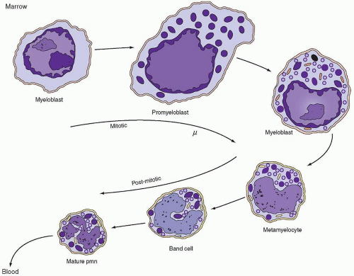
FIGURE 7.1. Appearance of granules during neutrophil maturation. Myeloblasts are undifferentiated cells with a large oval nucleus, large nucleoli, and cytoplasm lacking granules. They originate from a precursor pool of stem cells. Subsequently, there are two stages—the promyelocyte and the myelocyte—each of which produces a distinct type of secretory granule: azurophils (dark granules) are produced only during the promyelocyte stage; specific granules (light granules) are produced during the myelocyte stage. The metamyelocyte and band forms are nonproliferating stages that develop into the mature polymorphonuclear neutrophil characterized by a multilobulated nucleus and cytoplasm containing primarily glycogen and granules. Both nonspecific azurophilic granules and specific granules persist throughout these later stages. PMN, polymorphonuclear leukocyte. (Modified from Bainton DF, Ullyot JL, Farquhar MG. The development of neutrophilic PMN leukocytes in human bone marrow: origin and content of azurophil and specific granules. J Exp Med 1971;134:907.)

The neutrophil promyelocyte is somewhat larger on average than the myeloblast. In both light and EM preparations, it has a round or oval nucleus in which the nuclear chromatin is diffusely distributed, as in the myeloblast; in later stages, slight chromatin condensation is discerned around the nuclear membrane. Nucleoli are present, but as the cell develops, they become less prominent. Compared with the myeloblast, the endoplasmic reticulum in EM preparations is more prominent and takes on a dilated, vesicular appearance. The azurophilic, primary granules appear and accumulate in increasing numbers during this stage, but the specific or secondary granules are not yet present (Fig. 7.4).87,96,97,102,104 In early promyelocytes, the few granules present may be difficult to see by using light microscopy; they often lie over the nucleus and are evident only on examination at several focal planes.


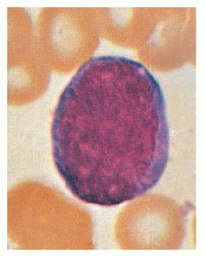
FIGURE 7.2. Myeloblast (×1,000, Wright stain).

Like the myeloblast, the promyelocyte is immobile in flat slide and cover glass preparations; only in the last stage is slight locomotion evident. Even then, the streaming of granules so characteristic of mature granulocytes is lacking.105 In hanging-drop preparations, however, promyelocytes are actively mobile.

The neutrophilic myelocyte may be defined as the stage in which specific (secondary) granules appear in the cytoplasm and the cell consequently can be identified as belonging to the neutrophilic series when stained and observed to have a pinkish ground-glass background color with the light microscope. As mentioned previously, earlier identification of a cell that will become neutrophilic can be made by EM examination of the azurophilic, or primary, granules.95 The nucleus of the neutrophilic myelocyte usually is eccentric and round or oval; one side may appear flattened. The nuclear chromatin is somewhat coarse, and nucleoli are small and often not visible, although they are seen clearly with the electron microscope.102 Primary granules
persist in myelocytes, but formation of new primary granules is limited to the promyelocyte, and each succeeding cell division leads to a decrease in their number in the daughter population (Fig. 7.1).97,104,105 The secondary granules of the neutrophil series are smaller than the primary granules; in the rabbit, cat, and human, they are formed in increasing numbers on the convex surface and lateral borders of the somewhat less prominent Golgi apparatus.97,102,104 The amount of granular endoplasmic reticulum is lower in the myelocyte than in earlier forms, so the cytoplasmic basophilia decreases and disappears. The mitochondria remain small and are few.


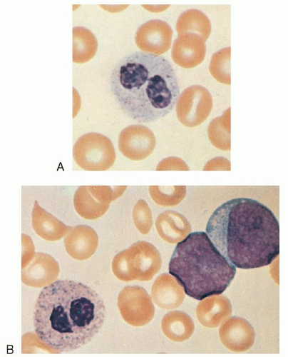
FIGURE 7.3. A, B: Pseudo-Pelger-Huët cells, the latter from the blood of a patient with acute myeloblastic leukemia (×1,000, Wright stain).


Neutrophil Granule Development

It has been suggested that the different neutrophil granules are formed because of temporal differences in gene expression of the granule contents82,106 and this model fits well with the data. Studies in rabbits, cats, and humans suggest that the primary granules are packaged and released from the inner, concave surface of the Golgi apparatus (Fig. 7.5A)—in contrast to the specific or secondary granules of the myelocyte and later granulocyte stages that appear to be formed and released from the outer, convex surface.97,102,103,104 Studies of membranes from rabbit azurophil and specific granules, although demonstrating similar ultrastructure, have shown them to be distinct and different in cholesterol-phospholipid ratios and protein components.107 In the mature neutrophil, a ratio of secondary to primary granules of approximately 9:1 is seen in the rabbit108 and 2 or 3:1 in humans.97,109

The mature primary granules of human neutrophils usually contain central crystalloids when lightly stained.103 They apparently bind neutral red dye and thus are seen easily as neutral red bodies in supravital preparations.110 These membrane-bound lysosomes contain enzymes and other substances (Table 7.1).87,97,111,112 Acid phosphatase activity varies considerably, as reported in different studies and in different species.102,103,113,114 This may be because of variation in the histochemical assays or to species variations.87 Peroxidase has been associated with primary granules by histochemical, cytochemical, and biochemical methods and is considered a marker enzyme for primary granules in mammals.87,115 Sulfated mucosubstance presumably accounts for the azurophilic staining of the primary granules; the uptake of radiosulfate by early-stage neutrophils may be the result of incorporation into this substance.116,117

The secondary granules of the neutrophil were once thought to be characterized by their content of alkaline phosphatase and lack of acid phosphatase.87,97,118 However, subsequent studies demonstrated that alkaline phosphatase is localized in a previously unrecognized organelle, the secretory vesicle (Table 7.1).119 Lysozyme is present in human neutrophil secondary granules,120 as well as in primary granules; approximately two thirds of this antibacterial basic protein is in the secondary granule. The standard marker enzymes for specific granules are lactoferrin115 and B12-binding protein.121

A third type of granule, the tertiary granule, also known as the gelatinase granule (Table 7.1),103,122 is synthesized mainly during the band and segmented neutrophil stages.103,123,124


Neutrophil Metamyelocytes

The metamyelocyte is characterized by a clearly indented or horseshoe-shaped nucleus without nucleoli, and the nuclear chromatin is moderately dense, with considerable clumping evident along the nuclear membrane. The cytoplasm is filled with primary, secondary, and tertiary93,116 granules, but the secondary granules predominate. The endoplasmic reticulum is sparse, as are polysomes, thus signifying the virtual completion of protein synthesis.

The boundary between the myelocyte and metamyelocyte compartments is best defined physiologically by the fact that myelocytes synthesize DNA, take up 3H-TdR into their nuclear chromatin, divide, and are actively involved in protein synthesis, as evidenced by the presence of nucleoli, abundant endoplasmic reticulum, and polysomes. Before such techniques became available, differentiation between myelocytes and metamyelocytes was defined mainly in terms of nuclear shape. This characteristic now is recognized as a poor criterion because it has been shown in time-lapse microcinematographic studies of human neutrophils that myelocyte nuclei may assume a markedly indented shape and may subsequently revert to an oval configuration and enter mitosis.95 Consequently, in classifying cells at this stage, the observer should pay particular attention to evidence in the nucleus and cytoplasm that protein synthesis has decreased or stopped. This determination is made on the basis of the fact that the nuclear chromatin is coarse and clumped and that the cytoplasm is faint pink and is essentially the color of the mature cell in stained preparations. These features are also helpful in differentiating metamyelocytes (Fig. 7.6A,B) from monocytes (Fig. 7.6C) because in monocytes, nuclear chromatin remains fine, and evidence of protein synthesis persists. Amoeboid movement is apparent in metamyelocytes, even in cover glass slide preparations, and it is at this stage that directional migration can regularly be demonstrated.125


Band Neutrophils

The band stage is characterized by further condensation of nuclear chromatin and transformation of nuclear shapes into sausage or band configurations that have approximately uniform diameters throughout their length (Fig. 7.1). Subsequently, one or more constrictions begin to develop and progress until the nucleus is divided into two or more lobes connected by filamentous strands of heterochromatin, the polymorphonuclear stage. Some workers require a clearly visible filamentous strand between lobes (Fig. 7.7A,B) before classifying a cell as a polymorphonuclear form; anything less clearcut, whether because of overlapping of nuclear lobes or incomplete constriction, is classified as a band form.126,127 Other investigators


have regarded a constriction greater than one half or two thirds of the nuclear breadth as adequate evidence of lobulation and classify such cells as polymorphonuclear128,129 or use slightly different criteria.130 Still others avoid the issue entirely. Because no clear difference has been shown between band and segmented stages other than nuclear shape and a slightly earlier appearance of 3H-TdR in the band forms, the distinction becomes arbitrary. However, a clear and easily recognizable separation is needed if one wishes to count nuclear lobes for diagnostic purposes, as in the early detection of folic acid deficiency131 or in assessing marrow release of young forms into the blood.132 For such purposes, we have chosen the clear separation of nuclear lobes as the criterion for inclusion in the polymorphonuclear category.127 Cells without this complete formation of distinct lobes (usually connected by a filamentous strand) are classified as band forms.


FIGURE 7.4. Early and late promyelocytes, a myelocyte, and a polymorphonuclear neutrophil (PMN) viewed by electron microscopy. (Courtesy of Bainton D, University of California, San Francisco.) A: Early neutrophilic promyelocyte (reacted for peroxidase, 10,500). The nucleus (N) with its prominent nucleolus (Nu) occupies the bulk of this immature cell. The surrounding cytoplasm contains a few azurophil granules (ag), a large Golgi complex (G), Golgi cisternae (Gc), several mitochondria (µ), scanty rough endoplasmic reticulum (er), and many free polysomes (r). A centriole (ce) is present in the Golgi region. All of the azurophil granules (ag) appear dense because they are strongly reactive for peroxidase. The secretory apparatus, i.e., the perinuclear cisterna (pn), rough endoplasmic reticulum (er), and Golgi cisternae (Gc), are also reactive, although less so than the granules. Specimen was fixed in glutaraldehyde for 16 hours at 4°C, incubated in the peroxidase medium of Graham and Karnovsky for 1 hour at 22°C, postfixed in osmium tetroxide, treated in block with uranyl acetate, dehydrated in ethanol, infiltrated with propylene oxide, and embedded in Araldite. Section stained for 1 minute with lead citrate. B: Late neutrophilic promyelocyte (reacted for peroxidase, 7,000). This cell is the largest (15 µm) of the neutrophilic series. It has a sizable, slightly indented nucleus (N), a prominent Golgi region (G), and cytoplasm packed with peroxidase-positive azurophil granules (ag). Note the two general shapes of the azurophil granules: spherical (ag) and ellipsoid (ag′). Most are spherical, with a homogeneous matrix, but a few ellipsoid forms containing crystalloids also are present. Many of the spherical forms (ag) have a dense periphery and a lighter core, presumably because of incomplete penetration of substrate into the compact centers of mature granules. Peroxidase reaction product is visible (under higher magnification) in less concentrated form within all compartments of the secretory apparatus (endoplasmic reticulum, perinuclear cisterna, and Golgi cisternae). No reaction product is seen in the cytoplasmic matrix, mitochondria, or nucleus. Specimen was fixed in glutaraldehyde for 10 minutes at 4°C and subsequently processed exactly as was the specimen in A. C: Neutrophilic myelocyte (reacted for peroxidase, 9,000). At this stage, the cell is smaller (10 µm) than the promyelocyte, the nucleus is more indented, and the cytoplasm contains two different types of granules: large, peroxidase-positive azurophils (ag) and the generally smaller specific granules (sg), which do not stain for peroxidase. A number of immature specific granules (is), which are larger, less compact, and more irregular in contour than mature granules, are seen in the Golgi region (G). Note that peroxidase reaction product is present only in azurophil granules and is not seen in the rough endoplasmic reticulum (er), perinuclear cisterna (pn), and Golgi cisternae (Gc), in keeping with the fact that azurophil production has ceased, and only peroxidase-negative specific granules are produced during the myelocyte stage. D: Mature PMN (reacted for peroxidase, 10,500). The cytoplasm is filled with granules; the smaller peroxidase-negative specific granules (sg) are more numerous, the azurophils (ag) having been reduced in number by cell divisions after the promyelocyte stage. Some small, irregularly shaped azurophil granule variants are also present (unlabeled arrow). The nucleus is condensed and lobulated (N1-N4), the Golgi region (G) is small and lacks forming granules, the endoplasmic reticulum (er) is scanty, and mitochondria (µ) are few. Note that the cytoplasm of this cell has a rather ragged, moth-eaten appearance because the glycogen, which is normally present, has been extracted in this preparation by staining in block with uranyl acetate.


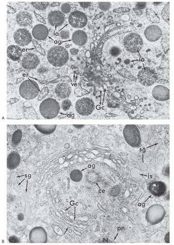
FIGURE 7.5. Granule formation in neutrophil precursors viewed by electron microscopy. (Courtesy of Bainton D, University of California, San Francisco.) A: Golgi region of a neutrophilic promyelocyte reacted for peroxidase (×40,000). At this stage, the peroxidase reaction product is present within the rough endoplasmic reticulum (er), the clusters of smooth vesicles (ve) at the periphery of the Golgi cisternae (Gc), in the Golgi cisternae, and in the immature (ia) and mature (ag) azurophilic granules. The immature granules are larger and less compact than the uniformly dense mature granules. B: Golgi region of a neutrophilic myelocyte reacted for peroxidase (×40,000). Peroxidase-reactive material is seen in the primary or azurophilic granules (ag) but not in the specific (secondary) granules (sg). At this stage (myelocyte), no peroxidase reaction product is seen in the endoplasmic reticulum, Golgi cisternae (Gc), or newly formed, immature specific granules (is). The stacked Golgi cisternae are oriented around the centriole (ce), and the outer cisternae (unlabeled arrow) contain material of intermediate density that is similar to the content of the specific granules, suggesting that the specific granules arise from the convex face of the Golgi complex as in the rabbit. pn, perinuclear cisternae.


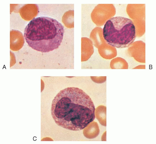
FIGURE 7.6. A: Late myelocyte or early metamyelocyte. B: Metamyelocyte. C: Monocyte (×1,000, Wright stain).


Polymorphonuclear Neutrophils

In the polymorphonuclear stage, the nucleus in Wright-stained preparations is a deep purplish color and contains coarse, condensed chromatin. The lobes are joined by thin filaments of chromatin, although the filaments may not be easily visible if the lobes are partially superimposed. The cytoplasm is faint pink and contains fine, specific granules that sometimes give only a ground-glass appearance. The azurophilic or primary granules have usually lost their dark-staining characteristics by this stage but can be seen with EM. With this technique, the granules exhibit considerable variation in density, presumably a reflection of variation in enzyme content, and they maintain a minimum distance of 100 nm from the cell membrane.133 Large masses of glycogen become evident for the first time in mature neutrophils; this finding may contribute to their capacity for anaerobic metabolism.


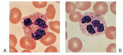
FIGURE 7.7. A, B: Polymorphonuclear neutrophils.

The mechanism and purpose of nuclear lobulation are the subject of speculation. Perhaps it enhances cell deformability and movement through vessel walls and into sites of inflammation, or perhaps nuclear segmentation results from nucleolar emptying and has no function.134 Thus, the mechanism and purpose of nuclear segmentation remain unclear.

It was suggested that granulocytes with three or four lobes are more mature than those with only two.135 However, the number of lobes a neutrophil develops appears to be determined in the band stage (or earlier), and the time of appearance of neutrophils in the blood after pulse labeling with 3H-TdR is unrelated to the number of nuclear lobes.136

In wet films, marked amoeboid activity of polymorphonuclear neutrophils at physiologic temperatures is characteristic.137 “Senile” polymorphonuclear leukocytes that are no longer motile and fail to take up neutral red stain have been identified in in vitro preparations.138 They are seen in small numbers in the blood, where their survival time is short.139


Pelger-Huët Anomaly

Pelger-Huët anomaly is a benign anomaly of leukocytes and is inherited as a non-sex-linked, dominant trait. It is characterized by distinctive shapes of the nuclei of leukocytes, a reduced number of nuclear segments (best seen in the neutrophils), and coarseness of the chromatin of the nuclei of neutrophils, lymphocytes, and monocytes. The nuclei appear rod-like, dumbbellshaped, peanut-shaped, and spectacle-like (“pince-nez”) with smooth, round, or oval individual lobes (Fig. 7.3), contrasted with the irregular lobes seen in normal neutrophils. Pelger-Huët cells appear to be normal functionally. Pseudo- or acquired Pelger-Huët anomaly may also be seen, in which cells with morphologic changes, such as described above, have been observed occasionally in association with myxedema, acute enteritis, agranulocytosis, multiple myeloma, malaria, leukemoid reactions secondary to metastases to the bone marrow, drug sensitivity, or chronic lymphocytic leukemia. More commonly, pseudo-Pelger-Huët cells are seen in patients with myeloid leukemia or myeloid metaplasia. Pelger-Huët anomaly is discussed further in Chapter 58.


Neutrophil Heterogeneity

Polymorphonuclear neutrophils were first thought to be a homogeneous population of end-stage cells incapable of protein synthesis and of essentially uniform size, granule content, and functional capability. Sabin first suggested potential heterogeneity among neutrophils when she reported that myelocytes were less motile than more mature neutrophils.138 A range of rates of motility among neutrophils from a single individual has been observed,140,141 and Harvath and Leonard suggested the existence of two neutrophil populations based on chemotaxis.142 Subsequently, several monoclonal antibodies were described that recognize subpopulations of neutrophils, including one that appears to recognize the classic NB1 (HNA-2a) neutrophil antigen, and one that recognizes an activation epitope on CD11/CD18.143,144 and 145 Another antibody, 31D8, appears to recognize a neutrophil subset that is more responsive to FMLP as determined by chemotaxis and respiratory burst activity.143 Neonates have a larger percentage of neutrophils that express low levels of 31D8 antigen.146 It has been reported that CR2 (CD21), the receptor for C3d, is present on immature neutrophils but not mature blood neutrophils.147,148 One report found that neutrophils from patients with localized juvenile periodontitis express CD21 (CR2) on their surface, whereas normal neutrophils do not.149
Neutrophil heterogeneity has also been demonstrated in the case of olfactomedin 4 expression. Olfactomedin 4 is highly induced in myeloid progenitors by G-CSF. Although olfactomedin 4 mRNA is expressed in all myelocytes and metamyelocytes, only ˜25% of neutrophils in peripheral blood contain olfactomedin 4, which is localized to the specific granules.150

Some studies of these different populations of polymorphonuclear neutrophils have been interpreted as reflecting maturation or environmental influences,151 in some cases possibly reflecting intravascular exposure to stimuli.152,153 The clinical significance of neutrophil subpopulations is unclear.


MACROPOLYCYTES

Macropolycyte is the name applied to giant polymorphonuclear neutrophils with a diameter greater than 16 µm and with 6 to 14 nuclear lobes.154 Such cells are seen only occasionally in healthy subjects (1.3%), but they are found in approximately 5% of people with infections of various types or with intoxications, usually in association with a neutrophilic leukocytosis and myelocytes in the blood.154 Macropolycytes are commonly seen in association with folic acid or vitamin B12 deficiency, as well as in patients recovering from the pancytopenia that attends treatment with cytotoxic agents, especially hydroxyurea.

Some authors describe cells with hypersegmented nuclei but of a normal size and call them polycytes126 or polylobocytes129; similar cells with complex nuclei but without hypersegmentation are called propolycytes.126 The latter forms are seen in approximately 10% of patients recovering from leukocytosis with a marked shift to the left and appear in increasing numbers when anticoagulated blood is allowed to stand in vitro.126,155

The mechanism of macropolycyte formation is unknown, but one suggestion is that the skipping of one of the usual cell divisions that occurs during maturation results in a hypersegmented cell.156


GENETIC SEX AS INDICATED BY LEUKOCYTES

Only one X chromosome is essential to the normal activity of a cell; the other in the normal XX female remains unextended and thus is visible as a chromatin body. Sex chromatin (Barr) bodies are present in 80% to 90% of the somatic cells of the normal female subject. The sex chromatin body of the neutrophil of females is a small mass, usually adjacent to the nuclear membrane, that stains deeply with hematoxylin and is approximately 0.7 to 1.2 µm in diameter. It takes the form of a drumstick projecting from one of the nuclear lobes of approximately 2% to 3% (extreme range, 1% to 17%) of the segmented neutrophils in the blood.157 They are well-defined, solid, round projections of chromatin connected to a lobe by a single, fine chromatin strand (Fig. 7.8). They must be distinguished from small clubbed or racket-structured, nonspecific nodules that may be smaller or larger as well as irregular in shape or lacking in chromatin, as well as from small (minor) lobes attached to the rest of the nucleus by two strands. Confirmation of the X chromosome in the drumstick has been provided by in situ hybridization.158 Sessile nodules are equally gender-specific but are more difficult to recognize. Drumsticks are not found in normal male subjects.159


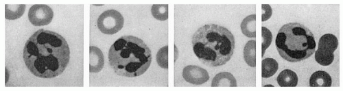
FIGURE 7.8. Granulocytes and sex chromatin patterns. Two cells on the left show the characteristic drumsticks found in the female subjects. The thin strand of chromatin joining the head to a nuclear lobe can be seen clearly. In the two cells on the right, small clubs, such as may be seen in male subjects, should not be confused with drumsticks (Wright stain, ×1,300).

The number of chromatin bodies seen in a cell is one less than the number of X chromosomes present. With the increased numbers of X chromosomes found in certain disorders of human development, the number of Barr bodies and drumsticks increases, and isochromosomes formed by duplication of the long arms of the X chromosome give rise to larger drumsticks than are found in the normal female subject.160 Drumsticks or sessile nodules are seen in chromatin-positive male patients with Klinefelter syndrome and are absent in chromatin-negative female patients with Turner syndrome. Eosinophils and probably basophils also have drumsticks. Drumsticks may be difficult to find in the presence of a marked shift to the left. Double drumsticks159 or a sessile nodule plus a drumstick in the same neutrophil are rare.161


DIFFERENTIAL CELL COUNTING AND NORMAL VALUES FOR LEUKOCYTES

Differential cell counting is the enumeration and classification of the leukocytes seen on the blood smear. The usual procedure is to count at least 100 consecutive leukocytes in an area of good cell distribution. A uniformly thin smear of blood on a cover glass is the best preparation for such examination. Differential cell counting in general is discussed in Chapter 1.

Distributional errors are reduced as more cells are counted. Confidence tables or curves can be used to estimate the probable error of a differential count when various numbers of cells are counted. Clearly, as more cells of a given type are counted and as the total number of cells enumerated increases, the accuracy of the differential count is greater. From the total leukocyte count and the differential count, the absolute concentration of each leukocyte type can be calculated. The accuracy of the result depends on the validity of the total leukocyte count and the differential count. With automatic cell counters, the major component of error now lies in the differential count.

The absolute leukocyte concentration provides a more accurate picture than the differential count because each leukocyte type is a separate cell system with its own functions, control mechanisms, and responses to disease processes; for example, a patient with chronic lymphocytic leukemia whose total leukocyte count is 100 × 109 cells/L, 7% of which are neutrophils and 93% are lymphocytes, does not have granulocytopenia. With a blood neutrophil concentration of 7.0 × 10⁹ cells/L, the problem is lymphocytosis.

Normal values for absolute leukocyte concentrations obtained by using a Coulter counter and differential counts are shown in Table 7.4. Similar values have been reported with the use of other methods.162 Some variation is evident in values obtained in different population groups and appears to depend on age, sex, pregnancy, time of day, activity level, and other factors.163 Racial variation has been reported, especially in Ethiopian Jews164
and black Africans, who have significantly lower neutrophil and monocyte counts.165 Lower counts are less evident in black Americans127 and in Africans eating a Western diet.163


TABLE 7.4 NORMAL BLOOD LEUKOCYTE CONCENTRATIONS (95% CONFIDENCE LIMITS)


Subjects (Age)


Sex


No.


Time of Day (hours)


WBC


Neutrophils


Lymphocytes


Monocytes


Eosinophils


Basophils


WBC Counter


Differential (No. Cells)


Reference


Neonates (day 4)


53


Variable, 96 h of life


1,981-7,553


2,200-7,100


421-2,022


200-1,900


Coulter ? model


200


109


Infants (9-12 mo)


Model S


105


White


50


5,400-24,200


1,062-10,890


2,178-11,718


Black


50


4,100-14,300


121-6,732


900-11,400


European adults


M


72


0930-1130


3,487-9,206


1,539-5,641


(median age, 25 y)


26


1430-1630


3,722-9,828


1,775-6,508


F


70


0930-1130


3,839-10,135


1,861-6,821


1,158-3,460


221-843


25-590


0-140


Model S


500


97


29


1430-1630


4,450-11,750


2,137-7,836


European adults


M


85


0900-1530


3,956-9,592


2,075-6,557


962-3,784


59-658


200


99


(age, 54-65 y)


F


76


0900-1530


3,423-8,258


1,833-5,476


776-3,455


59-732


Model B


Pregnant Europeans (third trimester)


F


50


0930-1630


5,915-13,962


3,656-10,769


1,023-3,128


349-1,140


22-330


0-90


Model S


500


97


American white adults


226


a.m. or early p.m.


4,550-10,100


2,050-6,800


1,500-4,000


220-950


30-860


0-160


Models A and F


200


102


(age, 16-44 y)


Black American adults


M


65


a.m. or early p.m.


3,600-10,200


1,300-7,400


1,450-3,750


210-1,050


30-720


0-100


Models A and F


200


102


(age, 16-49 y)


Black African adults


M


250


0900-1200


2,587-9,075


775-4,131


1,012-3,876


62-688


47-3,371


Counting chamber


200


106


(age, 20-45 y)


Mostly


M


109


3,363-8,977


Model S


108


WBC, white blood cell.


NOTE: Values obtained by using a Coulter counter and differential counts of at least 200 cells (except in one African study). Values are expressed in cells 109/L.


Various systems for differential counting have been used.166 Arneth, for example, painstakingly recorded and tabulated from left to right the number of neutrophilic leukocytes with 1, 2, 3, etc., lobes and made other subdivisions.135 The term shift to the left is derived from this practice and indicates an increase in the proportion of cells with only one or few lobes, whereas shift to the right represents an increase in the proportion of multisegmented forms.

From a clinical viewpoint, it is useful to determine whether young forms of neutrophils (band forms and younger) are increased and whether the proportion of multinucleated forms is increased. An increase of younger forms (band cells, metamyelocytes, and myelocytes) (shift to the left) suggests increased release of young neutrophils from the bone marrow, which is seen in association with acute infections132 and inflammation. If a shift to the right is suspected, a neutrophil lobe count may be useful. This process involves counting the total number of nuclear lobes in 100 or 200 neutrophils, calculating the average lobe number/neutrophil, and comparing the results with normal values. The chief difficulty associated with this count is clear definition of what constitutes a separate lobe (see “Band Neutrophils” earlier in this chapter). If complete separation of nuclear lobes with or without a connecting filament is the definition used, the normal mean neutrophil lobe count is 2.04, with 95% of normal values falling between 1.66 and 2.42. An increase in mean neutrophil lobe count suggests vitamin B12 or folic acid deficiency, congenital hypersegmentation of neutrophils, or renal disease.131 A ratio of five-lobed to four-lobed polymorphonuclear cells that is greater than 0.17 is said to be associated more regularly with B12 deficiency than is an increase in mean nuclear lobe count.129


Alterations in the total number of leukocytes and in their relative proportions are significant as measures of the reactions of the body to noxious agents. The reactions of leukocytes in association with certain diseases are discussed later in this book, as is the presence of abnormal inclusions, such as toxic granulation, Döhle bodies, and various inherited abnormalities in leukocyte morphology.


NEUTROPHIL KINETICS

The importance of leukocytes in the defense of the organism is well known. Basic to their roles are cell multiplication, maturation, storage, and delivery to the tissues and sites of infection or cell damage. These processes are called leukocyte kinetics and are different for each leukocyte type. To simplify the discussion, each type of leukocyte is considered as a separate system, but these systems constantly interact and complement one another in the defense of the body.


Neutrophil Kinetics in the Adult

In Chapter 5, the process of mitotic cell division, the cell generation cycle, and the origin of neutrophils and other cell types from a multipotent hematopoietic stem cell were discussed. The production, kinetics, and lifespan of the neutrophil have been the subject of a number of reviews.167,168,169,170,171,172,173,174,175,176,177,178,179,180 and 181 A model of these processes in adult humans is shown in Figure 7.9. The neutrophil system appears to be incompletely developed in premature babies and in early neonatal life; this topic is discussed in the section “Neutrophil Kinetics in the Fetus and Newborn” later in the chapter.


Mitotic and Maturation Compartments

Neutrophil production in normal adult humans appears to take place only in the bone marrow. The life cycle of the neutrophil can be divided conveniently into bone marrow, blood, and tissue phases. The assumption is that cells move through the system in an orderly manner as if in a pipeline; this view is supported by the progressive movement of isotopic tracers117,178,182,183 and azurophilic granules 87,97,104 through the system.


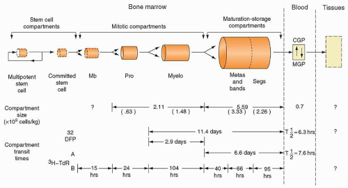
FIGURE 7.9. Model of the production and kinetics of neutrophils in humans. The marrow (555) and blood compartments (147) are drawn to show their relative sizes. In the lower one third of the figure, the compartment transit times as derived from di-isopropylfluorophosphate (DF32P) studies (138,147) and from tritiated thymidine (3H-TdR) studies (151,555) are compared. CGP, circulating granulocyte pool; MB, myeloblast; MGP, marginal granulocyte pool; myelo, myelocyte; pro, promyelocyte.

The myeloblast, promyelocyte, and myelocyte are capable of cell division, as judged by direct observation in cultures95 and by their ability to incorporate 3H-TdR into their nuclear DNA.183 These forms, therefore, constitute the mitotic compartment (Fig. 7.9). Simultaneously, they undergo differentiation, as evidenced by the appearance of azurophilic and specific granules in their cytoplasm. The more mature forms of the neutrophil series (metamyelocyte, band, and polymorphonuclear neutrophil) are usually considered incapable of cell division (except perhaps in unusual circumstances) 184 and do not incorporate 3H-TdR into their nuclei, but they do exhibit continuing maturational changes and thus constitute the maturation compartment. From the maturation compartment, cells flow into the blood and are distributed in two sites: the circulating blood granulocyte pool (CGP) and the marginal granulocyte pool (MGP). Cells in these two pools are in constant equilibrium. Eventually, the cells move through vessel walls to enter the tissues. The exact nature of the MGP is not clear. In the past, it was felt to represent transient adhesion to, and rolling along the surface of, endothelial cells, primarily in postcapillary venules. However, subsequent studies of a patient with leukocyte adhesion deficiency-2 (LAD-2) demonstrated the presence of a marginating pool,185 although the MGP in this patient with LAD-2 was reduced. Patients with LAD-2 lack the ligands for the selectins CD62P and CD62E and have a marked decrease in neutrophil rolling in postcapillary venules. Normally, the MGP is approximately equal in size to the CGP. Studies of this patient suggested that approximately 20% of neutrophils are in a selectin-independent MGP and approximately 30% are in a selectin-dependent MGP.185 Surprisingly, this patient’s neutrophils had a shorter than normal half-life in the circulation with an increased turnover rate. The inability of CD18 or CD62L antibodies to shift neutrophils from the MGP to the CGP also suggests that the transient adhesion to endothelial cells manifest as rolling does not account for the MGP.186,187


In this model, cell production can be estimated either by assessing the production rate in the mitotic compartment or by measuring cell flow through subsequent stages, such as the blood. Accuracy of these measurements is facilitated if the system is studied in the steady state when compartment sizes are constant and cell flow reflects net production and destruction.174

If one assumes a steady state in a scheme such as that shown in Figure 7.5, the flow of cells out of any pool (Kout) is equal to the flow of cells into that pool (Kin) plus any cells produced in the pool (Kb); thus,

Kout = Kin + Kb

Clearly, in pools other than those in the mitotic compartment, Kb is 0, and measurements of cell flow (Kin or Kout) equal effective cell production, provided no cell death occurs within the pool.


Production in the Mitotic Compartment

Because myeloblasts, promyelocytes, and myelocytes constitute approximately 0.9%, 3.3%, and 12.7% of the marrow cells, respectively, it has been assumed that the system has four or five divisions.180 From a review of blood neutrophil radioactivity curves obtained after di-isopropylfluorophosphate (DF32P) injection into humans, investigators raised the possibility of at least three divisions at the myelocyte stage.180 Another suggestion generated from marrow differential counts was that only four or five divisions occur in the entire neutrophil proliferation scheme.180 This supposition agreed with results from experiments in dogs188 and with data from model studies.189 In contrast, studies of myeloid islands in the rat thymus provided evidence for seven divisions during granulocytopoiesis: one in the myeloblast stage, two in the promyelocyte stage, three in the myelocyte stage, and a final one in the metamyelocyte stage.134,190

Calculations made from mitotic index (MI) data172,191,192 and 193 provide estimates of cell generation time (tg) and pool turnover time. MI191,192,194 is defined as

MI = Nm/N

in which MI is the mitotic index for any morphologic cell pool, Nm is the number of mitoses in that pool, and N is the total number of cells in the pool. MI can also be expressed as the ratio of the time spent in mitosis (tm) to the cell generation time (tg): MI = tm/tg

By combining both definitions,

MI = Nm/N = tm/tg

By providing determined values for MI and mitotic time (tm) in the last equation, the generation time (tg) for a particular cell pool can be approximated. From tg and the pool size (N), the birth rate, Kb, can be obtained if all cells in the pool are in cycle, because each mitosis gives rise to one new cell:

Kb = N/tg

In effect, the cell birth rate is equal to the number of mitoses occurring per unit time (t), or

Kb = Nm/t

Although the concept is simple, several problems arise.193,195 A major problem is that the morphologic boundaries of most cell pools are not clearly delineated in terms of the cell cycle.195 For example, to calculate cell production in the myelocyte pool, it must be assumed that all myelocytes are destined to divide; that is, no cells are recognized as myelocytes that are not going to divide again. Because the daughter cells of the last myelocyte mitosis almost certainly do not suddenly become metamyelocytes on completion of division, N in the preceding equation will be erroneously large, and thus estimates of tg will be erroneously long. If the fraction of nonmitotic cells in the myelocyte population

were known, the calculations could be corrected for this error, as has been attempted.170 A second major problem is the fact that values for the MI have varied considerably.191,193,196,197,198,199 and 200 In addition, a considerable diurnal variation exists in the MI in humans, as well as in animals.193,199,200,201 and 202

Finally, to calculate the absolute neutrophil production rate (in cells per unit of time), the size of the marrow mitotic compartment must be known. Methods for measuring the sizes of marrow myeloid pools have been developed203,204,205 and 206 (see “Size of Marrow Compartments and Their Morphologic Subdivisions,” later in the chapter), but to date no one has measured these sizes and MI in the same animal at the same time and then calculated neutrophil production rate. Nevertheless, values for the MI for each of the neutrophil precursors capable of mitosis have been determined,193,200 and within the assumptions inherent in such calculations,195 neutrophil production has been estimated.176,192

Similar calculations of neutrophil production can be made from 3H-TdR-labeling index data. After flash labeling with 3H-TdR, autoradiographs of the bone marrow are obtained, and the proportion of nucleated cells that have incorporated the label into their nuclei is determined.171,177 This labeling index (LI) represents the ratio of labeled cells, *N, or cells in DNA synthesis (Ns) to total cells (N) of a defined morphologic type:

LI = *N/N = Ns/N

The LI can also be defined in terms of DNA synthesis time (ts) and the cell generation time (tg), because 3H-TdR is taken into the cell only during the period of DNA synthesis; thus,

LI = ts/tg

By combining both definitions,

LI = Ns/N = ts/tg

and from determined values for LI and tg, the generation time and turnover of a given cell population can be estimated. As with MI data, birth rate is a function of the population turnover time, which can be approximated from the generation time or time spent in various phases of the cell cycle:

Kb = N/tg = Ns/ts

Some of the same problems arise with the 3H-TdR LI that are encountered in the use of the MI.195,207 In addition, the use of labeled compounds raises questions of label reuse 208,209 or elution207 and perturbation of the cell population by the compound168,188,210,211 and 212 or by its radioactivity.207,213,214

The LI reported for humans is myeloblast, 0.85; promyelocyte, 0.65; and myelocyte, 0.33.171 Somewhat different values have been reported in dogs177 and rats.200 By using the LI for humans and a value for ts of 5 hours (based on studies in dogs) and by determining relative compartment sizes for each cell type from the bone marrow differential count, the relative birth rates (Kb) of cells have been calculated.170,200

Some authors have found good agreement between neutrophil production as calculated from the MI and the LI,200 but considerable discrepancy has been reported by other authors.170 This difference may result from the fact that the MI values obtained were low, the studies were done in different subjects at different times, and too small a value for ts was used in the calculations.

The turnover time of a labeled compartment and neutrophil production rate also may be estimated by measuring the grain count halving time. The generation time is derived only if each cell in a given class divides and if no label feeds into the compartment from a labeled precursor class or as a result of label reuse.215 If any of these criteria are not met, the half-time for grain count decrease is longer than the true value, and the estimate of generation time is only a maximal value. Additional disadvantages of this method are that at least several bone marrow samples distributed
throughout several half-times are needed, and that grain counting is extraordinarily tedious and subject to considerable error. Nevertheless, estimates of compartment turnover time have been made with this method by using 3H-TdR and radiosulfate.122

After flash labeling with 3H-TdR, the cohort of cells labeled during DNA synthesis may be followed as it subsequently enters mitosis, and the time course of labeled mitoses can be recorded.215,216 Theoretically, such curves should permit measurement of the post-DNA synthesis gap (G2), mitotic time (tm), DNA synthesis time (ts), cell generation time (tg), and pre-DNA synthesis gap (G1) (see Chapter 5 and Fig. 7.10). In actual practice, biologic variation rounds off the percentage of labeled mitosis curves, and rapid damping of the waves of cells passing through mitosis (Fig. 7.10) renders such measurements less precise than ideal. However, estimates of myeloid DNA synthesis time obtained with this method are approximately 11 to 13 hours in humans215,216 and are in good agreement with estimates made in gastrointestinal mucosal cells. From the level of the damped plateau reached after a few hours, the ratio of ts to tg can be obtained (Fig. 7.10), and the generation time can then be calculated. If the generation time and compartment transit time are presumed to be the same or if the proportion of cells in a compartment that is actively proliferating is known, the neutrophil production rate can be calculated.


Neutrophil Production as Measured by Cell Flow in Other Compartments

Another method for approximating neutrophil production involves following the appearance of 3H-TdR-labeled cells in the metamyelocyte compartment. Because metamyelocytes do not divide or take up 3H-TdR, the appearance of labeled cells in this compartment should reflect the flow of cells into it from the myelocyte compartment; in the steady state, this influx of cells should also reflect the turnover of the metamyelocyte compartment and thus cell production. Approximately 3 hours pass after the injection of 3H-TdR before the label appears in metamyelocytes both in dogs177 and in humans215; this time interval is the minimum time for myelocytes taking up the label to pass through G2 and mitosis and become metamyelocytes. After this lag, the rate of labeled cell inflow into the metamyelocyte compartment is approximately 3% to 5%/hour in both species. In the dog, cell inflow into the metamyelocyte compartment measured in this fashion is less than 50% of that calculated from LI data,177 suggesting the existence of a major component of ineffective granulocytopoiesis, a myelocyte sink, in the normal animal. However, similar calculations in humans do not confirm the findings of such studies.170 The resolution of this enigma requires the simultaneous measurement of cell production by using several methods in the same animal at the same time.


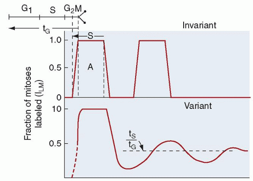
FIGURE 7.10. The percentage of labeled mitoses in the course of cell generation. At the top of the diagram is the theoretical configuration that would be seen if cells flowed through the proliferation cycle with no variation. The effects of moderate variation in time spent in the several cycle phases on the percent labeled mitosis curve are shown at the bottom. G1 is the pre-DNA synthesis resting phase (gap), S is the DNA synthetic period, G2 is the post-DNA synthesis gap, and M is mitosis. tS is the time spent in S, and tG is the duration of the entire generative cycle. (From Cronkite EP, the Brookhaven National Laboratory, and the National Cancer Institute. Kinetics of granulocytopoiesis. Natl Cancer Inst Monogr 30, Human Tumor Cell Kinetics 1969;51.)

Similarly, the marrow production of neutrophils has been estimated from the size of the postmitotic maturation storage compartment and the compartment turnover time.200,204,217 The compartment size is calculated from the marrow neutrophil-toerythroid (NE) ratio (determined in marrow sections) and the mean normal marrow normoblast pool size (calculated by multiplying the ratio of subjects’ erythroid iron turnover value over the mean normal value for this determination by the mean normal erythroblast population).204 The compartment transit time is estimated by injecting 3H-TdR and noting the time required for labeled neutrophils to appear in the blood. By this method, marrow neutrophil production in humans has been calculated to be 0.85 × 10⁹ cells/kg/day in the normal steady state.

Neutrophil production also can be approximated by measuring the flow of cells through the blood, the blood granulocyte turnover rate (GTR). DF32P binds irreversibly with a number of esterase enzymes and has been shown to label neutrophils primarily.218,219 By means of this agent, a subject’s own cells can be labeled, and the total blood granulocyte pool (TBGP) and the rate of disappearance of labeled neutrophils from the blood can be determined.175 Similar measurements can be made by transfusing 3H-TdR-labeled cells from a suitable donor.204 Because neutrophils leave the blood in a random manner (exponential disappearance curve), the GTR is calculated as follows from the TBGP and the t1/2:

GTR = 0.693/t1/2 TBGP

in which 0.693 is the natural logarithm of 2 and t1/2 is the blood neutrophil half-disappearance time.

All of these methods assume that the system is in a steady state during the entire course of the measurements. If neutrophil death in the marrow is not significant, the blood GTR equals total neutrophil production. If neutrophil death in the bone marrow is significant, the blood GTR measures effective neutrophil production, and the difference between this determination and total neutrophil production is ineffective granulocytopoiesis. Measurements of neutrophil production by compartment turnover methods have given values ranging from 62 to 400 × 107 neutrophils/kg/day in humans (Table 7.5) 204,220 and 150 to 560 × 107 neutrophils/kg/day in dogs.204,217,221

Of the methods for assessing neutrophil production just described, only the measurement of blood neutrophil turnover rate with DF32P or 3H-TdR can be performed easily enough to be of use in studying groups of patients in a clinical setting, and even this is possible in only a few research centers.


Size of Marrow Compartments and Their Morphologic Subdivisions

In all of the procedures described, with the exception of DF32P and 3H-TdR blood kinetic measurements, the number of marrow myeloid cells under study must be known in order to calculate neutrophil production. In the absence of such data, only calculations of relative cell production are possible.170 Direct measurements of the volume and cellularity of the marrow (isolation of skeleton and cell counting or bone biopsy and radioactivity measurement) have been made in dogs,204 rats,222 mice,223 and guinea pigs.224,225

Only gold members can continue reading. Log In or Register to continue

Stay updated, free articles. Join our Telegram channel

Oct 21, 2016 | Posted by in HEMATOLOGY | Comments Off on Neutrophilic Leukocytes

Full access? Get Clinical Tree

Get Clinical Tree app for offline access