Hematopoietic Cell Transplantation


Hematopoietic Cell Transplantation


Richard A. Nash

Vijayakrishna K. Gadi


Transplantation of hematopoietic cell grafts containing pluripotent hematopoietic stem cells (HSCs) after myeloablative or nonmyeloablative conditioning regimens reconstitutes immunohematopoiesis. Sites from which HSC can be harvested and then used for transplantation include bone marrow, peripheral blood, and the umbilical cord. Patients may serve as their own donors (autologous) or may receive HSC from other individuals (related or unrelated). Hematopoietic cell transplantation (HCT) is done for a variety of therapeutic indications: (1) to support hematopoiesis after myeloablative doses of total body irradiation (TBI) and chemotherapy, (2) to establish a graft-versus-leukemia or tumor (GVL or GVT) reaction, or (3) to replace diseased tissues of hematologic or immunologic origins. The advances in supportive care after transplantation have resulted in improved outcomes, and HCT has become more accepted as a therapeutic modality that can be successful in otherwise life-threatening diseases. In the 2003-2007 period as compared with the 1993-1997 period, a 60% reduction in day 200 nonrelapse mortality and a 41% reduction in overall mortality was observed.1 There were also significant reductions in severe graft-versus-host disease (GVHD), opportunistic infections, and organ damage.

The focus of this chapter is on the general indications for HCT, sources of stem cells, conditioning regimens, transplant biology issues, and complications of HCT; other chapters provide detailed discussions of the indications and results of HCT in relation to specific diseases (Table 102.1).


HISTORICAL PERSPECTIVE


Early Preclinical Studies

After the effects of radiation on hematopoiesis became evident during World War II, Jacobson and colleagues reported that mice survived an otherwise lethal exposure to TBI if the spleen was shielded (Fig. 102.1).2 Radiation protection was also conferred by infusion of bone marrow.3 A runting syndrome developed after recovery of hematopoiesis when the infused marrow was from a donor of a different mouse strain.4 This syndrome was due to GVHD, a complication that was soon recognized to limit the use of allogeneic marrow transplantation in humans. In further studies in mice, methotrexate and 6-mercaptopurine were found to be effective in inducing immune tolerance or ameliorating the graft-versus-host (GVH) reaction.5

The dog served as a random-bred large animal model for studies of principles and techniques of bone marrow transplantation applicable to humans. The dog was the first random-bred species in which it was demonstrated that the results of in vitro histocompatibility typing could predict the outcome of marrow transplantation.6 Littermates genotypically identical for the major histocompatibility complex (MHC) survived longer after marrow transplantation than did their MHC-nonidentical siblings. However, despite the MHC genotypic identity, GVHD was still potentially severe in many but not all dogs. This indicated that other factors identified as minor histocompatibility antigens (mHC) were involved in the development of GVHD. Pharmacologic immunosuppression with a calcineurin inhibitor
(i.e., cyclosporine or tacrolimus) or methotrexate for prevention of GVHD improved survival after allogeneic marrow grafting.7, 8 It was then established that methotrexate and cyclosporine in combination were more effective than either used alone.9 The efficacy of a calcineurin inhibitor and methotrexate combined for GVHD prevention was subsequently confirmed in clinical trials and remains the standard in many transplant programs.


TABLE 102.1 DISORDERS TREATED BY TRANSPLANTATION


Nonmalignant


Aplastic anemia


Fanconi’s anemia


Diamond-Blackfan syndrome


Sickle cell disease


Thalassemia


Paroxysmal nocturnal hemoglobinuria


Myelofibrosis


Congenital neutropenia


Chediak-Higashi syndrome


Chronic granulomatous disease


Glanzmann’s thrombasthenia


Osteopetrosis


Gaucher’s disease


Mucopolysaccharidosis


Mucolipidoses


Immune deficiencies


Malignant


Acute nonlymphoblastic leukemia


Acute lymphoblastic leukemia


Hairy cell leukemia


Myelodysplasia


Chronic myelogenous leukemia


Chronic lymphocytic leukemia


Hodgkin’s disease


Non-Hodgkin’s lymphoma


Multiple myeloma


Solid tumors


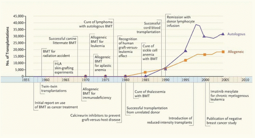
FIGURE 102.1. Timeline showing numbers of bone marrow transplantations and advances in the field, 1957-2006. BMT denotes bone marrow transplantation; HLA, human leukocyte antigen. Data are from the Center for International Blood and Marrow Transplant Research. (Reprinted with permission from Appelbaum FR. Transplantation of bone marrow as compared with peripheral blood cells from HLA-identical relatives in patients with hematologic cancers. N Engl J Med 2007;357:1472-1475. Copyright 2007 Massachusetts Medical Society. All rights reserved.)


Early Clinical Studies

Bone marrow was the first commonly used source of HSC for transplantation. Bone marrow transplantation from human leukocyte antigen (HLA)-identical sibling donors was first successfully used by two groups in 1968 to treat patients with immunologic deficiencies.10, 11 After extensive preclinical studies of GVHD, Thomas et al. then reported the successful transplantation of marrow from a HLA-identical sibling donor for aplastic anemia in 1972.12 Five years later, the same group reported their experience in 100 patients with end-stage leukemia treated with allogeneic marrow transplantation.13 Allogeneic HCT from an HLA-matched related or unrelated donor is now considered standard therapy for many malignant and nonmalignant hematologic diseases.


HEMATOPOIETIC STEM CELLS

The HSC is defined as a cell with the ability to achieve long-term reconstitution of both myeloid and lymphoid lineages. To fulfill these criteria, HSC must be able to self-renew and be pluripotent. In vitro colony-forming units (e.g., CFU-GM, CFU-Meg, CFU-E, BFU-E) are progenitor cells that cannot reconstitute hematopoiesis, whereas populations of cells enriched for long-term cultureinitiating cells can, at least in the mouse, rescue lethally irradiated recipients.14, 15 No accepted in vitro assays for human HSC are available currently. A population of small mononuclear cells enriched for HSC can be identified by (1) the presence of the CD34 and CD133 antigen, the absence of lineage-specific antigens, and high content of aldehyde dehydrogenase and (2) the exclusion of fluorescent vital dyes including Rhodamine123 and Hoechst 33 342 (Table 102.2).16, 17, 18, 19, 20 In humans and other species, successful sustained engraftment was achieved with isolated CD34-positive cells confirming that a true “stem cell” is contained within this population.21, 22, 23, 24

The true extent of the pluripotency of adult marrow-derived stem cells remains under investigation. Several studies have reported that populations of HSCs may contribute to regeneration of muscle, osteoblasts, hepatocytes, and neuronal and nonneuronal cell types of the brain.25, 26, 27 However, some of the
experimental observations may have resulted from cell fusion, technical artifact, or culture induced changes in cellular gene expression. Nevertheless, there is agreement that if “plasticity” of circulating HSCs occurs, it is likely to be a rare event.28


TABLE 102.2 CHARACTERISTICS OF HUMAN HEMATOPOIETIC STEM CELL (HSC)


A. Cell Surface Antigen Expression


Positive


aCD34-positive


CD133-positive


Aldehyde dehydrogenase


Low Positive


Thy 1 (CDw90), c-KIT


Negative


CD38, CD33, T and B cell markers, CD71, DR


B. Dye Exclusion


Rhodamine123, Hoechst 33 342


a CD34 negative HSC have been identified in mice.


Identification of Cell Populations Enriched for Hematopoietic Stem Cells

The CD34 antigen is a cell surface type 1 transmembrane protein which is highly O-glycosylated and expressed primarily on hematopoietic progenitor cells and vascular endothelium from many tissues.29, 30, 31 It is also expressed on stromal cell precursors identified by the STRO-1 antibody. Cell surface expression of CD34 is developmentally regulated in hematopoiesis and is inversely related to the stage of differentiation such that CD34 expression is absent beyond the committed progenitor stage. The functional significance of CD34 expression on hematopoietic progenitor cells, stromal cells, and developing blood vessels is unknown, except that CD34 on vascular endothelial cells binds to L-selectin.32 The CD34 antigen is expressed on 1% to 5% of normal human adult marrow cells, up to 1% of mobilized peripheral blood cells, and by 2% to 10% of normal fetal liver and marrow cells.29, 33 Approximately 90% to 95% of the CD34-positive cells express antigens indicating commitment to the lymphoid or myeloid lineages.34, 35 Purified populations of HSC can be obtained with strategies that lineage-deplete a CD34-positive population of cells using monoclonal antibodies specific for DR, CD33, CD38, CD71, and B and T cell markers (Table 102.2). Other work suggests that human HSC are Thy-1low, c-KITlow, Rhodamine123 low, and CD133-positive.36, 37, 38, 39, 40, 41 In vivo preclinical models for studying populations of purified human HSCs based on the aforementioned characteristics include transplantation into SCID-Hu mice or into fetal sheep.40, 42, 43

Enriched populations of autologous CD34-positive marrow cells or blood cells, in both animal and human studies, have been shown to protect from myeloablative doses of radiation or chemotherapy.22, 44, 45 Conversely, the CD34-negative subset of the marrow was not protective.46, 47 Complete and stable donor hematopoietic chimerism has also been shown in humans after transplantation with allogeneic CD34-enriched cells from the peripheral blood.24, 48 Thus, CD34-positive hematopoietic cells are capable of long-term stable reconstitution of multiple hematopoietic lineages.


Sites of Hematopoiesis and for Collection of Hematopoietic Stem Cells

In the developing human embryo, the production of hematopoietic cells shifts to the liver from the yolk sac after 6 weeks of gestation. At 16 weeks of gestation, the most active site of hematopoiesis is the fetal liver. At the end of gestation, essentially all hematopoietic production derives from the marrow with only small contributions from the liver and spleen. Umbilical cord blood is enriched for HSC. The number of progenitors in one cord blood unit were comparable to the number of progentiors from adult marrow that had been reported to achieve successful engraftment.49

Historically, the bone marrow served as the routine collection site for HSC.50 Marrow is a clinically reliable and easily accessible source of long-term reconstituting cells. The presence of HSC in peripheral blood was first documented in preclinical studies.51, 52 In early clinical studies, transplantation of autologous peripheral blood HSC resulted in reconstitution of hematopoiesis following myeloablative chemotherapy or chemoradiotherapy; however, obtaining sufficient HSC required a prolonged period of collection.53 To overcome this problem, granulocyte-colony-stimulating factor (G-CSF) can be administered to mobilize HSC from the marrow to the peripheral blood. Collections from G-CSF-mobilized peripheral blood yielded similar or greater numbers of HSC than that harvested from marrow.54 Combining chemotherapy and G-CSF administration for mobilization resulted in higher yield of CD34-positive cells than G-CSF alone.55 Plerixafor is a small molecule that reversibly inhibits chemokine stromal cell-derived factor-1α binding to its cognate receptor CXC chemokine receptor 4. In a randomized clinical trial, G-CSF with plerixafor was more effective than G-CSF alone for collection of stem cells in patients with myeloma.56 In this study, a total of 54% of plerixafor-treated patients collected a target number of 6.0 × 10⁶ CD34+ cells/kg after one apheresis, whereas 56% of placebo-treated patients required 4 daily aphereses to achieve this target. Other cytokines have been demonstrated to mobilize HSC into peripheral blood, including stem cell factor, granulocyte/macrophage-CSF, inter-leukin-6 (IL-6), IL-8, and flt-3 ligand.57, 58 While most normal donors receive only G-CSF for mobilization of HSC for allogeneic transplant, mobilization strategies including plerixafor or chemotherapy are now routinely used for the collection of autologous HSC from the peripheral blood of patients with hematologic malignancy.59, 60, 61 Plerixafor is not indicated for use in patients with acute leukemia.

Monoclonal antibodies specific to the adhesion molecule VLA-4 (very late antigen-4 or α4β1 integrin) and VCAM-1 (vascular cell adhesion molecule-1) can also mobilize hematopoietic progenitors in nonhuman primates.62, 63 An essential step contributing to the release of hematopoietic progenitors from the marrow may be the cleavage of VCAM-1 expressed on stromal cells by neutrophil proteinases following the administration of G-CSF.64 Hematopoietic progenitors reversibly downregulate VLA-4 expression and adhere significantly less to stroma and fibronectin during mobilization.65 VLA-4 integrin expression is restored after progenitors are removed from the in vivo mobilizing milieu, which may account for their homing properties after transplantation.


SELECTION OF STEM CELL SOURCE

Autologous, syngeneic, and allogeneic are the three general categories of HSC grafts (Table 102.3). In general, the origin of HSC used for HCT is based on both the availability of the donor and the type of disease for which the patient is being transplanted. While autologous HSC should be available for most patients, extensive prior cytotoxic therapy or heavy involvement of marrow with malignant cells may preclude the use of this source of HSC. Although the preferred allogeneic donor is an HLA-identical sibling, fewer than 30% of patients have access to this source. Availability of HLA-identical sibling donors for pediatric patients may be less than this because of the smaller family sizes now compared to previous generations. Syngeneic donors are available in less than 1% of cases, and phenotypically HLA-matched or one-antigen-mismatched haploidentical family donors are available in less than 5% of cases.66 Approximately 30% to 40% of patients may identify a phenotypically HLA-matched unrelated donor from the volunteer registries.67 The availability of unrelated (or related) umbilical cord blood banks increases the chances of successfully identifying a compatible allogeneic graft source for both pediatric and adult patients.68, 69, 70 HLA-haploidentical donors are also available for the majority of patients.

The disease for which transplantation is being considered is another important determinant for choice of stem cell source. Autologous, syngeneic, or allogeneic HSC can support hematopoietic recovery after myeloablative chemoradiotherapy for malignant hematologic and nonhematologic diseases. For acquired disorders of marrow function (e.g., aplastic anemia), syngeneic or allogeneic HSC are required.71 Patients with congenital hematopoietic or immunologic defects (e.g., thalassemia, severe combined immunodeficiency (SCID) syndrome) require transplantation with allogeneic stem cells or gene-modified autologous stem cells.72, 73, 74, 75


TABLE 102.3 POTENTIAL SOURCES OF HEMATOPOIETIC STEM CELL (HSC) FOR HEMATOPOIETIC CELL TRANSPLANTATION


Relationship of Donor


MHC (HLA)-Matching


Genetically Identical Haplotype


mHCa Matching


Site of HSC Collection


I. Syngeneic


Sibling


Identical


2


Identical


Marrow or peripheral blood


II. Allogeneic


Sibling


Identical


2


Shared


Marrow, peripheral blood, or umbilical cord blood


Sibling, Parent, Child


0-3 Antigenb Mismatch


1


Shared


Marrow, peripheral blood, or umbilical cord blood (sibling or child)


Unrelated


Identical


1 Antigen Mismatch or


2 Allele Mismatch


0


Divergent


Marrow or peripheral blood


Unrelated


0-3 Antigen Mismatch


0


Divergent


Umbilical cord blood


III. Autologous


Self


Marrow or peripheral blood


HLA, human leukocyte antigen; mHC, minor histocompatibility complex; MHC, major histocompatibility complex.


a Shared indicates a higher probability of sharing mHC antigens within the family. Divergent indicates that the probability of sharing mHC antigens is no better than what is expected by matching two unrelated individuals who were randomly chosen. There is currently no routine testing for mHC compatibility.

b Two or three antigen mismatched transplants of unmanipulated marrow grafts from adult or pediatric family donors have in general a higher transplant-related mortality.


Autologous Source of Stem Cells

Autologous marrow or mobilized peripheral blood stem cells are the grafts of choice for many patients. Autologous stem cell support is most commonly derived from “mobilized” peripheral blood stem cells instead of marrow because of faster hematopoietic recovery in the recipient.60 Peripheral blood stem cell transplants have decreased the time required for hospitalization. Autologous stem cell support after myeloablative therapy has been successful for treatment of acute myelogenous leukemia (AML), non-Hodgkin lymphoma, and Hodgkin lymphoma.76, 77, 78, 79 Disease-free survival was prolonged in patients with multiple myeloma receiving a single autologous or two sequential autologous transplants.80, 81 Significant transplant-related complications encountered most frequently include infections and organ toxicity of the liver or lung, caused in large part by the high-dose myeloablative cytotoxic therapy. A GVHD-like syndrome (also known as the engraftment syndrome or pseudo-GVHD) of the skin and gastrointestinal tract has been described, but it is generally not frequent or severe.82, 83


Allogeneic Source of Stem Cells


Transplantation from Related Donors

Hematopoietic cell grafts from related donors may be HLA-identical or HLA-haploidentical. The preferred allogeneic donor has been a genotypically HLA-identical sibling. A genetic match at the HLA loci between siblings is confirmed by the genotyping of 5 HLA loci including HLA-A, HLA-B, HLA-C, HLA-DRB1, and HLA-DQB1. If an HLA-identical sibling is available, patients with hematologic malignancies should be transplanted with peripheral blood stem cells rather than marrow. Two phase 3 studies have now shown an improved disease-free and overall survival with transplantation of peripheral blood stem cells (Fig. 102.2).84, 85 Ten year follow-up of the study by Bensinger et al. showed that the benefit persisted for disease-free survival, but the likelihood of overall survival was not significantly different between the 2 groups.86 The 10-year cumulative incidence of chronic GVHD and the duration of systemic immunosuppression were similar between the 2 groups. A third study concluded that peripheral blood was an equivalent source of HSCs compared with marrow if administered to patients with standard-risk leukemia, since a significant difference in survival could not be demonstrated.87 All 3 studies showed an accelerated recovery of neutrophil counts in the group receiving peripheral blood stem cells. Although most studies have not shown a significant increase in the incidence of acute GVHD in the peripheral blood stem cell group, the incidence of chronic GVHD was significantly greater.84, 85, 87, 88 Higher doses of CD34-positive cells in the peripheral blood stem cell graft (>8.0 × 10⁶/kg) have been significantly associated with the development of chronic GVHD.89 Since outcome is unlikely to be improved if there is an increased risk of chronic GVHD from the use of peripheral blood stem cells, patients with nonmalignant disorders are still being transplanted with marrow in some centers. With a median follow-up of 6 years, survival of aplastic anemia patients transplanted with marrow was 88% (Fig. 102.3).90 In a retrospective analysis by CIBMTR, rates of chronic GVHD and overall mortality were greater after transplantation with peripheral blood stem cells than marrow, especially in the younger patient population (<20 years of age).91 Similar survivals to those observed after HCT for aplastic anemia have been reported in patients transplanted with marrow for hemoglobinopathies.72, 92


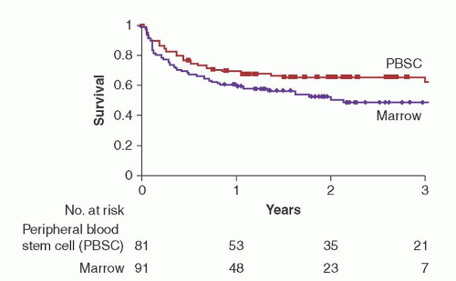
FIGURE 102.2. Probability of overall survival after myeloablative conditioning and transplantation with either peripheral blood stem cells or marrow. Survival at 2 years was improved in the peripheral blood stem cell group as compared to marrow (66% vs. 54%; P = 0.006). Disease-free survival was also improved in the peripheral blood stem cell group (P = 0.003). (Reprinted with permission from Bensinger WI, Martin PJ, Storer B, et al. Transplantation of bone marrow as compared with peripheral blood cells from HLA-identical relatives in patients with hematologic cancers. N Engl J Med 2001;344:175-181. Copyright 2001 Massachusetts Medical Society. All rights reserved.)


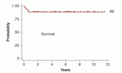
FIGURE 102.3. Overall survival among 94 patients with aplastic anemia who underwent transplantation from HLA-identical siblings after conditioning with cyclophosphamide (CY) and antithymocyte globulin. With a median follow-up of 6.0 (0.5 to 11.6) years, the Kaplan-Meier estimate of survival was 88%. Cyclosporine and methotrexate were administered after transplantation for graft-versus-host disease (GVHD) prophylaxis. (Reprinted with permission from Storb R, Blume KG, O’Donnell MR, et al. Cyclophosphamide and antithymocyte globulin to condition patients with aplastic anemia for allogeneic marrow transplantations: the experience in four centers. Biol Blood Marrow Transplant 2001;7:39-44.)

An HLA-haploidentical donor (parent, sibling, child) is available for almost all patients, but haplotype differences (genotypically identical at one HLA haplotype but nonidentical at the other HLA haplotype) are associated with a high risk of severe GVHD, graft rejection, and increased mortality with conventional posttransplant immunosuppression. However, transplants for hematologic malignancies from HLA-haploidentical family members mismatched for only one HLA antigen in the nonidentical HLA haplotype may have a similar overall survival to transplants from HLA-identical siblings.93 In this situation, although the higher incidence of GVHD results in an increased transplant-related mortality, relapse is less frequent, resulting in no overall difference in long-term survival compared to transplants from HLA-matched siblings. Patients transplanted from HLA-haploidentical family members mismatched for 2 or more HLA loci had significantly lower overall survivals compared to patients transplanted from phenotypically HLA-matched unrelated donors.94 In patients with advanced leukemia, transplantation with high-dose CD34-positive cell grafts which had been highly T cell-depleted resulted in 12 of 43 patients being alive and disease-free at 18 months.95 Immune reconstitution after transplantation was poor, however.96 High-dose cyclophosphamide (CY) early after HCT from an HLA-haploidentical donor appears effective in preventing the development of severe acute and chronic GVHD.97, 98 In a report of parallel phase 2 studies, transplantation of HLA-haploidentical marrow grafts appeared to have favorable outcomes at 1 year compared to transplantation of umbilical cord blood grafts.99 Patients who lack a closely matched family donor should be offered a phenotypically HLA-matched unrelated donor before considering a transplant from an HLA-haploidentical donor.


Transplantation from Unrelated Donors

After the initial success with transplantation of marrow from matched unrelated volunteers, large databanks were established around the world, including the National Marrow Donor Program (NMDP) in the United States. Available through the world-wide registries and the NMDP are approximately 20 million volunteer donors who have been typed for HLA-A and -B antigens, and many of these are also typed for HLA-DR (www.bmdw.org). Certain racial and ethnic groups are underrepresented in the registry and therefore there is a lower probability that an HLA-matched donor will be found.

Outcomes have improved after HCT from HLA-matched unrelated donors because of improved supportive care and high-resolution HLA typing. Outcomes after HCT from an unrelated donor are now comparable to what is observed after HCT from an HLA-identical sibling. In a study of 2,223 adult AML patients, the overall survival after transplantation from 8/8 HLA-matched donors was similar to that observed after transplantation from an HLA-identical sibling (RR = 1.03; P = 0.62).100 The risk of acute GVHD, however, was lower after transplantation from an HLA-identical sibling.

The relative importance of the various HLA loci has been defined for HCT from unrelated donors. In a study from CIBMTR of 3,857 transplants performed from 1988 to 2003, it was observed that high-resolution DNA matching for HLA-A, HLA-B, HLA-C, or HLA-DRB1 (8/8 match) was the minimal level of matching associated with the highest survival.101 A single mismatch at HLA-A, HLA-B, HLA-C, or HLA-DRB1 (7/8 match) was associated with an increased mortality compared to an 8/8 match. A single mismatch at HLA-B and HLA-C appeared better tolerated than a mismatch at HLA-A and HLA-DRB1. A mismatch at HLA-DQ or HLA-DP loci did not have an effect on survival. Ninety-four percent of the transplants in this study were performed with marrow grafts. In a follow-up study, the association between HLA matching and outcomes was investigated for transplantation of peripheral blood stem cells from unrelated donors.102 Survival was better with 8/8 HLA matching compared to 7/8 HLA matching. Single HLA-C antigen mismatches were associated with an increased risk of treatment-related mortality and grade III-IV acute GVHD. HLA-B antigen/allele mismatching was associated with an increased risk of grade III-IV acute GVHD with no effect on survival. No significant differences in outcomes were observed with HLA-C allele mismatches, HLA-A antigen/allele mismatches, or HLA-DRB1 mismatches compared to 8/8 HLA-matched pairs. The differences in the reported associations for HLA mismatches between marrow and peripheral blood stem cell grafts may result from differences in cell numbers in the graft as well as the graft composition.

Disease status is an important consideration in the search process. In patients with advanced malignancies, survival after transplantation from an HLA-matched or 1-antigen/allele HLA-mismatched unrelated donor was similar.103 Therefore, when transplants cannot be delayed because of disease status, selection of a donor with the fewest HLA mismatches may be an alternative choice for patients without a completely HLA-matched donor.

Because of the possible higher risk of acute GVHD, studies of peripheral blood stem cell transplantation from unrelated donors started later than those done from HLA-identical donors. A recently completed randomized clinical trial (BMTCTN 0201) comparing marrow to peripheral blood stem cells observed that there was no difference in overall survival, disease-free survival, nonrelapse mortality, relapse, or acute GVHD outcomes at 2 years between the 2 arms. There was a significantly increased risk of chronic GVHD.


Transplantation with Umbilical Cord Blood Grafts

There are several advantages to the use of umbilical cord blood when compared to unrelated donor peripheral stem cell or marrow product harvested from adults. First, umbilical cord blood represents a potentially nonlimiting donor source for transplantation. At present, over 550,000 HLA typed cord bloods are banked and are conceivably available with several days notice (www. bmdw.org). Many of the banked specimens are from underrepresented ethnic and racial groups, thus expanding the potential donor pool for individuals poorly served by the unrelated donor marrow registries. The total nucleated cell dose required for successful engraftment is 10-fold less than that required for conventional peripheral blood or marrow transplant. The transmission
of Epstein-Barr virus (EBV) or cytomegalovirus (CMV) is negligible compared to conventional allogeneic transplantation. HLA allele mismatches (up to 2 to 3 loci) are permissible in cord blood transplantation. Notable disadvantages to cord blood transplantation are the potential for prolonged pancytopenia and lower rates of overall engraftment.

The first successful cord blood cell transplant in a pediatric patient was done in 1988.69 Two studies were then successfully conducted of cord blood transplantation from HLA-matched or HLA-haploidentical allogeneic siblings (44 patients) and partially HLA-mismatched unrelated donors (25 patients).104, 105 In the first report of a large experience in 562 recipients (including 18% adults) after transplantation with umbilical cord blood grafts from unrelated donors, the probability of engraftment at 42 days and grade III-IV GVHD, was 81% and 23%, respectively.106 It was concluded from this study that umbilical cord blood grafts regularly engraft and cause a low rate of GVHD relative to the number of HLA mismatches, and produced survival rates comparable to those with transplantation of marrow from unrelated donors. This experience was confirmed in a retrospective analysis of 541 children with leukemia transplanted with stem cell grafts from unrelated donors of which 99 were from umbilical cord blood.107 Recipients of cord blood had an increased number of HLA mismatches but a lower risk of both acute and chronic GVHD compared to recipients of unmanipulated marrow from unrelated donors. The day 100 mortality was higher, however, in the cord blood group, possibly because of the significantly delayed recovery of hematopoiesis and immunity after transplantation. These results were later confirmed by another group.108 It was concluded that the use of umbilical cord blood was an option for children with acute leukemia lacking an acceptably matched unrelated marrow donor. In children who had received umbilical cord blood or bone marrow grafts from HLA-identical siblings, the umbilical cord blood group had a lower risk of acute and chronic GVHD (relative risk 0.41 [P = 0.001] and 0.35 [P = 0.02]), respectively.109 Survival was similar in both groups. The progenitor cell and CD34-positive cell content of the umbilical cord blood graft predicted the rate of neutrophil recovery after transplantation.110

The reported low incidence of severe GVHD after transplant of umbilical cord blood cells from unrelated donors (relative to the degree of HLA disparity) may be related to the decreased immunocompetence of the fetal blood cells compared to adult cells.111 Two years after transplantation, T cell receptor rearrangement excision circles (TRECs), a measure of recent thymic output, were greater in recipients of umbilical cord blood than in recipients of marrow grafts, suggesting complete immune recovery despite the low number of cells infused.112

Initially, recipients of umbilical cord blood cell transplants had been mostly children. Even though, at first, there was a concern about the low cell count for the larger body size, outcomes in adults were comparable to those reported in children.113 Prospective trials comparing cord blood transplantation to unrelated peripheral blood/marrow transplants are not available; however, two large registry or retrospective studies summarizing consortia experience for acute or chronic leukemias have been published.114, 115 Slower engraftment kinetics, reduced engraftment rates, and decreased acute and chronic GVHD rates and severity were observed in the cord blood transplantation groups. Rates of relapse reported for the two studies were similar between the 2 groups. Transplant-related complications, including delayed recovery of blood counts after cord blood transplantation, may be reduced in the adult population with reduced intensity conditioning.116

Transplantation of multiple cord blood units was investigated to determine if recovery of neutrophil and platelet counts after transplantation could be improved.117 After the infusion of two umbilical cord blood grafts into adult patients, the median day to neutrophil recovery was 24 (range 12 to 28) in 23 adult patients compared to 27 (range 13 to 59) in another study of 68 adult patients transplanted with a single cord blood unit.113 This observation of improved recovery times was not confirmed in a retrospective study at a single institution.118 After transplantation with 2 cord blood units, only one unit eventually dominates and engrafts long-term.119 Higher CD3+ cell dose and percentage of CD34+ viability were associated with unit dominance. Higher dominant unit total nucleated cells, CD34+ cells, and colony-forming unit doses were associated with higher sustained engraftment and faster neutrophil recovery. Mechanistically, the dominant cord mounts an allogeneic immune response mediated by CD8+ T cells against the nondominant unit.120 Outcomes after double cord blood transplantation were compared with outcomes after transplantation from HLA-matched related donors, HLA-matched unrelated donors, and 1-antigen HLA-mismatched unrelated donors.121 Leukemia-free survival at 5 years was similar for all donor types. The risk of relapse was decreased in the double cord blood transplantation group compared to the other donor types, but the risk of nonrelapse mortality was increased. The lower risk of relapse after transplantation from a double cord blood unit compared to a single cord blood unit had been previously seen in another retrospective study, although a significantly increased risk of acute GVHD was also observed.118 Sharing of one or more HLA-A, B, or DRB1 loci between the inherited paternal MHC alleles (IPA) of the donor and the recipient after unrelated cord blood transplantation leads to superior leukemia-free survival with no increase in acute GVHD, a result attributed to maternally derived anti-IPA elements persisting in the cord blood.122

Umbilical cord blood grafts should be considered for both pediatric and adult patients lacking a suitably matched unrelated donor or unable to wait for an unrelated search to be completed.


COLLECTION OF HEMATOPOIETIC STEM CELLS FOR TRANSPLANTATION


Marrow

Marrow is obtained by multiple aspirations from the posterior iliac crest under general or epidural anesthesia.123 The anterior iliac crest (or the sternum) may also be harvested if larger quantities of marrow are required. The target volume of marrow for transplant is 10 to 15 ml/kg of recipient or donor weight, whichever is the smaller individual. Marrow is collected with heparinized syringes through large bore needles and placed into small amounts of culture medium. The collected marrow must be filtered prior to intravenous transfusion into the recipient to remove small particles or clots. If the patient and donor are ABO-incompatible and there are high anti-A or -B titers, the marrow can be red blood cell-depleted or plasma-depleted, depending on whether it is a major or minor ABO mismatch.124 In some cases of a major ABO mismatch, plasmapheresis of the recipient may be effective in reducing the high anti-A or anti-B titers so that RBC depletion of marrow is not required. Marrow is infused immediately after harvesting, but delays of 24 hours may occur without adverse consequences. Such delays are common when marrow is shipped to a transplant program after harvest from an unrelated donor. In an analysis of marrow harvest characteristics of 1,549 donors, the median total nucleated cell count from the marrow was 2.5 (range 0.3 to 12.0) × 108/kg recipient weight.125 Life-threatening complications from marrow harvesting, usually related to the administration of anesthesia, were reported in 0.27% to 0.4% of the donors.126


Peripheral Blood

HSC circulate in the peripheral blood but the concentration is very low and it requires multiple aphereses to collect adequate
numbers. The number of aphereses may be reduced to one or two sessions when HSC are mobilized to the peripheral blood after the administration of cytokines alone or in combination with chemotherapy or plerixafor. An effective mobilization strategy of autologous HSC for patients with malignancy is chemotherapy in conjunction with G-CSF, 6 µg/kg/day.61 After chemotherapy, patients are apheresed when the total white blood cell count has recovered to 1,000/µl or the CD34-positive cell count in the peripheral blood is at least >10/µl. For patients not requiring chemotherapy or normal allogeneic donors, mobilization is with G-CSF alone (5 to 16 µg/kg) by daily subcutaneous injections for 5 to 8 days.48, 127, 128 These doses of G-CSF are generally well tolerated, with common side effects of bone pains, myalgias, and flu-like symptoms that are managed with acetaminophen or low-dose narcotics. Plerixafor in combination with G-CSF was effective for increasing the yields of circulating CD34+ progenitor cells and is indicated for patients with lymphoma or multiple myeloma who are being collected for autologous HCT.56 The recommended dose is 0.24 mg/kg/day administered subcutaneously 11 hours before the apheresis procedure. The maximum dose is 40 mg/day. Common side effects included diarrhea, nausea, fatigue, headaches, and arthralgias. Apheresis was performed as early as day 4 after the start of G-CSF using a continuous blood flow separation technique. Apheresed products may be cryopreserved in 5% dimethylsulfoxide (DMSO) for use after thawing on the day of transplant. A more rapid sustained hematopoietic recovery of both neutrophil and platelet counts occurs with increasing numbers of CD34+ cells in the hematopoietic cell graft (up to 5 × 10⁶/kg). Some investigators consider 2.5 × 10⁶/kg of recipient weight a minimum dose of CD34+ cells from the peripheral blood to achieve complete autologous recovery. Platelet recovery is more rapid at higher CD34+ cell doses.60, 61 Since the cell dose used in the autologous transplant setting yields consistent and prompt engraftment, it is also considered an appropriate target for collection of allogeneic HSC from the peripheral blood. Donors of peripheral blood avoid general anesthesia and other complications of marrow harvesting. If peripheral veins are inadequate, a large bore double lumen catheter for vascular access may be required. In a large analysis of safety from the NMDP (n = 2,408 donors), it was concluded that collection of peripheral blood stem cells was safe but that nearly all patients will experience bone pain and 1 in 4 will have headache, nausea, or citrate toxicity.129 Serious and unexpected toxicities were experienced by 0.6% of the donors, but complete recovery was universal.


Cord Blood

Umbilical cord blood cells are now being routinely collected and cryopreserved for storage in a cord blood bank.130, 131 Directed-donor banking of cord blood for siblings in a current good tissue practices environment has also now been reported.132 After the separation of the placenta, umbilical cord blood cells are collected into a closed system which utilizes a sterile donor blood collection set. The placenta and umbilical cord can be suspended on a frame and the blood drained as a “standard gravity phlebotomy” into CPD (citrate, phosphate, dextrose) anticoagulant. The median volume of umbilical cord blood collected in one study of 44 patients was 100 ml (range 42 to 282 ml).104 The median number of total nucleated cells per kilogram of recipient weight for banked umbilical cord blood is 2.5 × 10⁷ (range 1 to 33) and corresponds to a CD34+ cell dose of 1.5 × 10⁵ per kilogram of recipient weight.110


ABO INCOMPATIBILITY

ABO incompatibility between the donor and the recipient occurs in about 30% of cases. A major ABO incompatibility is considered to occur when the isohemagglutinins in the recipient plasma are directed against the donor red blood cell antigens. A minor ABO incompatibility is when the isohemagglutinins in the donor plasma are directed against recipient red blood cell antigens. ABO incompatibility between the donor and the recipient has no significant effect on the incidence of graft rejection, GVHD, or survival, although bidirectional ABO mismatches were associated with a higher risk of grade III-IV acute GVHD.133 ABO incompatibilities may result in severe hemolytic episodes after transplantation. For major ABO incompatibilities, if the recipient isohemagglutinin titers in the plasma are high, an acute hemolytic event can be prevented by red cell depletion of the graft. Conversely, plasmapheresis may be effective in reducing the anti-donor isohemagglutinin in the recipient plasma. If the latter approach is chosen, the goal should be to reduce the isohemagglutinin titer to 1:16 or lower.134 If there is continued production of anti-donor isohemagglutinins in the recipient plasma after transplantation, delayed erythropoiesis or even pure red cell aplasia and persistent hemolysis may result.135 Although plasmapheresis and erythropoietin may be of some benefit, the hemolytic episode may persist for months after transplantation. If there are no contraindications, early withdrawal of immunosuppression may result in a more rapid resolution of the delayed hemolytic event, possibly because of a GVH reaction against the isohemagglutinin-producing cells of the recipient.136 To prevent hemolytic events related to minor ABO incompatibilities, the isohemagglutinins can be removed from the stem cell graft if the anti-recipient titers are high. The risk of hemolysis from a minor ABO mismatch appears to be increased after peripheral blood stem cell transplantation, possibly related to the higher content of lymphoid cells (B cells) in the graft. The development of severe hemolysis may be prevented with the administration of methotrexate after transplantation.137


CONDITIONING REGIMENS


Myeloablative

Myeloablative conditioning regimens are sufficiently intense that recovery of hematopoiesis would not be expected without the support of transplanted hematopoietic progenitor cells. The ideal myeloablative conditioning regimen should fulfill the following criteria: (1) eliminate or reduce the tumor load; (2) suppress the host immune system to prevent graft rejection (not applicable to hematopoietic support with autologous cells); and (3) have tolerable nonhematopoietic toxicity. The first conditioning regimens consisted of TBI, alone or in combination with cyclophosphamide (CY).50 TBI is an effective antineoplastic modality that is both cell cycle nonspecific and immunosuppressive. CY is a chemotherapeutic agent with immunosuppressive properties that has few nonhematopoietic toxicities that are similar to TBI, and therefore can be used in combination. Other conditioning regimens which have since been developed include: (1) the replacement of CY with other chemotherapeutic agents (e.g., etoposide, Ara-C, and melphalan) in combination with TBI.138, 139; (2) other chemotherapeutic agents in combination with both CY and TBI140, 141; (3) chemotherapeutic agents used in combination with CY but without TBI, including busulfan (BU)/CY or carmustine-cyclophosphamide-etoposide (BCV)142, 143; and (4) replacement of CY with fludarabine and used in combination with BU or melphalan.144, 145 In one study, BCV was associated with a significant incidence of transplant-related complications and mortality.146 Other high-dose cytotoxic regimens have been used, especially with autologous stem cell support.147, 148

Most preparative regimens used for the treatment of malignant diseases have not been tested in Phase III studies, so it is generally unclear if any one regimen represents an improvement over those previously used. Two Phase III studies in allogeneic marrow transplantation have compared differing intensities of TBI
(1,200 cGy versus 1,575 cGy).149, 150 The relapse rate was reduced in the group of patients receiving the higher dose of TBI, but was associated with an increase in complications from regimen-related toxicity and GVHD which negated any improvement in disease-free survival compared to the group receiving the lower dose of TBI. In a more recent study, conditioning with TBI 800 cGy and fludarabine was compared with TBI 1,200 cGy and CY and there was no difference in overall survival, relapse, or treatment-related mortality.151 The combination of BU/CY was determined to be an acceptable alternative to CY/TBI for patients with leukemia in several studies.152, 153, 154 However in one study, patients in the BU/CY group had an increased risk of sinusoidal obstruction syndrome (SOS) of the liver and other transplant-related complications.155 Since this last study, it has been demonstrated that targeting of busulfan levels in the plasma may decrease the risk of relapse and severe regimen-related toxicities, contributing to an improved disease-free survival (Fig. 102.4).156 Monitoring levels of metabolites from cyclophosphamide may also be important to decrease the risk of liver toxicity and nonrelapse mortality.157, 158

In patients already profoundly immunosuppressed with SCID syndrome, engraftment of allogeneic stem cells from matched related siblings may occur without conditioning therapy.159 High-dose CY in combination with antithymocyte globulin (ATG) as a preparative regimen for patients with aplastic anemia was associated with graft rejection in less than 5% of cases.90 The actuarial survival rate was 88% at 6 years after transplantation (Fig. 102.3). For transplantation of patients with thalassemia or sickle cell disease, a myeloablative conditioning regimen, usually consisting of the combination of BU and CY, was thought to be necessary to prevent a high incidence of graft rejection, since most patients with these disorders had received multiple blood transfusions and were potentially sensitized to donor-derived mHC.160, 161 However, reduced intensity conditioning regimens have now been shown to successfully overcome the risk of graft rejection associated with these hematologic disorders.162

Only gold members can continue reading. Log In or Register to continue

Stay updated, free articles. Join our Telegram channel

Oct 21, 2016 | Posted by in HEMATOLOGY | Comments Off on Hematopoietic Cell Transplantation

Full access? Get Clinical Tree

Get Clinical Tree app for offline access