Microbiology Tools for the Epidemiologist


Microbiology Tools for the Epidemiologist


Nicole M. Parrish

Stefan Riedel


Microbiology is the study of microorganisms, which are a diverse group of unicellular and multicellular organisms. Viruses, although they are strictly speaking acellular organisms, are included in this field. Medical microbiology is the study of interactions between organisms and their human and animal hosts that result in infectious disease manifestations. Despite our concentration on microorganisms that cause disease, the vast majority of microorganisms that we continuously interact with do not result in disease. In fact, many of these organisms are beneficial and essential for human health and well-being. Conversely, pathogenic bacteria, fungi, viruses, and parasites result in disease in a specific host by interactions of their cellular structures, products, or toxins with host tissue and cells. These components of organisms, which are collectively referred to as virulence factors, can be classified according to the function they provide for the microorganism:


  • Colonize


  • Evade host defense mechanisms


  • Invade and disseminate

Because humans and other animals have an abundance of microorganisms that colonize the external and internal surfaces of the body, it is important to identify and define which microorganisms cause disease. Despite the vast numbers of bacteria that inhabit our mouth, teeth, gastrointestinal tract, urogenital tract, and skin, only a small number of microorganisms cause disease. In addition to this normal flora, many pathogenic microorganisms can be found on body surfaces, whether as temporary colonizers or as a more permanent carrier state.

The prevention of infection and disease is in large part attributable to an array of defense mechanisms that have evolved to deal with these ongoing intimate interactions. Mechanical barriers are typically the first line of defense. Examples include skin and mucous membranes as a physical barrier, ciliated cells and mucus in the respiratory tract, and the washing action of tears and urine. Chemical barriers are produced (often by resident normal flora) at these sites that prevent colonization by pathogens; these barriers include fatty acids and propionic acid. Other compounds produced by the host that are inhibitory include lysozyme in tears, blood, urine, and sweat; acid in the stomach, vagina, and skin; basic polyamines and complement components in plasma; and acute-phase proteins, such as β-antitrypsin, fibrinogen, C-reactive protein, and β2-microglobulin. In addition to mechanical and chemical mediators, defensive cells such as macrophages, polymorphonuclear neutrophils, monocytes, and eosinophils are all components of nonspecific host resistance or innate immunity. The fourth type of defense in humans and other vertebrates is specific immunity, which is a function of both the humoral immune system, involving antibody-mediated B-cell functions, and the cellular immune system, involving T-lymphocyte-mediated functions. A compromise in any of these defense mechanisms can lead to infection and disease in the host.

At times it can be difficult to determine that an organism is the cause of an infection. In 1884, Robert Koch, a German microbiologist, outlined his famous postulates for proving an organism is the cause of an infection (Table 8-1). Although Koch’s postulates are not foolproof, they do describe a framework for understanding causation. The advent of modern and molecular microbiology has provided further methods to better determine the link between
a microorganism and disease. In the absence of a clearly identifiable microorganism, the host immune response to a specific organism can be a useful diagnostic tool.


Table 8-1 Guidelines Establishing the Etiology of Infectious Disease


Koch’s Postulates


1. The microorganism must be found in all cases of the disease, but must not be found in those not suffering from the disease.


2. The microorganism should be isolated from a diseased body and should be grown in vitro and in pure culture for several generations.


3. When the organism from a pure culture is introduced into a healthy body or animal, the typical disease must be the result.


4. The same organism must then be reisolated from the lesion/animal of such an experimentally caused disease.


Pathogenic microorganisms may cause disease either directly or indirectly. The pathology they cause can be another means of identifying them. The presence of a potentially pathogenic microorganism results in a host response (e.g., antibody production), which, in combination with the organism’s virulence factors, can result in toxicity and host tissue damage characteristic of the specific infection. In other cases, microorganisms produce toxins that can be the sole cause of the pathology observed. In addition to true pathogenic microorganisms, some organisms of low virulence or pathogenicity can cause infections when the host is immunocompromised. Microorganisms that are only pathogenic in compromised hosts are referred to as opportunistic pathogens.

This chapter provides a broad overview of the classification, taxonomy, and structure of the various microorganisms. It also briefly describes the current and commonly used diagnostic tests for infectious diseases available to the clinical microbiologist. Although by no means comprehensive, the information presented here provides a basis of understanding for a further in-depth study of microbiology as it relates to infectious disease epidemiology.


Table 8-2 Comparison of Eukaryotic and Prokaryotic Cells


Eukaryotic Cell


Prokaryotic Cell


Form


Multicellular structures


Single cells


Nucleus


Nuclear membrane


DNA and chromosome(s) free in cytoplasm


Mitosis


Yes


No


Organelles


Membrane-bound organelles (e.g., mitochondria) present


No organelles present in cytoplasm


Ribosomes


80S = 40S + 60S


70S = 30S + 50S


Cell wall


Absent or cellulose


Peptidoglycan


Sterols


Yes, always


Only in Mycoplasma spp.


TAXONOMY, CLASSIFICATION, AND STRUCTURE OF INFECTIOUS AGENTS

Early biologists realized that microorganisms such as algae, protozoa, fungi, and bacteria did not readily fit into the already-established plant and animal kingdoms. This recognition led to the proposal by Haeckel in 1886 of a third kingdom, the Protista. As viruses were still unknown at the time, they were not included in this classification system. Subsequent advances in the biologic sciences, specifically microscopy, led to a further subdivision of the Protista into eukaryotic cells, which included the algae, fungi, and protozoa, as well as members of the plant and animal kingdoms, and prokaryotic cells that represent the bacteria. The terms eukaryotic and prokaryotic reflect only the presence of a true nucleus (eukaryotic) or the absence of a well-delineated nucleus (prokaryotic). Significant structural and other biologic differences are described in Table 8-2, although the presence of transition forms can result in a blending of some of the listed characteristics.

The taxonomy of infectious agents is based on three concepts: classification, nomenclature, and identification. During the past 300 years of microbiology and since the invention of the microscope, a variety of characteristics have been used to name and identify microorganisms. Classification of microorganisms is a process by which microorganisms
are systematically divided into groups; this process of grouping refers to the organization of organisms from (at the lowest level) phylum, class, order, and family, to genera and species (the highest level). Much of the organization of microorganisms continues to be based on phenotypic properties and morphology, such as size and shape, Gram stain and staining characteristics, and biochemical properties. Other criteria that may be useful for classification describe physiologic properties of specific organisms, their metabolism, or a unique ecologic niche (e.g., in the case of cyanobacteria). Recent advances in molecular technologies have also affected the traditional taxonomy of microorganisms, including molecular methods using DNA and RNA homology analysis, DNA sequencing, polymerase chain reaction (PCR) and micro-array technology, proteomic analysis, and nucleic acid base sequence similarity (e.g., pulsedfield gel electrophoresis). These methods are used to evaluate relatedness of microorganisms. Evidence from established phenotypic analyses, together with information from molecular and genetic analyses, is used to establish new taxa and reorganize existing groupings of microorganisms.

The naming of microorganisms follows a well-defined set of rules. With the exception of the viruses, nomenclature of microorganisms is typically binomial and includes the genus and species name—for example, Staphylococcus aureus. Beyond the species level, microorganisms can be further subdivided based on characteristics that do not warrant a separate species designation but do differentiate a specific member of a species or strain from other members of that particular strain or species. Differences or similarities used for subspecies or strain designation within a species can include a variety of parameters, such as structural or functional differences, phenotypic differences, antigenic differences in surface or subsurface structures, and genomic polymorphism. Strain differentiation is an important component of epidemiologic studies, as the significant diversity within species may result in different clinical manifestations or may be useful in defining a cluster of cases.


VIRUSES

Viruses are the smallest of the infectious agents, with the exception of prions, the agents of spongiform encephalopathies, such as scrapie and Creutzfeldt-Jakob disease. Viruses range in size from 20 to 200 nm and, as such, are not readily visible by light microscopy. They contain a single form or type of nucleic acid, either DNA or RNA, which functions as their genome. In addition to a single form of nucleic acid, viruses compositionally may contain proteins, lipids, and glycoproteins as structural components, depending on their level of complexity. Viruses are obligate intracellular parasites, and their replication is hostcell dependent, directed by their DNA or RNA. Viral subversion of the host’s cellular machinery favors the synthesis of viral nucleic acid and structural proteins.

Viral infection is host-cell specific and depends on the presence of specific surface receptors (attachment molecules) for successful entry. Viruses specific for almost every organism have been identified. Even bacteria may be infected by phage viruses—an interaction that has proved useful in the laboratory for introducing genes into bacteria. The outcome for cells infected by a virus can vary and is often virus specific. For instance, some viruses cause rapid cell death (e.g., influenza), whereas others induce continued cellular growth with concomitant release of new virus particles (e.g., adenoviruses). Some viruses are capable of integrating their nucleic acid into the host cell’s genome, thereby establishing a latent or quiescent state (e.g., herpesviruses). Latency can continue for long periods of time before reactivation followed by initiation of viral replication and subsequent lysis of the host cell (herpesviruses). Other viruses carry specific oncogenes capable of promoting cellular growth that can lead to transformation and immortalization of the cells. Such changes have been associated with human papillomavirus, the cause of cervical cancer.

In terms of classification, viruses are divided into families, genera, and species, as are other microorganisms. However, a typical binomial classification using genus and species is not used in a standardized fashion. Instead, viruses are referred to by their single names. Characteristics used for viral classification include the following:


  • Viral genome: DNA or RNA; single- or double-stranded, linear or circular, segmented or nonsegmented; and genome capping with a protein or polynucleotide


  • Size and shape of the capsid and whether it is enveloped or non-enveloped


  • Method of replication


  • Pathophysiology of the virus, such as host range, antigenic composition, vectors, and tissue tropism


  • Physical/chemical features, such as susceptibility to acid or lipid solvents

DNA and RNA viruses of medical importance and some common disease associations are listed in Table 8-3 and Table 8-4. These tables provide a broad overview of the more common viruses and some of the diseases they cause. Table 8-5 lists the predominant hepatitis viruses, grouped the nature of the disease they cause, rather than by virus family or genome type and structure. The reader is referred to the references list at the end of this chapter for additional information.


Table 8-3 Selected DNA Viruses of Medical Importance and Common Disease Associations


Family


Virus


Common Disease Associations


Papillomaviridae


Papillomavirus


Common warts, genital warts, laryngeal papilloma, cervical cancer


Papovaviridae


BK virus


Ureteral stenosis, hemorrhagic cystitis


JC virus


Progressive multifocal leukencephalopathy


Adenoviridae


Adenovirus


Pharyngoconjunctival fever, pharyngitis, respiratory infections, gastroenteritis


Herpesviridae


HSV-1


Cold sores, encephalitis


HSV-2


Genital herpes


Varicella zoster virus (VZV)


Chickenpox, shingles


Epstein-Barr virus (EBV)


Infectious mononucleosis, Burkitt’s lymphoma, nasopharyngeal carcinoma


HHV-6


Roseola


Cytomegalovirus (CMV)


Wide spectrum of disease in newborns and immunocompromised hosts


HHV-7


Occasional roseola


HHV-8


Kaposi’s sarcoma


Poxviridae


Variola virus


Smallpox


Parvoviridae


Parvovirus (B19)


Fifth disease


Table 8-4 Selected RNA Viruses of Medical Importance and Common Disease Associations


Picornaviridae


Poliovirus


Polio


Coxsackie A virus


Undifferentiated fever, respiratory tract infections, herpangina, aseptic meningitis, acute hemorrhagic conjunctivitis, conjunctivitis


Coxsackie B virus


Undifferentiated fever, respiratory tract infections, aseptic meningitis


ECHO virus


Aseptic meningitis


Rhinovirus


Common cold


Coronaviridae


Coronavirus


Common cold, severe acute respiratory syndrome (SARS)


Caliciviridae


Norwalk virus


Gastroenteritis


Paramyxoviridae


Measles virus


Measles


Parainfluenza virus


Respiratory tract infections


Mumps


Mumps


Respiratory syncytial virus (RSV)


Lower respiratory tract infections, pneumonia, bronchiolitis


Metapneumovirus


Respiratory tract infections


Orthomyxoviridae


Influenza virus


Influenza


Rhabdoviridae


Rabies


Rabies


Filoviridae


Ebola


Marburg and Ebola hemorrhagic fever


Reoviridae


Rotavirus


Gastroenteritis


Coltivirus


Colorado tick fever


Togaviridae


Rubella virus


German measles


Venezuelan encephalitis virus


Meningoencephalitis


Eastern equine encephalitis virus


Meningoencephalitis


Western equine encephalitis virus


Meningoencephalitis


Flaviviridae


Dengue fever virus


Dengue fever, dengue hemorrhagic fever


Yellow fever virus


Yellow fever, hemorrhagic fever, hepatitis


Japanese encephalitis virus


Encephalitis


West Nile virus


Febrile illness, meningoencephalitis


St. Louis encephalitis virus


Febrile illness, meningoencephalitis


Bunyaviridae


Hantavirus


Hemorrhagic fever with renal syndrome, hantavirus


pulmonary syndrome


California encephalitis virus


Meningoencephalitis


Arenaviridae


Lymphocytic choriomeningitis virus


Undifferentiated febrile illness


Retroviridae


Human immunodeficiency virus


AIDS


Table 8-5 Predominant Hepatitis Viruses of Medical Importance


Family


Virus


Genome


Picornaviridae


Hepatitis A virus (HAV)


RNA


Hepadnaviridae


Hepatitis B virus (HBV)


DNA


Flaviviridae


Hepatitis C virus (HCV)


RNA


Hepeviridae


Hepatitis E virus (HEV)


RNA


Structure

The complete infectious virus is termed a virion. It is composed of its specific nucleic acid, DNA or RNA, surrounded by a protein coat known as the capsid. The capsid is further subdivided into capsomers, repeating identical morphologic protein subunits. Each capsomer subunit is composed of one or more polypeptides.

Viruses construct several different capsid shapes, depending on how these proteins combine. Arrangement of capsomers in the shape of an icosahedron (polygon with 20 faces), for example, results in cubic symmetry. In a cubically symmetrical virus, some capsomers are surrounded by five (penton) capsomers, whereas others are surrounded by six (hexon) capsomers. In helical viruses, the capsid proteins are arranged around a helical nucleic acid core. In addition to icosahedral and helical viruses, complex viruses have more intricate structural elements. The viral genome can have an associated protein complex that is referred to as the nucleocapsid and forms the viral core. Enveloped viruses possess a lipoprotein coat that surrounds the virion and is acquired from the infected host cell’s membrane. Non-enveloped viruses are also referred to as naked viruses. Finally, viruses may possess glycoprotein spikes, known as peplomers, which protrude from the envelope and function in the attachment of the virus to target host cells.


BACTERIA


Classification

Classification of bacteria into taxonomic groups requires the use of both experimental and observational laboratory methods and techniques. As they were discovered earlier in history, bacteria have been subjected to human classification schemes for much longer than the viruses. Prokaryotic bacteria are named using the binomial system of genus and species (Table 8-6). Further subdivision can then occur among species, such as subspecies, serotype, and so on.


Table 8-6 Examples of Bacteria of Medical Importance


Organism


Clinical Features/Disease


Epidemiologic Features


Aerobic and Facultatively Anaerobic Gram-Positive Cocci


Staphylococcus aureus


Cutaneous infections (e.g., abscess, impetigo, wounds); pneumonia; bacteremia; osteomyelitis; arthritis; toxic shock syndrome; food poisoning


Colonize human skin and mucosal membranes (e.g., nares); resistance development (healthcare- and community-associated MRSA)


Staphylococcus, coagulase negative (e.g., S. epidermidis)


Opportunistic pathogen: bacteremia, endocarditis; prosthetic joint infects


Colonize human skin and mucosal surfaces


Enterococcus faecalis and E. faecium


Bacteremia, endocarditis; urinary tract infections; intraabdominal abscesses


Elderly patients; patients with long-term hospitalization and broad-spectrum antibiotic therapy


Streptococcus pyogenes (group A)


Pharyngitis; scarlet fever; sinusitis; impetigo, erysipelas, cellulitis, necrotizing fasciitis; glomerulonephritis; rheumatic fever; toxic shock syndrome


Diverse populations; carrier state


Streptococcus agalactiae(group B)


Neonatal disease (bacteremia, meningitis, pneumonia); urinary tract infections in adults


Diverse populations; carrier state


Viridans group streptococci


Abscess formation; subacute endocarditis; dental caries


Patients with abnormal or prosthetic heart valves


Streptococcus pneumoniae


Pneumonia and other respiratory tract infections; meningitis; bacteremia; endocarditis


Diverse populations: neonates, children, adults with chronic pulmonary disease, elderly


Aerobic or Facultatively Anaerobic Gram-Positive Rods


Bacillus anthracis


Anthrax (cutaneous, inhalational, gastrointestinal)


Animal workers; laboratory accidents; bioterrorism; consumption of infected meat


Bacillus cereus


Gastroenteritis, bacteremia, ocular infections


Contaminated food; traumatic eye injury; IV drug use


Corynebacterium diphtheriae


Diphtheria (respiratory or cutaneous)


Spread by respiratory droplets


Erysipelothrix rhusiopathiae


Erysipeloid (cellulitis)


Occupational disease (e.g., butchers, meat processors, farmers)


Listeria monocytogenes


Early-onset and late-onset neonatal disease (meningitis, sepsis); bacteremia in pregnant women


Ingestion of contaminated food (nonpasteurized dairy products); immunocompromised hosts; elderly women


Mycobacteria


Mycobacterium tuberculosis


Tuberculosis


Opportunistic pathogen; emerging multidrug resistance


Mycobacterium avium complex


Localized pulmonary disease; also disseminated disease with multiorgan disease


Localized in patients with chronic pulmonary disease; disseminated in patients with HIV/AIDS


Mycobacterium leprae


Leprosy (Hansen’s disease)


Spread by close contact with infected patients


Other Gram-Positive, Partially Acid-Fast Bacteria


Nocardia species


Bronchopulmonary disease


Commonly found in soil and water; opportunistic pathogen in patients with underlying pulmonary disease


Rhodococcus equi


Lung abscess; opportunistic infections in immunocompromised patients


Commonly found in immunocompromised patients (e.g., AIDS, transplant recipients)


Aerobic Gram-Negative Cocci


Neisseria meningitides


Meningitis, bacteremia


Carrier state; aerosol transmission


Neisseria gonorrheae


Gonorrhea, pelvic inflammatory disease, arthritis


Sexual transmission; asymptomatic carriers


Aerobic and Facultatively Anaerobic Gram-Negative Rods


Escherichia coli


Urinary tract infections; bacteremia; meningitis


UTI: sexually active women Meningitis: neonates


Escherichia coli, enterotoxic (STEC), enterohemorrhagic, and entoaggregative strains


Watery diarrhea, hemorrhagic colitis; watery diarrhea with mucus; hemolytic-uremic syndrome (HUS)


Foodborne or waterborne outbreaks; travelers’ diarrhea; infants in developing countries


Klebsiella pneumonia Serratia marcescens Enterobacter species


Pneumonia; urinary tract infections Pneumonia; urinary tract infections; wound infections


Nosocomial infections; alcoholism Nosocomial infections


Proteus species


Urinary tract infections; wound infections


Structural abnormalities in the urinary tract


Vibrio cholera


Cholera; severe watery diarrhea


Children and adults in developing countries


Vibrio vulnificus


Wound infections; primary septicemia


Immunocompromised patients; preexisting (chronic) hepatic disease


Vibrio parahaemolyticus


Watery diarrhea


Seafood-borne outbreaks


Aerobic and Facultatively Anaerobic Gram-Negative Rods


Salmonella species


Diarrhea; enteric fever (serovar typhi)


Contaminated food; immunocompromised patients are at risk for bacteremia


Shigella species


Bacillary dysentery


Contaminated food or water; person-to-person transmission


Yersinia pestis


Plague


Natural reservoir: rodents; transmitted to humans by rodent flea bites; bioterrorism


Yersinia enterocolitica and Y. pseudotuberculosis


Enterocolitis; mesenteric lymphadenitis


Worldwide distribution; contaminated food and water


Aeromonas species


Wound infections; gastroenteritis


Widely distributed in aquatic reservoirs; contaminated drinking water


Acinetobacter baumaniicomplex


Pneumonia; septicemia; wound infections; opportunistic infections


Nosocomial infections; patients with large wounds or burns are at increased risk; elderly


Pseudomonas aeruginosa


Pulmonary infections; primary skin infections; infections of urinary tract, ear, eye, and wounds; bacteremia


Nosocomial infections; ubiquitous organism in nature, in aquatic reservoirs, and on surfaces


Stenotrophomonas maltophilia


Wide variety of serious disseminated infections


Nosocomial infections


Burkholderia cepacia complex


Pulmonary infections; opportunistic infections


Compromised patients, especially those with cystic fibrosis and chronic granulomatous disease


Burkholdreia pseudomallei


Melioidosis


Opportunistic pathogen


Moraxella catarrhalis


Ear, eye, and respiratory tract infections


Children; patients with underlying pulmonary disease


Fastidious and Other Gram-Negative Bacilli


Eikinella corrodens


Subacute endocarditis; wound infections


Human bite wounds; opportunistic pathogen in patients with damaged heart valves


Kingella kingae


Subacute endocarditis


Opportunistic pathogen


Haemophilus influenzae


Encapsulated type b strains: meningitis; septicemia; epiglottitis Nonencapsulated strains: otitis media; sinusitis; bronchitis; pneumonia


Aerosol transmission in young and unimmunized patients; possible spread from upper respiratory tract of elderly patients with chronic respiratory disease


Legionella pneumophilia


Legionnaires’ disease (pneumonia) and Pontiac fever (flu-like illness)


Waterborne transmission; elderly and immunocompromised patients at increased risk


Francisella tularensis


Tularemia (pulmonary or skin infections)


Human infections via contact with wild rabbits, ticks, or deerflies; bioterrorism


Fastidious and Other Gram-Negative Bacilli


Bordetella pertussis


Pertussis (whooping cough)


Aerosol transmission; pertussis toxin; severe disease in infants, milder in adults


Brucella species


Brucellosis


Exposure to infected goats, sheep, and cattle


Pasteurella multocida


Wound infections; pulmonary and disseminated infections


Cat bites; commensal in upper respiratory tract of animals, fowl, and perhaps humans


Campylobacter jejuni


Gastroenteritis


Contaminated food, milk, or water


Helicobacter pylori


Gastritis; peptic and duodenal ulcers; gastric adenocarcinoma


Infections are common (people in lower socioeconomic classes and in developing countries)


Anaerobic Gram-Positive Organisms


Clostridium perfringens


Cellulitis, necrotizing fasciitis, myonecrosis; food poisoning; septicemia


Ubiquitous in environment (e.g., soil, water, sewage)


Clostridium difficile


Antibiotic-associated diarrheal disease; pseudomembranous colitis


Colonizes human GI tract; nosocomial, hospital pathogen


Clostridium tetani


Tetanus


Ubiquitous in environment (e.g., soil, water, sewage)


Clostridium botulinum


Botulism (foodborne, infant, wound type)


Ubiquitous in environment (e.g., soil, water, sewage) and GI tract of animals; bioterrorism


Anaerobic Gram-Negative Organisms


Bacteroides fragilis


Polymicrobial infection of the abdomen, skin, soft tissue, and female genitourinary tract


Colonizes human mucosal surface (oropharynx, intestine, vagina)


Spirochetes


Treponema pallidum


Syphilis


Worldwide distribution; sexually transmitted disease


Borrelia burgdorferi


Lyme disease


Reservoir: rodents, deer, domestic pets, hard-shelled ticks Vector: hard-shelled ticks


Borrelia recurrentis


Epidemic, louse-borne relapsing fever


Reservoir: humans Vector: body louse


Leptospira


Leptospirosis (asymptomatic to influenza-like illness with yalgia); Weil syndrome (jaundice)


Worldwide distribution; rodents are common reservoir; soil is contaminated by rodent urine


Mycoplasma and Gram-Negative, Obligate Intracellular Bacteria


Mycoplasma pneumoniae


Tracheobronchitis, pharyngitis, pneumonia


Strict human pathogen; asymptomatic carrier state


Chlamydophila pneumoniae


Asymptomatic respiratory tract infections to “atypical” pneumonia


Human pathogen


Chlamydophila psitacci


Psittacosis/ornithosis; pulmonary disease with hematogenous dissemination to liver and spleen


Exposure to dried excrement, urine, or secretions from psittacine birds (parrots, parakeets, macaws, cockatiels)


Chlamydia trachomatis


Trachoma and lymphogranuloma venerum (LVG)


Direct transmission by droplets via hands, eye-to-eye, or contaminated clothing; sexually transmitted


Ehrlichia chaffiensis


Human monocytic ehrlichiosis


Region: southeastern, central, and midwestern United States Reservoir: white-tailed deer, foxes, coyotes, wolves, domestic dogs Vector: Lone Star tick (Amblyomma americanum)


Anaplasma phagocytophilum


Human granulocytic ehrlichiosis


Region: northern United States central Midwest Reservoir: small mammals (mouse, chipmunks, voles) Vector: Lone Star tick and Ixodes tick


Rickettsia prowazekii


Epidemic typhus


Human body louse (Pediculus humanus) More than 90% of infections in the United


Rickettsia rickettsia


Rocky Mountain spotted fever


States occur from April to September Vector: dog tick (Dermacentor variabilis) and wood tick (Dermacentor andersoni)


Coxiella burnetii


Asymptomatic to flu-like symptoms; also pneumonia and hepatitis


Wide variety of hosts: cattle, sheep, goats, dogs, cats, and rabbits; exposure occurs via contaminated soil and milk


Originally, the taxonomic classification of bacteria was solely based on morphologic and biochemical characteristics. Today, the majority of microbiology laboratories continue to use bacterial microscopy, a variety of selective and nonselective growth media, and biochemical and immunologic tests for the identification and classification of bacterial organisms. In recent years, improvements in molecular technologies have afforded microbiology laboratories the opportunity to enhance, improve, and/or expedite the process of bacterial identification. Nevertheless, the use of established morphologic characterization of bacteria and basic phenotypic and biochemical tests remain valuable tools in the hands of experienced microbiologists.

Morphologic classification of bacteria is based on staining characteristics, shape, and size of the microorganism when visualized by light microscopy. The Gram stain reaction, which highlights a variation in cell wall structures of bacteria, has been used extensively for classification. Most of the clinical specimens for which bacterial culture is requested are smeared onto a glass slide, Gram stained, and reviewed at high-power magnification with a light microscope. The Gram stain procedure utilizes first gentian or crystal violet (purple dye) as the primary stain, then iodine, which serves as a mordant to bind the dye. These two steps are followed by decolorization with 95% ethanol or acetone-alcohol, and final counter-staining with safranin (a red dye). Crystal violet and iodine form large aggregates within the cell, which, depending on the nature of the cell wall, will be either retained or washed out by the action of alcohol. The decolorization is presumably due to the higher lipid content in the cell walls of such bacteria. Cells that retain the crystal violet-iodine complex will appear blue/purple when observed under the microscope and are called gram-positive. Conversely, bacteria that are decolorized by the alcohol and pick up the final safranin counter stain appear red/pink; they are called gram-negative. While the Gram stain procedure can be performed easily and quickly, specimen preparation and the interpretation of the stained slides under the microscope can be difficult.

Special stains are required for the evaluation of specimens that are submitted for the detection of mycobacteria, as these organisms have a different cell wall structure that resists regular staining methods; more importantly, once stained, their cell walls resist decolorization with strong organic solvents (e.g., acid-alcohol). Such stains are known as acid-fast stains, and the two most commonly used methods are the Ziehl-Nielsen stain and the Kinyoun stain. Acid-fast stains can differentiate members of the genus Mycobacterium, including the etiologic agents of tuberculosis (M. tuberculosis) and leprosy (M. leprae), from other bacteria. This differentiation is based on the ability of mycobacteria to retain a particular primary stain (carbolfuchsin) when decolorized with 95% alcohol containing 3% hydrochloride (hydrochloric acid). All other bacteria become decolorized during the acid wash. When these stains are applied, mycobacteria appear red against a green or blue background, depending on the counter stain used. Some other bacteria, such as Nocardia species and Rhodococcus species, exhibit a partial acid-fast staining characteristic appearance.

In addition to their differential staining characteristics, bacteria are morphologically classified on the basis of their shape and arrangement. Three general shapes have been identified:


  • Cocci: round or spherical cells


  • Bacilli: rod-shaped cells


  • Curved, spiral forms

Further descriptive terms include coccobacilli, which are short, rod-shaped cells, and pleomorphic cells, which demonstrate variable morphologies.

Morphologically, bacteria are also categorized based on their arrangement when grown in cell culture. For example, Streptococcus pneumoniae is frequently described as gram-positive diplococci (pairs of cocci) because it grows in characteristic pairs, whereas Staphylococcus aureus is most often described as gram-positive cocci in clusters.

The ability of most bacteria to grow in vitro has allowed for their biochemical and morphologic classification. These taxonomic characteristics are based on the metabolic and physiologic differences between different groups of organisms and are most commonly referred to as phenotypic characteristics. A variety of methods have been developed to test for the presence or absence of particular enzymes, for example. Commonly used taxonomic classification tests based on this principle include determining whether bacteria can utilize specific nutrients for growth or whether they metabolize particular substrates, such as carbohydrates. Other phenotypic methodologies directly test for the presence of enzymes, such as catalase or oxidase. Catalase is an enzyme that breaks down H2O2 to water and oxygen; the test based on it involves applying hydrogen peroxide to a growing colony. If O2 bubbles arise from the colony, the organism possesses the catalase enzyme. The presence of particular enzyme systems is most commonly detected by colorimetric assays; that is, if the enzyme is present, a pH change or production of a colored product in the culture medium will be evident. Similarly, antigen-antibody reactions have been used to distinguish between species, subspecies, or serotypes within a genus. In these tests, antibodies
that bind to specific bacterial surface antigens can be used to identify bacteria that produce these proteins.

The most definitive classification and taxonomic organization of bacterial organisms is the latest edition of Bergey’s Manual of Systematic Bacteriology. First published in 1923, this classification was solely based on morphologic and phenotypic characteristics of bacteria. Advances in genetics during the past 30 years have since resulted in reclassification of many of the bacteria to allow for more accurate reflection of evolutionary and phylogenetic relationships. Through the application of not only earlier serotyping methods but also molecular-based technologies (e.g., PCR, plasmid analysis, restriction endonuclease analysis, ribotyping), many of the bacterial organisms have been reclassified or renamed and bacterial taxonomy is more solidly based on nucleic acid profiles. These molecular methods themselves have gone through an evolution: whereas earlier tests used the measurement of DNA homology of the entire chromosome as the determinant of genetic relatedness, newer hybridization techniques using cloned or amplified portions of the genome have been developed to look for the presence of specific DNA sequences. These methods are now commonly used in diagnostic microbiology laboratories.

In particular, current genetic taxonomy has evolved to incorporate sequence analysis of highly conserved genes for determination of phylogenetic relationships. The most commonly used genes are the ribosomal RNA genes, which include the 16S, 23S, and 5S genes. These genes are found in all prokaryotes and contain both highly conserved as well as variable regions. Sequence, or base, changes in these genes reflect evolutionary and phylogenetic relationships among bacteria. As newer information will undoubtedly continue to emerge, the bacterial taxonomy and Bergey’s Manual should always be understood as a “work in progress.”


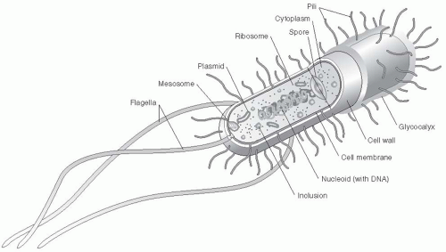
Figure 8-1 Structure and organelles of a basic bacteria. Note that they lack a nucleus.

Bacterial microorganisms are present in all environments (e.g., soil, water, and air). They are part of all vital life functions of more complex lifeforms and are found in association with both living and nonliving environments. Thus organisms of interest to the medical community represent only a part of the taxonomic classification of all bacteria. The work of Robert Koch and Louis Pasteur had dispelled many misconceptions about the causes of infectious diseases by the end of the nineteenth century. Subsequently, as the knowledge base grew, a clinically driven approach to classification of bacterial organisms emerged that relied on the concepts of pathogenicity and virulence.


Structure

Figure 8-1 illustrates the basic structure of a bacterial cell. Although all of the structures shown in the figure are involved in the life and survival of prokaryotic,
bacterial cells, many are also important virulence factors and are briefly described here with emphasis on their roles in pathogenesis.

Only gold members can continue reading. Log In or Register to continue

Stay updated, free articles. Join our Telegram channel

Jul 8, 2016 | Posted by in INFECTIOUS DISEASE | Comments Off on Microbiology Tools for the Epidemiologist

Full access? Get Clinical Tree

Get Clinical Tree app for offline access