Mast Cells and Basophils: Ontogeny, Characteristics, and Functional Diversity


Mast Cells and Basophils: Ontogeny, Characteristics, and Functional Diversity


A. Dean Befus

Kelly M. McNagny

Judah A. Denburg


INTRODUCTION

In 1965, Selye1 reviewed the literature on two populations of basophilic leukocytes, namely mast cells and basophils. These cells have many similarities, but they also exhibit several intriguing differences. Recent contributions to the literature documenting advances made in our understanding of these cells, their development, contents, biosynthetic activities, activation, and functions in physiology and pathophysiology2,3,4,5,6,7 are helping us begin to understand the nature of their relationship in ontogeny and immunity,2 and factors that govern their elicitation and contributions to a wide variety of inflammatory disorders, including allergic inflammation. Mast cells and basophils contain electron-dense cytoplasmic granules and stain metachromatically with selected basic dyes. They produce numerous inflammatory mediators, many—such as histamine—that are common to both cells, and others that are cell-specific. Both cells express a tetrameric isoform of the high-affinity receptor for immunoglobulin E (IgE) and are best known for their ability to release a plethora of factors and inflammatory mediators in response to receptor cross-linking. When the tetrameric high-affinity IgE receptor is cross-linked by sensitizing allergen or by anti-IgE antibodies, mast cells and basophils can be activated, mediator synthesis and secretion are induced, and gene expression altered, with consequences on many immune and inflammatory events.


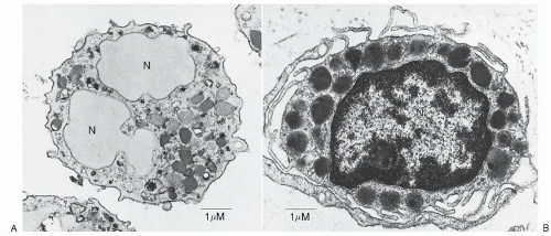
FIGURE 9.1. A. Basophils are “cells with the kinetics and natural history of granulocytes that mature in bone marrow, circulate in the blood, and retain certain characteristic ultrastructural features, even after migrating into the tissues during inflammatory or immunologic processes. The ultrastructure of mature basophils varies according to species but generally includes electron-dense cytoplasmic granules, prominent aggregates of cytoplasmic glycogen, and short, blunt, irregularly distributed plasma membrane processes. There is no convincing evidence that mature basophils, whether in the circulation or in the tissues, retain mitotic capability, or that basophils metamorphose into mast cells upon entering the tissues.” (Galli SJ, Dvorak AM, Dvorak HF. Prog Allergy 1984;34:1-141). As shown in Figure 9.1A, human basophils are round and have irregular, short surface projections, cytoplasmic secretory granules, and aggregates of cytoplasmic glycogen. N, nucleus; bar, 1 µm. (Reproduced with permission from the authors and Blackwell Science Ltd., Dvorak AM, Warner JA, Fox P, et al. Recovery of human basophils after FMLP-stimulated secretion. Clin Exp Allergy 1996; 26:281-294.) B. Mast cells “ordinarily mature outside of the bone marrow or circulation, generally in the connective tissues or serous cavities. Cells in this lineage(s), wherever distributed, apparently retain at least limited or latent proliferative capacity…(I)mmature and mature granules of mast cells and basophils differ distinctively in ultrastructure. Mast cells also differ from basophils in lacking electron-dense aggregates of cytoplasmic glycogen, and in having a plasma membrane surface with uniformly distributed, thin, elongate folds and processes. Mast cell nuclei may appear bilobed in an individual photomicrograph, but they generally lack the pattern of peripherally condensed nuclear chromatin characteristic of basophils and other granulocytes.” (Galli SJ, Dvorak AM, Dvorak HF. Prog Allergy. Basophils and mast cells: morphologic insights into their biology, secretory patterns, and function. 1984;34:1-141). A human skin mast cell is shown with monolobed nucleus with partially condensed chromatin, numerous cytoplasmic granules containing crystalline structures and regularly distributed, narrow, thin surface projections. Bar, 1 µm. (Reproduced with permission from the author and Springer-Verlag. Dvorak AM. Human mast cells. In: Beck F, Hild W, Kriz W, et al., eds. Advances in anatomy, embryology and cell biology. Leicester: Springer-Verlag, 1989;114:1-107.)

Classical ontogenic and ultrastructural descriptions for mast cells and basophils are shown in Figure 9.1 A,B; however, new information and emerging technologies have greatly expanded our understanding of the roles of basophils and mast cells in both innate and acquired immunity,2,4,8,9,10,11,12,13 notably including: (a) a burgeoning literature and controversy on basophil involvement as antigen-presenting cells in Th2 immune responses2,8,9,10,11,12,13; (b) new insights into the role of gut microbiota and thymic stromal lymphopoietin (TSLP) in basophil hemopoiesis and in mast cell localization and function14,15; (c) expression of the hemopoietic antigen, CD34, on mast cells involved in a variety of inflammatory reactions16,17,18; (d) novel roles for mast cells in tissue remodeling, angiogenesis, and tumor growth19,20,21,22; and (e) the capacity of mast cells to be derived from a compartment of multipotential hemopoietic progenitors, distinct from the myeloid lineage.23,24,25 In many ways the original statement by Ehrlich in 18791 that basophils are “blood mast
cells,” and the corollary, that mast cells are “tissue basophils,” although imprecise from a strictly developmental standpoint, is still of some value in thinking about the nature of these two cell types. The striking inverse relationship between the numbers of circulating basophils and the numbers of tissue mast cells has been used for decades to infer similarities in function.1

This chapter provides an overview of the developmental biology of these two cell types, comparing and contrasting basophil and mast cell physiology, phenotype, activation, and function; lineage commitment and differentiation; and roles in innate and acquired immunity. Anticipated increases in the understanding of basophil and mast cell function will lead to effective new diagnostic and therapeutic strategies for the betterment of those suffering from allergic and other related diseases.


GENERAL MORPHOLOGY, MEDIATOR SECRETION, AND RECOVERY


Morphology

Histochemical staining of blood smears or cytocentrifuge preparations of enriched basophils or mast cells with Wright or May-Grünwald-Giemsa stain show many similarities in these cell types (Fig. 9.2 A,B). The cytoplasm of the cells generally stains pink, the nucleus is purplish or blue, and the cytoplasmic granules are dark blue to purple or even blackish. Basophils in peripheral blood or tissues range in size from 10 to 15 µm, whereas mast cells in tissue sites may appear irregular in shape and up to 20 µm in a long dimension. Ultrastructural analyses demonstrate many similarities between mast cells and basophils, but also identify some distinct differences (Fig. 9.1A,B).26 In the blood, basophils are round, whereas in the tissues they can acquire various shapes. Mast cells can appear to be round, oval, or elongate-spindle shaped in the tissues. The surface of basophils exhibits blunt processes of variable shape and size, whereas mast cells often possess long fingerlike processes that extend from the surface. The nucleus of mast cells can be round or lobed, whereas that of the basophil is generally multilobed. Nucleoli are often not apparent or are absent from normal mast cells and basophils. Basophils have an abundance of condensed chromatin positioned at the periphery of the nucleus, whereas mast cells have little condensed chromatin, possibly reflecting their capacity for continued proliferation. The cytoplasm of normal mature mast cells has few mitochondria and a relatively inconspicuous Golgi apparatus; and ribosomes, rough endoplasmic reticulum, and aggregates of glycogen are rare. In normal basophils, mitochondria and aggregates of glycogen are more abundant than in mast cells, but as with mast cells, Golgi apparatus, ribosomes, and rough endoplasmic reticulum are rare in normal basophils. The most prominent cytoplasmic elements in both cell types are the membrane-bound, electron-dense granules.

Basophils generally possess fewer granules than mast cells and the granules exhibit a more homogeneous ultrastructural morphology than those of mast cells. Basophil granules are often homogeneously electron-dense, although dense particles may be interspersed with membrane aggregates and whorls. Charcot-Leyden crystals can be formed in basophils as well. Mast cell granules may be homogeneously electron-dense or may exhibit electron-dense particles, membrane or complex scroll-like patterns, highly organized crystalline structures, or combinations of these. The relationship of these different granule patterns to the tissue site, phase in development, or mediator content is not clear. Interestingly, Dvorak has reported that mast cell granules, in addition to storing mediators, are also sites of RNA metabolism and protein synthesis activity. Mast cells produce 30 to 100 nm extracellular membrane vesicles of endocytic origin, called exosomes, that possess mRNA, microRNAs, proteins, and lipids, and have been shown to transfer these components to other cells and influence their functions.27 Moreover, the composition and cell-to-cell communication functions of exosomes are influenced by activation of mast cells.28 Basophils have not been shown to produce exosomes, but they do contain numerous electron-lucent vesicles of 50 to 70 nm that often have contents similar to granules. These may be associated with a form of mediator exocytosis (see below).


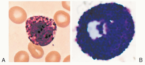
FIGURE 9.2. A. Human peripheral blood basophil stained with Wright’s. (From Lee, Bithell, Foerster, et al., eds. Wintrobe’s clinical hematology, 9th ed. Philadelphia, PA: Lea & Febiger, 1993.) B. Rat peritoneal mast cell stained with May-Grünwald-Giemsa.

Mast cells and basophils also contain rounded, non-membrane-bound, electron-dense structures called lipid bodies, a rich store of arachidonic acid. Lipid bodies increase in number during cell activation, and are thought to be derived from membrane catabolism and the rapid synthesis of lipid mediators such as the cyclooxygenase and lipoxygenase derivatives of arachidonic acid.29


Mediator Secretion

Mast cells and basophils undergo distinct patterns of granule mediator secretion or synthesis, depending on the stimuli involved in their activation.26,30 Degranulation or regulated exocytosis, whereby granules or their contents are released, occurs following stimulation through the IgE receptor or numerous other receptors, such as those for fragments of the complement cascade. This degranulation can be extensive, involving the majority of the granules, or it can be more restricted. The numbers of granules involved appear to correlate with the proportion of the total cellular histamine that is released. The granules rapidly exhibit signs of swelling, then demonstrate membrane fusion with adjacent granules; interconnecting chains and channels form that ultimately fuse with the plasma membrane, creating pores or larger openings and subsequently granule contents or larger granule structures can be seen outside the cell. Prominent cytoplasmic microtubules can be seen close to the granules, and actin complexes appear outside the cell in association with granules or their contents. The process is generally similar for basophils. Electron-dense granules appear to evolve into electron-lucent vacuoles, many of which communicate with other vacuoles and the cell surface through pores within 5 to 10 minutes of activation. Apparently intact granules can be seen outside the cell. This degranulation does not lead to prominent cell death, and cells can recover and degranulate again.26,31

Another form of secretion of stored mediators is piecemeal degranulation, involving production of small vesicles arising from granules that shuttle selected granule components to the extracellular milieu.30 Piecemeal degranulation also occurs in both mast cells and basophils, as well as in eosinophils,26,32 and is the most prevalent morphologic expression of mast cell and basophil secretion in nonallergic inflammatory conditions in human biopsy material. It is postulated that intragranular vesiculotubular networks fuse with the plasma membrane, and discharge their granule contents to the extracellular space. This form of mediator secretion appears to be associated with secretion of selected mediators, rather than the entire contents of the granules. It has been reported in communications between mast cells and neurons
in the brain,33 in psychosocial stress,34 and in acute gastritis,35 and may be a component of responses to Toll-like receptor (TLR) activation.4 In inflammatory reactions in which mast cell and basophil infiltration occurs, such as in cutaneous delayed hypersensitivity, piecemeal degranulation can occur. As with anaphylactic degranulation, piecemeal secretion is associated with the ability of mast cells to recharge their granules and function again.

Cytokines and chemokines are also secreted by mechanisms independent of degranulation, as are arachidonic acid metabolites, prostaglandins, and leukotrienes. Newly synthesized cytokines and chemokines can be secreted by a process called constitutive exocytosis. The pathways of secretion of arachidonic acid metabolites as well as those for exosome release are poorly known.


Recovery after Activation

Dvorak26 summarized the evidence for the ability of mast cells and basophils to recover and regranulate following activation. During this process, mast cells and basophils appear to conserve membranes and other components and to resynthesize granule and other components, such as rough endoplasmic reticulum, Golgi, and microtubules. This recovery generally occurs within 1 to 2 days; whether death occurs by necrosis or apoptosis as a critical feature of mast cell or basophil activation in vivo needs to be more carefully evaluated.


ONTOGENY AND DEVELOPMENTAL BIOLOGY OF MAST CELLS AND BASOPHILS

The tissue mast cell and the blood basophil are not normally derived directly from a common progenitor, but share their origins from a CD34+ hemopoietic stem cell regulated closely by various marrow and tissue stromal factors (Fig. 9.3A, B). A unique role is played by CD34 itself—which can be found on some mast cell subpopulations, and is involved in mast cell migration as well as tissue inflammatory responses—in these processes.18,36 Recent evidence suggests that mast cells are derived from a separate, nonmyeloid lineage-committed progenitor,23 and that, for the most part, basophils and mast cells have distinct ontogenic derivations and markers,23 although evidence from leukemic cell lines raises the possibility of some lineage pathway commonalities.37,38 and 39,40 Phylogenetically, there appears to be an inverse relationship between the presence of basophils and mast cells, as well as supporting evidence for a “common origin” by analysis of the evolution of a mast cell progenitor from an ancestral leukocyte involved in innate immunity.1,2,41 Major advances in understanding human basophil and mast cell growth and differentiation have come from rodent models,14,42,43,44 which have recently included an appreciation of the pivotal roles played by epithelial factors such as TSLP, IL-33, IL-25, and receptors such as the TLRs 45,46 in basophil, eosinophil, and mast cell differentiation.


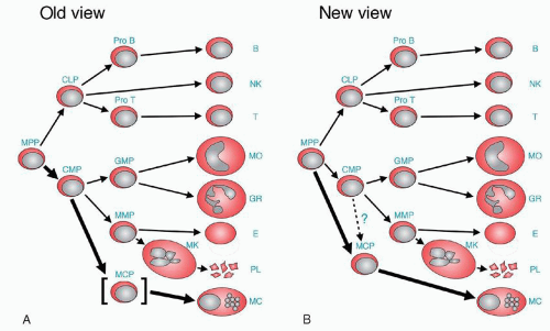
FIGURE 9.3. Change of lineage model of hematopoiesis. A. In the old view, the presence of mast cell committed progenitors (MCP) was not clear, but mast cells were considered to be the progeny of common myeloid progenitors (CMP) without convincing evidence. Therefore, MCP is put in brackets. B. In the new view proposed by Chen et al.23 the presence of MCP is clear, and MCP is directly derived from multipotential progenitors (MPP). The thick line shows the main differentiation route from MPP to mast cell. CLP, common lymphoid progenitor; GMP, granulocyte/macrophage progenitor; Pro B, progenitor B cell; Pro T, progenitor T cell; B, B cell; NK, natural killer cell; T, T cell; MO, monocyte/macrophage; GR, granulocyte; E, erythrocyte; PL, platelet; MC, mast cell. (Reproduced with permission, Proc Natl Acad Sci U S A 2005;102:11129-11130 © 2005 National Academy of Sciences, U.S.A74).

Identification of the mast-cell hemopoietin and ligand, stem cell factor (SCF), and its receptor, c-kit (CD117), has provided a wealth of insight into the functional role of mast cells in a variety of processes. Mutations in either the ligand (“Steel” locus) or its receptor (“W” locus) render mice largely mast-cell-deficient, facilitating evaluation of the role of mast cells in a variety of animal models.47,48 Mast cells have thus been revealed to be key players not only in allergic inflammation but also in angiogenesis and tumor growth, tissue remodeling, graft tolerance, and some autoimmune diseases.19,20,21,22,49,50,51,52 However, inasmuch as mutations of the SCF/c-kit loci also affect other hematopoietic lineages and gut function, these studies must be interpreted with caution; novel studies with more selective mutations have suggested that some of the functions previously ascribed to mast cells might be artifacts of the “W” strains.53,54 Nonetheless, advances in isolation and analysis of mast cell and basophil populations3,9,10,11 have provided novel insights into their functions and transcriptional regulation of their hemopoietic development.


Mast Cell Growth and Differentiation

Rodent and human mast cells can be grown in vitro from lineage-committed, unipotent, or multipotent progenitors. Although
interleukin-3 (IL-3) is known to contribute to murine mast cell and to both human and rodent basophil development,55,56,57 it is SCF or c-kit ligand58 which uniquely drives human mast cell differentiation, and not other cytokines that may help promote mast cell development in rodents, such as IL-3, IL-4, IL-9, IL-10,59 and nerve growth factor (NGF).60 SCF is produced by murine and human fibroblasts, epithelial cells, endothelial cells, and tumor cell lines,61 and must bind to c-kit to effect differentiation. Mutations in c-kit can result in mast cell deficiency in vivo and in vitro (“loss-of-function”) or, alternately, in autonomous mast cell growth (“gain of function”), generally leading to autophosphorylation.62 The latter mutations, especially the D-816-V-associated mastocytosis (which can be transferred with similar consequences to the mouse62) have formed the basis of newly designed tyrosine kinase inhibitors in some proliferative mast cell and hypereosinophilic disorders.

Recently, it has been shown that both Th1 and Th2 cytokines (interferon-g [IFN-g], IL-4, and IL-5), the latter induced in rodents via TSLP and IL-25 or IL-33 acting in concert, induce a distinct population of multipotent mucosal progenitors (“nuocytes”)14,42,43,44,63 and thus may exert differential modulatory effects on SCF-dependent mast cell numbers in vitro and in vivo.64 Table 9.1 lists cytokines, growth factors, transcription factors, and signaling molecules that regulate primate/human and rodent mast cell differentiation.51,65,66,67,68


Mast Cell and Basophil Progenitors

In humans and in rodents, mast cell differentiation proceeds from an immature CD34+, CD38+, CD13+, c-kit+, FcεRI, FcγRII/III+ cell, or, as recently shown, from a previously uncharacterized immature cell in mouse marrow.23,24,25 More specifically, a multipotent hemopoietic progenitor of both mucosal and serosal mast cell phenotypes can be identified, and is characterized as Linc-kit+Sca-1-Ly6cFcεRICD27β7+T1/ST2+69 (Fig. 9.4).

Mast cell differentiation, heterogeneity, abundance, and functional responses are also regulated in vitro and in vivo in rodents by CD34 itself and through the actions of several transcriptional and nucleosomal regulators such as PU.1, GATA factors, C/EBP factors, and MITF, as well as through M-Ras, and RabGEF1 type signaling molecules.39,66,68,70,71,72 Phenotypical alterations of mast cell populations can be governed by the tissue milieu, and they also exhibit switching and “trans-differentiation,” 1,73 reflecting stochastic processes in progenitor differentiation. Primitive cord blood CD34+ c-kit+ progenitors may respond quite differently to hemopoietic cytokines than more mature FcεRI+, c-kit+ cells.23,74 Thus, tissue-dependent stages of progenitor commitment and of growth and differentiation signals may ultimately predict differences in mast cell phenotype in vivo.75 Mast cell progenitors can be identified in blood, bone marrow, and various other tissues, especially in relation to mast-cell-inducing stimuli, such as viruses or nematodes.69

Leukemic or other self-sustaining cell lines with basophilic or mast cell phenotype have also been used to study progenitors and immunophenotypic markers of lineage commitment.76 Although these findings may represent aberrant pathways activated during malignant transformation, the recent identification of a novel antigenic marker of mast cells, basophils, and their progenitors,77 and the presence in peripheral blood of basophilic cells that express mast cell proteases78 have revived earlier postulates79 that these cell types share some lineage characteristics under conditions of aberrant cell growth,37,38 although rodent eosinophil lineage-committed progenitors may not express basophil/mast cell-related proteases.80

New and exciting information has emerged on the role of gut microbiota in regulating a TSLP-induced, IgE-dependent Th2 response, involving basophil progenitors in rodents, and in humans with hyper-IgE syndrome.12 Profound decreases in gut flora lead to a B-cell driven overproduction of IgE with binding to its high-affinity receptor, and thus to increased marrow basophilopoiesis, with Th2 skewed inflammatory responses. Previous work on normal, atopic, or leukemic/mastocytotic human blood or marrow had identified pure or mixed basophil colonies in semisolid cultures,79,81,82 thus defining a basophil progenitor (termed “CFU-baso” or “CFU-baso/eo”).81,83 The phenotype and lineage commitment of the basophil progenitor, including recent controversial notions of possible lineage pathway commonalities with mast cells and/or megakaryocytes,84,85 are depicted in Figure 9.4.


TABLE 9.1 CYTOKINES AND OTHER FACTORS INVOLVED IN BASOPHIL AND MAST CELL GROWTH AND DIFFERENTIATION


Cytokine


Effect


GM-CSF


Basophil growth and differentiation; promotes in vivo basophilia and increases in circulating CFU-baso/eo (primates); basophil activation/survival; down-regulates human mast cell differentiation.


IL-3


Human basophil growth and differentiation; basophil activation/survival; promotes in vivo basophilia (in primates); mast cell differentiating activity in rodents.


IL-5


Primarily eosinophil, but also basophil growth and differentiation; basophil and eosinophil activation/survival.


TSLP/IL-33/IL-25


Co-factors in rodent nuocyte (multipotent mucosal progenitor) differentiation and tissue basophilic responses.


IL-4/IL-13


Produced by basophils with minimal activity on differentiation.


IL-9/IL-10/THPO


Co-factors in rodent mast cell phenotype switching.


TGF-β/IFN-α


Negative regulators of basophil differentiation.


SCF


Primary mast cell growth factors in mouse and rat; little known effect on basophil differentiation.


NGF


Induces mast cell hyperplasia (rodents), human mast cell line (HMC-1), and basophil-eosinophil differentiation in vitro.


RA


A mutation in the RA receptor allows for expression of basophil differentiation; may concurrently down-regulate human mast cell differentiation.


v-erb


Associated with the development of lethal mastocytosis in the rodent.


FcγRIIB


Down-regulates mast cell growth.


Stat5


An essential regulator in vivo of mast cell development.


IL-6/TNF/IFN-γ


May play a role in regulating phenotypic direction and lineage commitment of human basophils and mast cell subtypes.


GM-CSF, granulocyte-macrophage colony stimulating factor; INF, interferon; IL-, interleukin-; NGF, nerve growth factor; RA, retinoic acid; SCF, stem cell factor; TSLP, thymic stromal lymphopoietin; TNF, tumor necrosis factor.


Basophil Differentiation-inducing Cytokines

IL-3 is the main cytokine involved in human basophil growth and differentiation,86 with some evidence for a co-factor role for TSLP. Granulocyte-macrophage colony stimulating factor (GM-CSF),79,81,82,87 IL-4,88 IL-5,83,88 and SCF89 may also play roles. A mutation in the retinoic acid (RA) receptor permits basophil differentiation; studies on basophil crisis in chronic myeloid leukemia and the in vitro suppressive effects of RA on basophil-eosinophil differentiation support this notion.90 Cytokines and other factors that modulate basophil or mast cell differentiation are listed in Table 9.1.


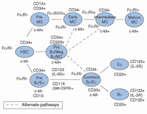
FIGURE 9.4. Mast cell and basophil ontogeny. An orderly sequence of differentiation is depicted, beginning with a primitive CD34+, FcεRI, c-kit hemopoietic stem cell (HSC), and proceeding through various stages of commitment to either mast cell or basophil/eosinophil lineages. In reality, the differentiation process is stochastic, and multiple cytokines binding to their receptors permit specific, end-stage differentiation to proceed. For more detailed phenotypic analyses of basophil and mast cell progenitors, see Wang et al. 37


Clinical Relevance of Basophil and Mast Cell Differentiation


Allergic Diseases

The relationship to disease activity, severity, and response to therapy of fluctuations in numbers of human basophil-eosinophil, basophil-mast, or mast cell progenitors—including CD34+ cell subpopulations—in the blood and bone marrow of patients with a variety of allergic disorders, including allergic rhinitis, nasal polyposis, asthma, atopic dermatitis, and drug allergies, has been extensively documented.59,91 Indeed, therapies targeting basophil-eosinophil progenitors with anti-IL-5 monoclonal antibodies have had some efficacy in causing maturation arrest of these shared lineages, accompanied by some clinical benefits, in patients with eosinophilic bronchitis and asthma92,93; targeting of basophil or mast cell progenitors in these and some hematologic disorders may likewise prove to be effective treatments. For example, the IL-3R is expressed at high levels by basophil progenitors and subsets of dendritic cells; therefore, development of high-affinity monoclonal antibodies to this receptor would be predicted to dampen allergic disease.

Only gold members can continue reading. Log In or Register to continue

Stay updated, free articles. Join our Telegram channel

Oct 21, 2016 | Posted by in HEMATOLOGY | Comments Off on Mast Cells and Basophils: Ontogeny, Characteristics, and Functional Diversity

Full access? Get Clinical Tree

Get Clinical Tree app for offline access