8. Infectious Disease


“Bacteria, just like any other life form, need energy


to survive, multiply, and colonize. Patients with uncontrolled diabetes have high sugar content in their sweat and connective tissues, which can become a perfect energy source for bacterial colonization and subsequent infection. To make matters worse, diabetic patients also frequently have immunosuppression and microvasculopathy.”


8.1 Infection and Sepsis


Systemic infection with any microbe can present with nonspecific features of fever, tachycardia, leukocytosis, altered mental status, etc. As infection progresses, patients can start looking sicker, and develop decompensation in various organ systems, as following:


Circulatory


Persistent hypotension, despite adequate IV fluids resuscitation → vasopressor requirement


Lactic acidosis


Kidney


Oliguria or anuria (renal failure), acute tubular necrosis


Lung


Tachypnea → acute lung injury → acute respiratory distress syndrome


CNS


Agitation, confusion, coma


Hematologic


Anemia, thrombocytopenia, DIC, etc.


Liver


Ischemic or septic liver injury


To prevent this, prompt diagnosis and aggressive treatment are of utmost urgency.


Abbreviations: CNS, central nervous system; DIC, disseminated intravascular coagulation.


Clinical pearl: any patient who presents with fever of unknown source, think of an infectious source from head to toe: CNS → upper respiratory tract → lower respiratory tract → heart → GI → urinary tract → joints → skin. This helps in history taking and to make the diagnosis.


8.2 A Simplified Approach to Antibiotics for Empiric Therapy


Antibiotics


Gram positive (staphylococcus/ streptococcus, except MRSA)


Gram negatives


Anaerobes


Pseudomonas


  • B-lactamase resistant narrow spectrum penicillins (oxacillin, cloxacillin, dicloxacillin, nafcillin)a


  • First generation cephalosporinsb


  • Third generation cephalosporinc


  • Ciprofloxacin (IV, PO)


  • Aztreonam (IV)


  • Ceftazidime (IV)


  • Amikacin, Gentamicin, Tobramycind


  • Metronidazole


  • Cefepime (IV)


  • Amoxicillin-clavulanate (oral)


  • Ampicillin-sulbactam (IV)


  • Piperacillin- tazobactam (IV)


  • Ticarcillin-clavulanate (IV)


  • (IV Meropenem, IV Doripenem)e


  • Clindamycinf


aOther penicillins (such as Penicillin V or G, amoxicillin, ampicillin) are good against the Streptococcus group (S. viridans, S. pneumoniae, S. pyogenes) but not against staph, as staph usually have B-lactamases which deactivate penicillins.


bFor example, oral cephalexin, IV cefazolin.


1 Cephalosporins can be given in patients with mild penicillin allergy (e.g., rash), but are contraindicated in patients with severe penicillin allergy (e.g., anaphylaxis, Stevens-Johnson syndrome, allergic interstitial nephritis).


cIV forms are ceftriaxone, cefotaxime, and ceftazidime. Oral forms are cefpodoxime and cefdinir. These drugs have good Streptococcus/Staphylococcus coverage as well, but not as good as 1st generation cephalosporins or B-lactamase resistant narrow-spectrum penicillins.1


dAminoglycosides for pseudomonas is usually not used as monotherapy, but as an adjunctive treatment for serious infections.


eUnlike piperacillin-tazobactam, carbapenems cover extended spectrum beta-lactamase (ESBL)-producing superbugs, such as Acinetobacter baumannii or ESBL E. coli/Klebsiella.


2 Meropenem or imipenem may be the right answer in a patient with prolonged hospitalization and evidence of active gram-negative infection who does not improve despite adequate antibiotic therapy. The only difference of coverage in between (meropenem, doripenem, imipenem) and ertapenem, is that ertapenem does not cover Pseudomonas.


fThe old dictum of treating above-diaphragm infection (e.g., lung abscess) with clindamycin and below-diaphragm infection (e.g., intestinal infection or perforation) with metronidazole no longer applies. Clindamycin is no longer preferred due to increased risk of C. difficile. The preferred antibiotics for treatment of aspiration pneumonia now are amoxicillin-clavulanate, or third-generation cephalosporin + metronidazole.


Abbreviations: IV, intravenous; MRSA, methicillin-resistant Staphylococcus aureus


8.2.1 Other Bacteria and Their Antibiotic Coverage


Bacteria


Antibiotic coverage


MRSA
(methicillin-resistant Staphylococcus aureus)


Intravenous agents: vancomycin (DOC) Second line—daptomycin (IV only), linezolid (oral or IV), IV ceftaroline (fifth-generation cephalosporin), IV quinupristin, or IV dalfopristin.
Oral agents: doxycycline, clindamycin, and sulfamethoxazole-trimethoprim.


All MRSA antibiotics have good gram-positive coverage, except doxycycline which does not have good Streptococcus coverage.


Atypical bacteria
(Legionella, Chlamydia, Mycoplasma, Coxiella, etc.)


Macrolides (azithromycin)
Doxycycline
Respiratory fluoroquinolones (levofloxacin)


Listeria monocytogenes


Ampicillin


Bacillus Anthracis


Ciprofloxacin or Doxycycline ©MRS ABCD


Syphilis


Penicillin or doxycycline


Abbreviations: DOC, drug of choice; IV, intravenous.


8.2.2 Example Clinical Cases


Condition


Coverage needed for


Examples of empiric coverage


Cellulitis without MRSA risk factorsa


Gram positives (staph and strep)


Cephalexin
or
IV cefazolin or nafcillin


  • Diverticulitis


  • Fistulating Crohn’s disease


GI flora (anaerobes and gram negatives)


Ceftriaxoneb + metronidazole
or
Ampicillin-sulbactam


Infections with risk factor for Pseudomonasc


Pseudomonas


IV cefepime
or
IV piperacillin-tazobactam


Hospital-acquired catheter infection


MRSA


IV vancomycin


Community-acquired pneumonia requiring hospitalization


Streptococcus spp., gram negatives (Moraxella catarrhalis, Haemophilus influenzae), and atypicals.


Ceftriaxone + azithromycin
or
Cefpodoxime + doxycycline


a MRSA risk factors: IVDA (intravenous drug abuse), human immunodeficiency virus (HIV) positive, recent prior antimicrobial therapy, hemodialysis, recent hospitalization, residence in long-term care facility such as nursing homes, etc.


bNowadays third-generation cephalosporins are preferred over quinolones (e.g., ciprofloxacin), because quinolones are associated with higher risk of C. difficile and other side effects.


cRisk factors for Pseudomonas infection include the following:


Abbreviations: GI, gastrointestinal; IV, intravenous; MRSA, methicillin-resistant Staphylococcus aureus.


MRS

Cephalosporins are LAME because they do not cover:


  • Listeria


  • Atypicals


  • Mycobacteria


  • Enterococcus


Clinical pearl: Do not use ceftriaxone or cephalexin to treat UTI caused by Enterococcus.


  • Neutropenic fever (always look at WBC count in a question about empiric antibiotic therapy)


  • Burns


  • Hospital-acquired pneumonia, especially in intubated patient


  • Chronic granulomatous disease


  • Severe pulmonary infection in a patient with cystic fibrosis


  • Diabetes mellitus with certain infections such as malignant otitis externa


  • Cellulitis and osteomyelitis associated with a punctured sole wound


8.2.3 Indications for Antibiotic Prophylaxis in Adults


Clinical situation


Drug of choice


Close contacts of patients with Neisseria meningitidis


Rifampin or fluoroquinolone


Significant bite wounds


(Ampicillin-sulbactam) or (amoxicillin-clavulanate)


High-risk cardiac defects undergoing high-risk procedures require endocarditis prophylaxis


Amoxicillin


8.2.4 Notable Side Effects of Antibiotics


Antibiotic


Notable side effects


Penicillin


All types of hypersensitivity reaction (type I, II, III, and IV)


An uncommon but unique side effect of oral pen-VK is black hairy tongue


Trimethoprim-sulfamethoxazole


  • Rash, angioedema, allergic interstitial nephritis, serum sickness, etc.


  • Folate deficiency


  • Hyperkalemia


  • Hemolysis (in patients with G6PD deficiency)


Doxycycline


Pill esophagitis, tooth discoloration in children


Linezolid


Thrombocytopenia, serotonin syndrome


Fluoroquinolones


Tendinitis, Achilles tendon rupture, irreversible neuropathy, and bony growth abnormalities (use with caution in children)


Cefotetan (second-generation cephalosporin with anaerobic coverage) and metronidazole


Disulfiram-like effect (with alcohol)


Vancomycin


  • Renal toxicity


  • Red man syndrome: itching and erythematous flushing reaction during infusion due to histamine release. In rare situation chest pain and hypotension can occur. This is not an allergic reaction and is related to rate of infusion.


Rx: use antihistamines (diphenhydramine + ranitidine), and slow down the infusion rate.


8.3 Skin Infections


Condition (given below in order of severity from superficial to deep infection)


Presentation


Microbial cause


Empiric therapy


Infection of epidermis = Impetigo a


No Image Available!


Source: Impetigo. In: Laskaris G, ed. Color Atlas of Oral Diseases. Diagnosis and Treatment. 4th ed. Thieme; 2017.


Papules, vesicles, pustules and/or bullae containing yellow fluid, surrounded by erythema.
When pustules/ bullae break, they form a honey-colored crust.
(Look for weepy, oozy, crusty, dirty looking skin rash)


Staph. aureus
or
B-hemolytic streptococcus


  • If mild and localized, use topical mupirocin or retapamulin.


  • If severe or widespread, use oral semi-synthetic penicillins (e.g., cloxacillin) or cephalexinb


Infection of dermis and epidermis = Erysipelas


No Image Available!


Source: Erysipelas. In: Sterry W, Paus R, Burgdorf W, ed. Thieme Clinical Companions -Dermatology. 1st ed. Thieme; 2006.


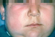
Shiny red, edematous, patch found particularly on face, arms or legs. May be painful.
(Look for raised, clearly demarcated borders)


B-hemolytic streptococcus


  • If mild, use oral amoxicillin or penicillin (usually staph coverage isn’t required).


  • If severe, use IV ceftriaxone or cefazolin.


Infection of dermis and subcutaneous tissue =
Cellulitis


No Image Available!


Footnote: Facial cellulitis on the right side due to dental abscess.


Source: Cellulitis. In: Laskaris G, ed. Color Atlas of Oral Diseases. Diagnosis and Treatment.


4th ed. Thieme; 2017.


Redness, swelling, increased temperature and tenderness over the involved skin area.
Borders are not raised or clearly demarcated


S. aureus
or
B-hemolytic Streptococcus


  • Management is discussed on next page


aThis is contagious, and outbreaks can occur.


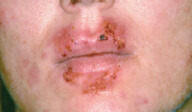
3 D/Dx (differential dx) of impetigo is eczema herpeticum


Causative organism: HSV infection


Presentation: Painful rash with clear vesicles with underlying erythematous area. When the vesicles rupture, they leave behind punched-out ulcers. Vesicles might be hemorrhagic, which form dark red crusting after rupture.


Rx: Systemic acyclovir


bIn culture proven S. pyogenes, penicillin or amoxicillin alone should suffice.


In patients with immunosuppression, morbid obesity, or uncontrolled diabetics, soft-tissue infection can cross the fascial planes and involve muscles causing necrosis = necrotizing fasciitis.


Abbreviations: IV, intravenous; MRSA, methicillin-resistant Staphylococcus aureus.


Potential autoimmune complication of strep infection (e.g., erysipelas, cellulitis, strep throat) is Rheumatic fever and postinfectious glomerulonephritis (glomerulonephritis can also occur with staph infection).


Cellulitis management depends on the type of cellulitis:


1. Management of non-purulent cellulitis


Severity


MRSA risk factora


Empiric therapy


Cellulitis with signs of severe infection (e.g., fever, tachycardia)
– requires hospitalization. NSIM is blood culture and IV antibiotics


Present


IV vancomycin or ceftaroline
– If no improvement, initiate gramnegative coverage


Absent


IV cefazolin
– If no improvement, initiate MRSA and/or gram-negative coverage


Mild (outpatient treatment)


present


  • Trimethoprim-sulfamethoxazole, or


  • Amoxicillin (for strep) + doxycycline or minocyclineb, or


  • Oral clindamycin (last choice)


Absent


Cephalexin or dicloxacillin


aRisk factors for MRSA include – IVDA (intravenous drug abuse), HIV positive status, recent prior antimicrobial therapy, hemodialysis, recent hospitalization, residence in long-term facilities such as nursing homes, etc.


bUnlike clindamycin or trimethoprim-sulfamethoxazole, doxycycline doesn’t have good streptococcus coverage (hence it is combined with amoxicillin or cephalexin).


2. Management of – Purulent Cellulitis or – abscess with significant cellulitis, systemic symptoms or immunosuppression


These cases are almost always due to Staph and MRSA coverage is needed.


  • Send wound cultures


  • I&D if there is an abscess


Clindamycin is increasingly falling out of favor due to increased risk of C difficile infection.


High risk population for Infective endocarditisa


Empiric therapy


YES (patient needs strep coverage)


Severe infection: IV Vancomycin or Daptomycin
Mild infection :
doxycycline + amoxicillin
or
sulfamethoxazole-trimethoprim


NO (doesn’t warrant strep coverage)


Doxycycline alone should suffice.


aExamples of high risk conditions include prosthetic cardiac valve (including bioprosthetic heart valve), previous infective endocarditis, unrepaired cyanotic congenital heart disease, etc. Please refer to infective endocarditis section discussed later in this chapter.


Clinical tip : Note in all the above situations if Cellulitis is associated with periodontal disease, perirectal location, pressure ulcer or with prominent necrosis , it warrants anaerobic coverage.


8.3.1 Necrotizing Fasciitis and Differential Diagnoses


The following severe soft tissue infections usually have signs of systemic toxicity (Toxic shock syndrome is included in this table as sometimes cellulitis can lead to toxic shock syndrome and cause severe systemic toxicity).


Necrotizing fasciitis


Gas gangrene


Toxic shock syndrome


Causative organism


Polymicrobial (gram positive, gram negative, anaerobes, MRSA, etc.)
-or
Group A ß-hemolytic streptococci


Clostridium perfringens or Clostridium histolyticum
(gram positive bacilli)


Staph, aureus a


History


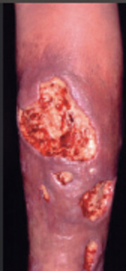
May not have bacterial point of entry at all, or may have coexistent groin ulcer or colonic fistula.


More likely to have hx of trauma. Trauma can be as minor as intramuscular injection, or as major as surgery.


Look for foreign body in situ (tampons or prosthesis), or an active site of Staph. infection, (e.g., osteomyelitis, or even cellulitis)


Clinical feature


They do have overlapping features


  • Crepitus (more prominent in gas gangrene)


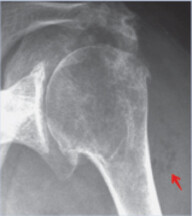
  • Gas in tissue seen in imaging (mostly along fascial planes in Necrotizing fasciitis)


  • Skin is dark blue to black necrotic


  • Severe pain out of proportion to clinical exam findings


  • Elevated creatine kinase


  • Sick looking patient with severe systemic features (as in necrotizing fasciitis and gas gangrene)


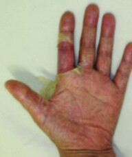
  • Generalized red sunburn like rash (diffuse erythroderma) involving palms and soles and/ or maculopapular rash


  • This rash can later desquamate after about a week or so (shown below in hands)


No Image Available!


Source: Necrotizing Fasciitis. In: Sterry W, Paus R, Burgdorf W, ed. Thieme Clinical Companions – Dermatology. 1st ed. Thieme; 2006.


No Image Available!


Footnote: multiple lucent gas collections deep to the deltoid muscle.


Source: Pathology. In: Gunderman R, ed. Essential Radiology. Clinical Presentation, Pathophysiology, Imaging. 2nd ed. Thieme; 2000.


No Image Available!


Source: Toxic Shock Syndrome Caused by Methicillin-Resistant Staphylococcus aureus (MRSA) After Expander-Based Breast Reconstruction. In: Suga H, Shiraishi T, Takushima A, Harii K. Eplasty; 2016.


Treatment of choice


Surgical exploration. Surgical specimens’ wound gram stain/ culture can differentiate in between them.


Removal of foreign body


Adjunctive empiric antibiotic treatment (Intravenous immunoglobulin, which neutralizes toxins, can be tried in severe cases.)


One broad spectrum antibiotic (meropenem or piperacillin/tazobactam or ampicillin-sulbactam) +
Clindamycinb
+
MRSA coverage-Vancomycin or daptomycin


Penicillin
+
clindamycinb
+/- hyperbaric oxygen therapy


Clindamycinb
+
Oxacillin, or vancomycin (if MRSA suspected)


aThis is due to colonization or infection with Staph. aureus that produces toxic-shock-syndrome-toxin (TSST). This toxin is unique in that it non-specifically activates T-helper cells, which release excess inflammatory mediators into the bloodstream causing SIRS, sepsis and even death.


bClindamycin also decreases exotoxin production


8.3.2 Diabetic Foot Ulcer Infection


Type


Features


Treat


Nonlimb-threatening mild infection


Minimal cellulitis (erythema) that extends not more than 2 cm from a superficial ulcer that does not go beyond superficial fascia and lacks systemic toxicity


Treat as cellulitis (as above)


  • No risk factor for MRSA, use any one of the following: cephalexin, dicloxacillin, amoxicillin-clavulanate, or clindamycin


  • If risk factor for MRSA present, use either clindamycin, or trimethoprim-sulfamethoxazole alone
    or
    combination of cephalexin + doxycycline


Moderate infection


Any one of the following present:
erythema extends >2 cm, lymphangitic streaking or infection goes beyond superficial fascia (to involve muscle, tendon, etc.)


Mostly this requires coverage for staph, strep, gram negatives, and anaerobes. Use antibiotics that cover MRSA, if risk factors present, e.g.,
Amoxicllin-clavulanate (covers anaerobes, GNR, gram positives) + trimethoprim-sulfamethoxazole (covers MRSA)


Severe infection


Infection with systemic toxicity or limb-threatening infection


NSIM is to admit the patient, do careful wound debridement, and start broad-spectrum IV antibiotic that covers gram-negative bacilli, anaerobes, and MRSA. Urgent consultation and a multidisciplinary approach are needed.


aCiprofloxacin and levofloxacin have good pseudomonal coverage. Use them if macerated looking wound or hx of significant water exposure.


Abbreviations: IV, intravenous; MRSA, methicillin-resistant Staphylococcus aureus.


In patients with diabetic foot ulcer infection, workup for osteomyelitis is recommended if one or more of the following are present:


  • Elevated ESR > 70 mm/h.


  • Bone exposure or probe to bone positive.


  • Ulcer size > 2 cm2.


  • Duration of ulcer >1–2 weeks.


Clinical Case Scenarios

24 y/o F with history of diabetes presents with raised, erythematous, weepy rash with clear border on her face. Patient reports of malaise.


  1. What is the likely diagnosis?


  2. What is the likely causative organism?


  3. How do you treat it?


8.4 CNS Infections


In a nutshell: clinical presentation and likely Dx


Fever + headache ± seizures +


Likely Dx


+ Stiff neck or other signs of meningismusa


Meningitis


+ Focal neurological deficit (e.g., right lower extremity weakness)


Brain abscess or toxoplasmosis


+ Altered mental status ± personality change


Encephalitisb


aSigns of meningeal inflammation include photophobia, Kernig’s sign (inability to straighten the leg when hip is flexed), and Brudzinski’s sign (neck flexion causes reflexive flexion of hips and knees).


4 Remember that subarachnoid hemorrhage can present similarly with signs of meningeal infl ammation and fever.


bViral causes of encephalitis include:


  • HSV-1.


    5 HSV-2 does not cause encephalitis, but may cause meningitis.


  • Arboviruses: western equine encephalitis, eastern equine encephalitis, Japanese encephalitis, West Nile virus, Venezuelan encephalitis, St. Louis encephalitis, California encephalitis, etc.


  • Enteroviruses.


Encephalitis can also be associated with Systemic Lupus Erythematosus (SLE), acute disseminated encephalomyelitis, etc.


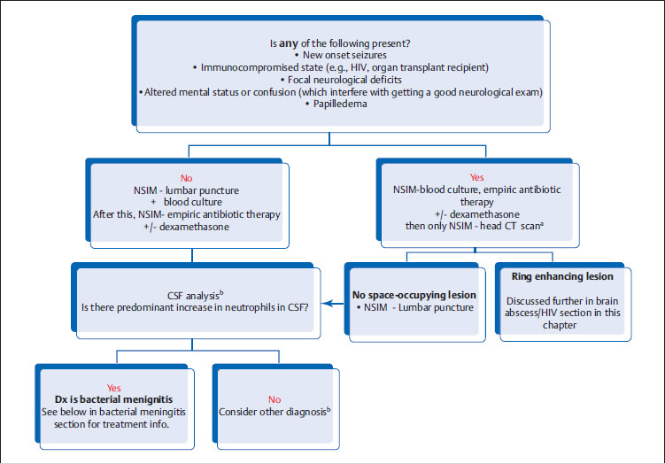
8.4.1 General Management of Suspected CNS Infection


No Image Available!


aIf there is a suspicion for space-occupying lesion, perform computed tomography (CT) scan prior to lumbar puncture as lumbar puncture can precipitate brainstem herniation in patients with elevated intracranial pressure (ICP). Also give antibiotics, as diagnostic workup should not delay life-saving antibiotic therapy.


b CSF analysis table


Abbreviation: CSF, cerebrospinal fluid.


Bacterial


Viral


Fungal or Lyme/rickettsia


Tuberculosis


Cell count


↑↑↑↑


↑↑


↑↑


↑↑


Differential


Neutrophils


Lymphocytes


Lymphocytes


Lymphocytes


Protein


↑↑


Can be ↑↑↑↑


Glucose


Low


Normala


Normal or low


Low


aAs opposed to viruses, organisms like bacteria, fungi, and mycobacteria directly use glucose for energy production.


8.4.2 Bacterial Meningitis


Microbiology


6 Know the MCC (most common cause) of bacterial meningitis in each age group.

:


Age


MCC in this age group


Neonate (<1 month)


Streptococcus agalactiae


  • 2nd MCC is E. coli.


1 month to 2 years


S. pneumoniae


2 years to 18-21 years, adults in military, or living in dormitories


N. meningitidis
2nd MCC is S. pneumoniae


>18-21 years (after one leaves college or dormitories)


S. pneumoniae


Other groups:


HIV


MCC is Streptococcus (NOT Cryptococcus)


Ventriculoperitoneal shunt


MCC is coagulase-negative Staph (S. epidermidis)


Empiric Treatment of Bacterial Meningitis


Age group


Empiric IV antibiotics of choicea


< 7 days of age


Gentamicin (or some-other aminoglycoside). Ampicillin b + Cefotaximec


7 days to < 1 month


Community-acquired Meningitis
– as above Gentamicin (or some-other aminoglycoside) + ampicillina + cefotaximec


If neonate never left the hospital, consider this hospital acquired – vancomycin + aminoglycoside+ cefotaximec


1 month to 50 years old (immunocompetent)


Vancomycin b + Ceftriaxone or cefotaxime


> 50 years of age, or
patients with alcoholism, diabetes mellitus (DM) or pregnancy


Vancomycind + Ampicillinb + ceftriaxone or cefotaxime


Community-acquired meningitis with risk factor for Pseudomonas, such as neutropenia, severe immunocompromised state (e.g., lymphoma, high-dose steroids, chemotherapy


Vancomycin d + ampicillin b + antipseudomonal cephalosporin
(e.g., cefepime) or meropenem


Hospital acquired meningitis (penetrating trauma or postsurgery meningitis)


Vancomycin d + ceftazidime/cefepimee


aConsider adding dexamethasone (except in neonates < 6 weeks old). In children, the greatest benefit of dexamethasone is in suspected H. influenzae meningitis; in adults in pneumococcal meningitis. It should be given concurrently with or immediately before the first dose of antibiotics, there’s probably no benefit if the corticosteroid is given after antibiotics are initiated. It reduces inflammation and decreases risk of neurologic complications, such as hearing loss.


7 MC sequela of untreated bacterial meningitis is eighth cranial nerve palsy.


bAmpicillin covers Listeria.


cCeftriaxone is contraindicated in this age group because of increased risk of biliary stasis.


dVancomycin is added in countries where pneumococcal resistance rate to ceftriaxone is > 1% e.g., USA, Canada.


eNo reason to cover Listeria, if not community-acquired.


MRS

The first exposure of a baby is to maternal vaginal flora, which may include S. agalactiae and E. coli.


In infants, community-acquired Streptococcus is most prevalent.


When a child starts attending day care, kindergarten and then school, there is increased crowding and hence increased risk of Neisseria.


As one becomes an adult and is no longer living in dorms or exposed to classmates, the incidence of Neisseria decreases, and community-acquired Streptococcus again becomes more prevalent.


MRS

GAC= Gentamicin + Ampicillin+ Cefotaxime


Coverage for vancomycin-resistant S. pneumoniae is not needed.


VCS: S = steroids


VACS


Age > 50, or with risk factors, give VACS therapy: Vancomycin, Ampicillin, Cephalosporin, and Steroids (dexamethasone)


Additional notes on N. meningitis


  • Additional unique clinical features include:


    • Petechia and purpura.


    • It can also cause hemorrhagic infarction of adrenal glands leading to acute adrenal crisis (severe hypotension).


  • N. meningitis prophylaxis is indicated (with either rifampin or ciprofloxacin) in the following situations:


    • Close contacts of N. meningitis patients—all household members, childcare center, military recruits in training center, and roommates/dorm mates. (Note: classmates or coworkers are not automatically considered as close contact.)


    • Health care providers who were exposed to respiratory secretions of patients with N. meningitis, e.g., a clinician who performed endotracheal intubation.


8.4.3 Lymphocytic Meningitis


Meningitis + lymphocytosis in CSF +


Causative organism


NSIM


Treatment of choice


+ Hx of camping trip in endemic areas


Lyme disease due to Borrelia burgdorferi


  • Serology for Lyme disease


  • CSF for Lyme antibody and PCR


Ceftriaxone


Rickettsia causing Rocky Mountain spotted fever (RMSF)a


Serology and CSF PCR


Doxycycline


+ Hx of or risk factors for HIV


Cryptococcus neoformans


If India ink test is negative in a HIV-positive patient with lymphocytic meningitis, NSIDx is cryptococcal antigen test in CSF


Amphotericin B + flucytosine


+ Hx of tuberculosis


TB meningitis


AFB stain of spinal fluid obtained daily for 3 days, and CSF nucleic acid testing.


Anti TB regimen + steroids


Viral meningitis
Enteroviruses are the most common viral cause of meningitis.


  • Dx of exclusion


  • In patients with coexistent genital lesions, suspect HSV-2 meningitis. Consider IV acyclovir therapy and send CSF for HSV PCR.


  • Symptomatic treatment is the mainstay and hospitalization is not routinely required unless patient has defective humoral immunity or overwhelming infection.


Early bacterial meningitis or partially treated bacterial meningitis


Consider empiric antibiotics, especially in very young, elderly, or immunocompromised patients, or who have received antibiotics prior to presentation.


Hx of over-the-counter medication (NSAIDs) overuse


NSAID-induced aseptic meningitis


aUnlike lyme meningitis, RMSF may have neutrophilic predominant CSF, but cell count is usually below <100 cells/μL. Differential diagnosis of RMSF is N. meningitis:


Abbreviations: CSF, cerebrospinal fluid; HSV, herpes simplex virus; NSAIDs, nonsteroidal anti-inflammatory drugs; PCR, polymerized chain reaction.


Meningitis + petechia + purpura = N. meningitidis


Meningitis-like symptoms (fever, headache) + ONLY petechiae = also suspect Rocky Mountain spotted fever


8.4.4 HSV-1 Meningoencephalitis


Background: This can be due to primary infection or latent reactivation of HSV-1. (This is the same virus that causes cold sores)


Presentation: Affects predominantly temporal lobes, hence patients may present with symptoms of visual, auditory, or olfactory hallucinations or even seizures.


Workup/Management:


  • Usually presents with acute confusion and personality changes, thus CT scan is done prior to lumbar puncture. CT scan may reveal unilateral or bitemporal lobe abnormalities or hemorrhage. With clinical and/or CT findings suggestive of HSV-1; NSIM is IV acyclovir.


    8 This can be life-saving; do not wait for further diagnostic procedures if HSV CNS infection is suspected.


  • NSIM is lumbar puncture with HSV-1 PCR (most accurate test). CSF fluid typically shows increased lymphocytes + increased RBCs. After that, brain magnetic resonance imaging (MRI) is recommended to assess temporal lobe involvement.


    9 The pathological process also extends into the left thalamic area.


Clinical Case Scenarios

A 25-year-old male presents to the hospital with fever and seizures. Patient is diagnosed with herpes viral encephalitis. After few days, patient develops hypersexuality and is seemingly very apathetic to other issues. He is also found to be putting things in his mouth a lot (hyperorality, which is tendency to examine objects by mouth).


  1. What is the likely dx?


  2. What specific brain structure is involved?


8.5 Upper Respiratory Tract Infection (URTI)


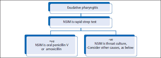
8.5.1 Acute Exudative Pharyngitis or Tonsillitis


Background: Two most common causes of exudative pharyngitis/tonsillitis are Epstein–Barr virus (EBV) and strep.


10 Most common etiology of nonexudative pharyngitis/ tonsillitis, however, is viral.

Ruling out S. pyogenes infection is important because if not treated promptly, it carries an increased risk of rheumatic fever or glomerulonephritis.


No Image Available!


Streptococcal pharyngitis


EBV


Acute HIV syndrome


Viral


Tonsillar exudate


Present


Present


Usually absent


Usually absenta


Upper respiratory symptoms


-ve


May be present


Usually absent


Presentb


Generalized lymphadenopathy


-vec


May be present


Present


-vec


Hepatosplenomegaly


-ve


Present


Present


-ve


Rash


-ve


May be presentd


May be presente


-ve


aHowever, adenovirus or herpes virus pharyngitis may have exudates.


bCoexistent symptoms such as nasal congestion, dry cough, conjunctivitis pointing toward viral origin.


cUsually associated with local cervical lymphadenopathy, but not generalized.


dMaculopapular rash, if present, signifies that patient might have been exposed to antibiotics (e.g., amoxicillin) prior to presentation.


ePatients with acute retroviral syndrome may have painful mucocutaneous ulcerations (oral, esophageal, penile, etc.) and generalized well-circumscribed salmon-colored macules or papules.


Other causes of sore throat include the following:


  • Mononucleosis-like illness; see below.


  • Corynebacterium diphtheriae: sticky dense gray pseudomembrane covering tonsils.


  • Gonococcal pharyngitis: due to oral sex.


8.5.2 Infectious Mononucleosis


Background: EBV is transmitted by salivary secretions, e.g., during kissing (hence, especially prevalent in college students) .


Diagnostic evaluation:


  • NSIM/NSIDx is rapid strep throat test. If negative, NSIDx is CBC with peripheral blood smear, and heterophile antibody test (a.k.a. monospot test).


    11 Monospot test detects the presence of antibody against RBC antigen of sheep and horse. This test is both very sensitive and specific; however, it can be negative in early infection.


  • If monospot test is negative, NSIDx is either repeating monospot test or serologic testing for antibodies against EBV-specific proteins (viral-capsid antigen or early nuclear antigen).


    12 If both monospot test and antibodies against EBV-specific proteins are negative, then consider the following causes:


    CMV: Known as heterophile-negative infectious mononucleosis


    HIV: Acute retroviral syndrome


    Toxoplasmosis


  • Peripheral smear may reveal atypical lymphocytes.


Treatment: Supportive (acetaminophen ± NSAIDs).


Consider steroids if any of the following is present:


  • Impending airway obstruction


  • Liver failure


  • Aplastic anemia


  • Significant autoimmune hemolytic anemia or thrombocytopenia.


Complication: Splenic rupture (patients are advised to avoid non-contact sports for 3 weeks and contact sports for at least 4 weeks).


Clinical Case Scenarios

A 24 y/o college-going male patient presents with exudative pharyngitis, posterior cervical lymphadenopathy, and fatigue for the last 10 days.


  1. What is the immediate NSIDx?


  2. If rapid strep test is negative, what test should be done next?


  3. What test would likely confirm the dx?


  4. Heterophile antibody test comes back positive. CBC reveals platelet count of 18,000. What treatment would you consider?


MRS

They CHAT like EBV. They can also have atypical lymphocytes. CHAT or EBV infection can cause Glandular fever syndrome = triad of fever, pharyngitis, and generalized lymphadenopathy.


8.5.3 Influenza


Presentation: Fever, myalgias (generalized muscle pain), headache, and upper respiratory symptoms, such as runny nose and nonproductive cough (due to the postnasal drip). Chest X-ray (CXR) may show increased interstitial marking due to viral bronchitis.


Workup: Rapid molecular DNA-based testing is preferred (e.g., RT-PCR). Antigen-based detection has lower sensitivity, but can also be used.


Rx: Antivirals (Oseltamivir or Zanamivir). Indication for antiviral treatment has been broadened. The only group that might not be treated is healthy patients < 65 years of age with uncomplicated influenza who present after >48 hours of symptoms, or who are already feeling better or pediatric population.


Complications: Adult respiratory distress syndrome, severe necrotizing pneumonia due to S. Aureus.


In a patient presenting with fever and cough +/-CXR infiltrates, order rapid COVID-19 PCR.


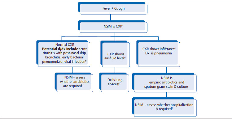
8.6 Lower Respiratory Tract Infection


Common differential dignosis of “fever + cough” are acute bacterial sinusitis with postnasal drip, bronchitis, pneumonia, and lung abscess.


No Image Available!


aDo not choose CBC as the NSIM, because it does not help to make diagnosis.


bIn hospitalized patients with hx of sick contacts or coexistent upper respiratory tract symptoms, a PCR testing that detects common viruses (e.g., RSV, parainfluenza, influenza) can be done.


cYoung patients without comorbid conditions and low-grade fever may not need antibiotics.


Empiric antibiotics used for treatment of bronchitis are similar to sinusitis (same microbiologic cause): amoxicillin-clavulanate or doxycycline.


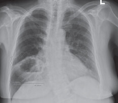
No Image Available!


dCXR above shows air-fluid level (lung abscess).


No Image Available!


eLobar pneumonia with multiple air bronchograms and absence of significant tracheal deviation (If there was lung opacity similar to this but with tracheal deviation to the same side and no air bronchogram, the diagnosis would be more likely atelectasis).


Source: Imaging. In: Gunderman R, ed. Essential Radiology. Clinical Presentation, Pathophysiology, Imaging. 2nd ed. Thieme; 2000.


fLung abscess is commonly a sequelae of aspiration (look for risk factors of aspiration, e.g., advanced dementia, alcoholism). NSIM is sputum gram stain/culture and CT thorax to rule out obstructive pathology. Empiric antibiotics should cover anaerobes (e.g., amoxicilin-clavulanate). Antibiotics is continued until chest imaging is clear (might take weeks).


gIf you think the patient is sick (e.g., septic, hypoxemic, and/or immunocompromised), then NSIM is to hospitalize the patient and give IV antibiotics (after taking blood cultures). There is a CURB-65 criteria to help you make that decision too.


If any of the following present, consider hospitalization:


Confusion


Urea ≥ 20 mg/dL


Respiratory rate ≥ 30/min


Blood pressure—systolic BP < 90 mmHg


• 65 or older aged patient (particularly if patient lives alone).


MRS

CURB-65


8.6.1 Pneumonia


Classification of pneumonia


Definition


Likely organism


Community-acquired


Patient living in community and with no risk factor for infection with multidrug-resistant organisms


Streptococcus, H. influenzae, mycoplasma, etc. (see table below for empiric therapy)


Hospital-acquired pneumoniaa


Patient develops pneumonia after more than 48 hours of hospitalization


Empiric treatment:


  • Cover gram-negative organisms and Pseudomonas, e.g., cefepime, or piperacillin-tazobactam.b


  • If facility has higher rates of MRSA, add coverage with IV vancomycin or linezolid.


  • No need to routinely cover atypical organisms.


Ventilator-associated pneumonia


Intubated patient develops new-onset fever and has increased secretions, more than 48 hours after intubation.


aHealth care-associated pneumonia (HCAP) is no longer included in recent guidelines. Treatment depends upon individual risk factors.


bConsider double pseudomonal coverage in very sick patients (e.g., Tobramycin + cefepime).


Empiric Antibiotic Treatment for Community-Acquired Pneumonia


No recent antibiotic use, CURB-65 is absent, AND patient has no comorbidities


Hospitalized patient, recent antibiotic use, OR presence of comorbidities (e.g., diabetes, alcoholism, chronic liver disease, immunosuppression)


  • Azithromycin or clarithromycin (DOC)


    or


  • Doxycycline


  • A third-generation cephalosporin + either doxycycline or macrolides.


    OR


  • β-Lactam + β-lactamase (e.g., amoxicillin-clavulanate) + either doxycycline or macrolides.


    OR


  • New-generation quinolones like moxifloxacin or levofloxacin.a


aOlder generation quinolones like ciprofloxacin, norfloxacin, or ofloxacin do not cover Streptococcus.


Test commonly done for hospitalized patients with pneumonia


  1. Sputum gram stain and culture


  2. Chest X-ray


  3. Respiratory pathogen panel (a DNA/RNA based PCR test) which detects multiple respiratory pathogens including influenza, RSV, chlamydia and mycoplasma pneumonia.


  4. Urine legionella and strep-tococcal antigen


Specific Clinical Situations Involving Pneumonia


Streptococcus pneumonia is the MCC of pneumonia.


Community-acquired pneumonia +


Causative organism


Additional points


Lobar pneumonia (CXR reveals infiltrates localized to a specific region)


S. pneumoniae


  • Rusty colored sputum.


  • Urine Streptococcus antigen testing


Klebsiella (gramnegative bacteria)


  • Risk factors: alcoholism, advanced age, significant comorbid conditions (e.g., ESRD, chronic liver disease), etc.


  • Klebsiella has a very thick sputum which is classically called currant jelly sputum.


  • Culture of sputum may show mucoid colonies.


Presence of the following coexistent features:


  • Symptoms of upper respiratory tract infection– pharyngitis, rhinitis, and/or bullous otitis media.


  • Intravascular immunoglobulin M (IgM)-mediated hemolysis.


  • Target-like skin rash (erythema multiforme).


Mycoplasma pneumonia


CXR usually shows diffuse bilateral streaky interstitial infiltrates


No Image Available!


Source: Clinical Aspects. In: Galanski M, Dettmer S, Keberle M et al., eds. Direct Diagnosis in Radiology. Thoracic Imaging. 1st ed. Thieme; 2010.


Most patients are treated empirically with antibiotics that cover atypical organisms (e.g., azithromycin). If diagnosis is sought, may use PCR of respiratory secretions or serum antimycoplasma antibody (Note: Cold agglutinins are neither sensitive nor specific).


MRS

Think of old drunk smoking legionnaires


Community-acquired pneumonia +


Causative organism


Additional points


Presence of the following co-existent features:


  • Confusion, headache


  • Diarrhea, abdominal pain


  • LFT abnormalities


  • Hx of going to a cruise or attending a convention (Legionella is frequently found in infected water resources such as in air-conditioning systems)


  • Hyponatremia (due to SIADH)


Legionella pneumonia


Risk factors: smokers, alcoholics, old age
CXR: single or multifocal infiltrates (usually involving bilateral lower lung fields)
NSIM/NSIDx: urine Legionella antigen test or sputum PCR. Sputum culture on special media is the most specific test.
Rx: DOC is respiratory quinolone (e.g., levofloxacin) or azithromycin.


Recent hx suggestive of influenza; now patient feels sicker and is having high fever.


S. aureus pneumonia


Chest imaging may reveal nodular cavities and pneumatoceles (multiple thin-walled cavities).


No Image Available!


Source: Differential Diagnosis. In: Galanski M, Dettmer S, Keberle M et al., eds. Direct Diagnosis in Radiology. Thoracic Imaging. 1st ed. Thieme; 2010.


Other causes of post-flu pneumonia include Streptococcus pneumonia and H. influenzae.


  • Recurrent pneumonia in the same lobe or segment


  • Pneumonia that fails to respond to adequate antibiotic therapy


Postobstructive pneumonia.


NSIM is Chest CT scan to rule out obstructive mass.


Hx of bird exposure


Chlamydia psittaci


DOC is doxycycline


Hx of animal exposure (e.g., sheep, cattle)


Coxiella burnetii


DOC is doxycycline


Abbreviation: LFT, liver function test.


Clinical Case Scenarios

10. A 65-year-old alcoholic, active smoker patient who recently went to a convention has pneumonia. What is the likely cause?


  1. Strep. pneumonia


  2. Legionella


  3. Klebsiella


Additional Clinical Pointers


No Image Available!


8.6.2 Aspiration Pneumonia/Pneumonitis


Pathophysiology


Typical clinical case progression


Aspiration pneumonitis


Aspiration of sterile gastric acid contents and subsequent transient inflammatory response, without development of infection


25 y/o M presents with acute gastroenteritis and significant vomiting. Day 2, he develops low-grade fever and has small new infiltrates on CXR. Day 4, patient feels great and wants to go home.


Aspiration pneumonia


Aspiration of oropharyngeal contents with accompanying anaerobic flora of oral cavity, resulting in infection of lung parenchyma


65 y/o M, day 2 after surgery, develops productive cough and fever. CXR shows lobar opacity. Day 4, he still looks sick and is continued on IV antibiotics.


Conditions that can predispose to aspiration:


Impaired swallowing and epiglottic reflex lead to increased risk of aspiration of oropharyngeal and gastric contents into the lung

Dec 11, 2021 | Posted by in INFECTIOUS DISEASE | Comments Off on 8. Infectious Disease

Full access? Get Clinical Tree

Get Clinical Tree app for offline access