(1)
State University of New York at Buffalo Kaleida Health, Buffalo, NY, USA
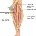
The arm has two compartments, anterior and posterior. The medial aspect of the arm is the area occupied by the neurovascular bundle of the arm, proceeding from the area of the axilla to that of the elbow. Tumors can arise from the tissues within the neurovascular bundle, or tumors in the anterior or posterior compartment can encroach upon the area of the neurovascular bundle. It is therefore important to be familiar with the dissection and exposure of the neurovascular bundle in medial arm.
Case 1: A Benign Neurilemmoma
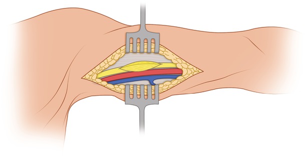
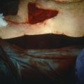
In this patient, a nodule was noted in the medial aspect of the arm with symptoms referable to the distribution of the median nerve. The incision over the swelling was longitudinal (Fig. 9.1), as longitudinal incisions generally are preferable in extremities because they provide greater versatility in achieving satisfactory exposure. The deep fascia was incised (Fig. 9.2), preferably using a scalpel or light cautery. With further dissection after the deep fascia was incised, the structures of the neurovascular bundle were exposed by incising their sheath, revealing the median nerve, the brachial artery, and the basilic vein (Fig. 9.3). In this case, there was a distinct, localized swelling of the exposed median nerve. In an attempt to dissect between the sheath of the nerve and the swelling, the nerve sheath was incised longitudinally where the nodule was closest to the surface. A gelatinous nodule was exposed and dissected around on its surface so as to preserve the adjacent nerve fibers of the median nerve (Fig. 9.4). The use of a nerve stimulator is important in these cases so as to be able to identify and preserve any strands that contain motor nerve fibers and separate them from the nodule, which the pathology report showed to be a benign neurilemmoma. (It is important to note that some of the presumed nerve sheath may contain motor nerve fibers.) Simple excision of this benign neurilemmoma was sufficient for control of the tumor in that area (Fig. 9.5). Neurilemomas encountered in other anatomical areas are treated similarly. They are truly encapsulated neoplasms and are almost always solitary. Closely related benign tumors, the neurofibromas, are not usually encapsulated and may result in diffuse, tortuous enlargement of the involved nerve (plexiform neurofibroma). They are either solitary or multiple (neurofibromatosis). Occasionally, neurofibromas may contain a microscopic focus of neurofibrosarcoma in the center of a mass, but these seem to do well on long-term followup if the bulk of the mass is benign.
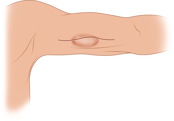
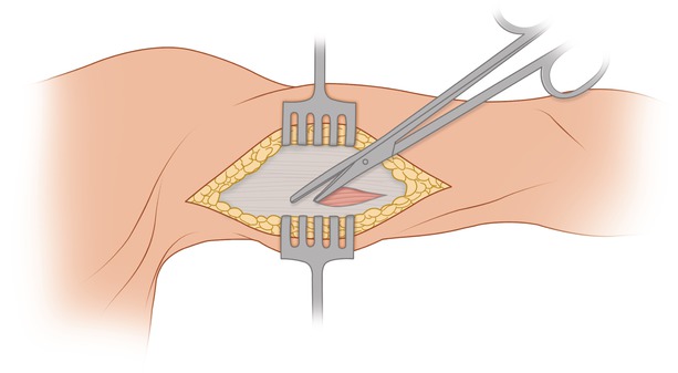
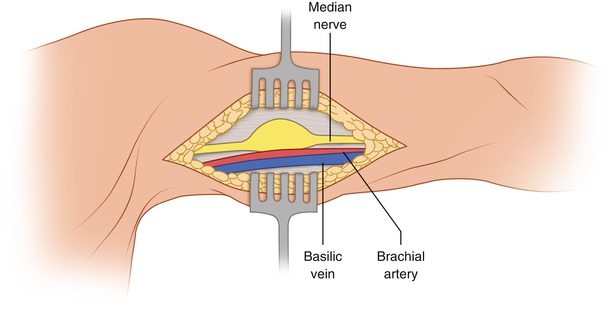
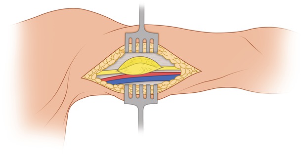

Fig. 9.1
Swelling in the medial aspect of the arm is observed. The proposed longitudinal incision is centered over the protuberant portion of the mass

Fig. 9.2
The investing fascia is incised

Fig. 9.3
The median nerve and brachial artery are exposed. The swelling due to the nodule involves the median nerve

Fig. 9.4
The nerve sheath is opened and the gelatinous lump is easily dissected free and removed

