(1)
State University of New York at Buffalo Kaleida Health, Buffalo, NY, USA
The most superficial and bulkiest muscle in the buttock area, the gluteus maximus, is more often involved by sarcoma than the other glutei. This muscle originates from a small area in the posterior part of the ilium, the aponeurotic layers covering the sacrum on its lateral side, and the intermuscular septum separating it from the gluteus medius. It inserts in the iliotibial band and in the gluteal tuberosity on the posterior surface of the femur, extending from the upper end of the linea aspera to the lower end of the greater trochanter.
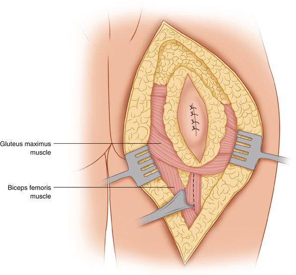
The patient is placed in a lateral or a prone position. The preferred incision is one that extends from the posterior superior iliac spine obliquely toward the middle of the space between the greater trochanter and the ischial tuberosity and then continues into the posterior thigh for several centimeters below the gluteal crease. In patients who have had a previous biopsy, the incision is made elliptical in order to circumscribe the previous biopsy incision (Fig. 46.1). Some authors have described an incision for resection of tumors in the gluteus maximus along the same line as the direction of its fibers, centering the incision over the middle of the muscle. This produces a nearly transverse incision extending from the posterior superior iliac spine obliquely to the greater trochanter [1]. This incision is not sufficient to resect the entire gluteus maximus, nor does it provide for the early identification and dissection of the sciatic nerve.
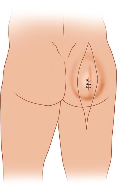

Fig. 46.1
An elliptical incision is made around a previous biopsy incision, with the longitudinal diameter slightly oblique from the posterior superior iliac spine, soon to become vertical into the upper posterior thigh
Flaps are then developed anteriorly to the surface of the gluteus medius and posteriorly to the lateral edge of the sacrum. Anteriorly, the fascia is incised, the gluteus medius is exposed, and the plane between the gluteus medius and maximus is entered. Of course, if the tumor extends to involve the gluteus medius, the appropriate portion or the entire gluteus medius may also be resected en bloc with the gluteus maximus. The incision is deepened between the ischial tuberosity and the greater trochanter and below the edge of the gluteus maximus through the fascia in the midline of the posterior thigh, exposing thus the posterior compartment of the thigh. At this level, the hamstring muscles are located medially, arising from the ischial tuberosity (Fig. 46.2). The greater trochanter, of course, is located laterally, and between the tuberosity and trochanter the sciatic nerve is identified and dissected so that it can be traced proximally. The posterior cutaneous nerve of the thigh is exposed and usually is divided. It is a sensory nerve, which issues from the sciatic nerve and proceeds on the deep surface of the gluteus maximus and then along the middle of the posterior aspect of the thigh (under and close to the fascia lata) to the popliteal fossa, providing sensory supply to the skin. Often it is sacrificed in order to obtain exposure and adequate margin around the tumor mass occupying the gluteus maximus. Having exposed the sciatic nerve proximally for as far as is possible by raising the inferior edge of the gluteus maximus, one then is free to start dividing the gluteus maximus at its origin from the ischial tuberosity and the edge of the sacrum medially and laterally off the gluteal tuberosity and the insertion to the iliotibial band (Fig. 46.3). As the lower edge of the gluteus maximus is thus mobilized, it is possible to dissect the sciatic nerve further, all the way to the point where it goes beneath the piriformis to enter the pelvis. The gluteus maximus continues to be divided medially next to the edge of the sacrum and off the ischial tuberosity and sacrotuberous ligament (Fig. 46.4). On the lateral side, it is dissected off the gluteus medius muscle, and superiorly the muscle is detached off the posterior portion of the iliac bone, from which a small portion of the gluteus maximus arises. In the middle of the gluteus maximus in relation to its deep surface, one encounters the inferior gluteal nerve from the sciatic, as well as the inferior gluteal vessels (branches and tributaries of the internal iliac vessels), which must be ligated and divided. They both issue from the inferior edge of the piriformis muscle and enter the deep surface of the gluteus maximus. Above the piriformis, through the suprapiriformal opening, the superior gluteal vessels enter the area of the buttock and supply primarily the gluteus medius and minimus. However, a small branch, which proceeds in a posterior direction to supply the gluteus maximus, must be ligated and divided. When the tumor is close to the origin of the gluteus maximus from the sacrotuberous ligament, the sacrotuberous ligament is divided medially at the sacral border and more laterally off the ischial tuberosity. A portion of this tuberosity is also removed if the tumor is close to its surface. Removal of the sacrotuberous ligament does not cause a problem in terms of postoperative function. However, on entering the ischiorectal fossa, one can see the pudendal nerve and vessels on the surface of the fascia covering the obturator internus. Usually, two suction drains are placed in the wound and the skin edges of the flaps are trimmed, in order to avoid postoperative necrotic edges where they are thinnest, provided that the edges can still be approximated without undue tension.
