(1)
State University of New York at Buffalo Kaleida Health, Buffalo, NY, USA
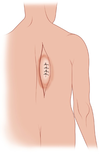
For tumors located in the back that are close to the vertebral spine, a longitudinal, vertical, paraspinal elliptical incision (which includes a biopsy incision) is preferable, particularly when the tumor mass is longitudinally disposed (Fig. 18.1). (A T incision consisting of a vertical and horizontal component may occasionally be required.) The medial flap is raised to beyond the spinous processes. Laterally, the flap is raised to well beyond the palpable extent of the tumor. The deep fascia is incised (Fig. 18.2). Superiorly and laterally, the trapezius muscle is encountered as the first layer and is divided. Depending on the location of the tumor in the inferior part of the wound, the latissimus dorsi is exposed and divided around the tumor (Fig. 18.3). Medially, the aponeurotic layer next to the supraspinous ligament is divided in the plane between the spinous processes and the sacrospinalis muscle (Fig. 18.4). With further dissection, the sacrospinalis muscle is separated from the spinous processes to the surface of the lamina. The rhomboid muscles at the level medial to the scapula are divided (Fig. 18.5). Dissection on the surface of the intercostal muscles and ribs then is carried out to some extent, in order to evaluate the proximity of the tumor to the fascia covering the intercostal muscles. If there is any evidence of fixation of the tumor to the rib surface laterally, the involved ribs are divided and the pleura is entered. To assess the situation, a 2-cm segment of each divided rib is removed, allowing digital exploration of the inner side of the chest wall (Fig. 18.6). The sacrospinalis muscle is divided both above and below the tumor. The involved ribs, which have already been divided laterally, are disarticulated medially and the specimen is removed (Fig. 18.7).
