Tuberculosis


Tuberculosis


Jonathan E. Golub

Jacqueline S. Coberly

Richard E. Chaisson


INTRODUCTION

The ancient Greeks called it phthisis, the Romans tabes, and the Hindus rajayakshma; in Victorian England, it was known as consumption.1 All of these names referred to the wasting illness that is characteristic of the disease we now call tuberculosis (TB). Tuberculosis is a complex communicable disease of humans caused by the tubercle bacilli, a group of genetically related mycobacteria also known collectively as the Mycobacterium tuberculosis complex. The tubercle bacilli are a group of slow-growing mycobacteria that include M. tuberculosis, M. africanum, M. canettii, M. bovis, and M. microti. The first three members of this group are, at least to date, strictly human pathogens.

M. bovis causes illness in a variety of animals as well as in humans.2, 3 and 4 Similarly, M. microti generally causes disease in rodents, but has been linked retrospectively to infections in llamas, ferrets, and cats. It has also been implicated as the cause of pulmonary tuberculosis in a small number of humans.5 With the advent of effective drug treatment in the 1950s and preventive therapy (or chemoprophylaxis, as it was then called) in the 1960s, many in the medical and public health communities, particularly in industrialized countries, assumed tuberculosis was conquered. This hubris led to several decades of neglect by the biomedical community, during which control efforts were ignored or deliberately weakened.6 Unfortunately, the economic, social, and public health factors that foster the propagation of tuberculosis had not been eliminated, not even from the industrialized nations. Thus, in the 1980s and 1990s, as the deterioration of control programs coincided with the burgeoning epidemic of human immunodeficiency virus (HIV), tuberculosis rebounded. Numerous outbreaks were seen in the larger cities of the United States that had the highest incidence of HIV.7 More serious, however, was the fact that in some areas of the developing world where tuberculosis and HIV are both endemic, the incidence of tuberculosis doubled, and healthcare facilities became overwhelmed by the dual epidemic.6

The World Health Organization (WHO) declared tuberculosis to be a global emergency in 1993.6 Eighteen years later, considerable progress has been made in managing this infection, but tuberculosis remains a leading cause of premature death in young adults around the world.8 Roughly one third of the world’s inhabitants are latently infected with M. tuberculosis. WHO has estimated that 8.8 million people developed tuberculosis and 1.45 million died from it in 2010, representing a significant decline from the 9.4 million cases reported in 2009.8 Efforts to promote tuberculosis control have more than doubled since 1993, and the tuberculosis incidence rate has continued to decline annually since 2002 when it peaked at 142 cases per 100,000 population. Moreover, TB mortality has fallen by more than a third since 1990 and all WHO regions, other than the African Region, are on track to halve their 1990 mortality rates by 2015. Control efforts, highlighted by the DOTS (directly observed therapy, short course)/Stop TB Strategy, have saved almost 7 million lives by successfully treating 46 million of 55 million TB patients treated in DOTS programs.8 The global success has been driven to a significant extent by China’s overwhelming achievements since 1990. That country has seen an annual 3.4% decline in its TB incidence, a halving of prevalence, and an 80% reduction in TB mortality.8

Despite this success, China and India account for one third of the world’s TB cases, with the primary drivers of the epidemic in these countries being social determinants such as tobacco exposure, diabetes, malnutrition, alcohol abuse, and indoor air pollution.9 Meanwhile, in Africa, the high prevalence of
latent TB infection and HIV infection, a high prevalence of multidrug-resistant TB, and the complex epidemiology and natural history of tuberculosis continue to make control of the disease on this continent particularly challenging.10


THE ORGANISM

The family Mycobacteriaceae, of the order Actinomycetales, is composed of a number of slow-growing, acid-fast bacilli. Most are saprophytes— useful inhabitants of soil and water that fix nitrogen and help degrade organic material. Some are pathogens in animals and occasionally cause opportunistic infection in humans.11, 12 Only four species are highly pathogenic in humans: Mycobacterium leprae (which causes leprosy) and three of the tubercle bacilli. The tubercle bacilli, or Mycobacterium tuberculosis complex, comprise a group of five closely related mycobacteria that cause tuberculosis (Table 18-1). M. tuberculosis, M. africanum, and M. bovis are the most common cause of human tuberculosis, although M. bovis is also known to cause disease in a variety of animal species. The other two members of the complex, M. canettii and M. microti, do cause tuberculosis in humans, albeit infrequently.1, 3, 4 and 5, 12 In fact, M. microti has been identified as a human pathogen only relatively recently.5 Although they vary widely by favored host, phenotype, and pathogenicity, the
bacteria that make up the M. tuberculosis complex share more than 90% of their genome and have identical 16S rRNA sequences.2, 13 With the advent of the HIV epidemic, several other mycobacteria—most notably, M. avium complex (MAC)— have emerged as common opportunistic pathogens and, in people infected with HIV, cause illness that is clinically similar to disseminated tuberculosis.1, 13


Table 18-1 Species of Mycobacteria


Microbe


Reservoir


Clinical Manifestation


Always pathogenic in man


M. tuberculosis


Man


Pulmonary and disseminated TB


M. bovis


Cattle, man


TB-like disease


M. leprae


Man


Leprosy


M. africanum


Man, monkey


Rarely TB-like pulmonary disease


Potentially pathogenic in man


M. avium complex


Soil, water, birds, fowl, swine, cattle, and environment


Disseminated and pulmonary TB-like disease


M. canettii


Man, possibly others


Rarely TB-like pulmonary disease


M. microti


Rodents, llamas, cats, ferrets, and possibly man


Rarely TB-like pulmonary disease


M. kansasii


Water, cattle


TB-like disease


Uncommon or rarely pathogenic in man


M. genavense


Possibly man and pet birds


Blood-borne disease with AIDS


M. haemophilum


Unknown


Subcutaneous nodules and ulcers primarily with AIDS


M. malmoense


Environment, possibly others


Adults: TB-like pulmonary children: Lymphadenitis


M. marinum


Fish, water


Skin infections


M. scrofulaceum


Soil, water


Cervical lymphadenitis


M. simiae


Monkey, water


TB-like pulmonary and disseminated disease with AIDS


M. szulgai


Unknown


TB-like pulmonary disease


M. ulcerans


Man, environment


Skin infections (Buruli ulcer)


M. xenopi


Water, birds


TB-like pulmonary disease


Adapted from Brooks GF et al, editors. Jawetz, Melnick & Adelberg’s Medical Microbiology, 21st Edition. Stamford: Appleton & Lange: 1998; and Niemann S et al. (2000). Two cases of Mycobacterium microti-derived tuberculosis in HIV-negative immunnocomptent patients. Emerging Infectious Diseases 6(5): 539-42; and Pffyer, G et al (1998). Mycobacterium canettii, the smooth variant of M. tuberculosis, isolated from a Swiss patient exposed in Africa. Emerging Infectious Diseases 4(4): 631-4.


The bacteria that make up the M. tuberculosis complex are slender, slightly curved, rod-shaped bacteria averaging 4 by 0.3 µm in size.1, 3, 13 M. tuberculosis is strictly aerobic; in contrast, M. bovis is microaerophilic and adapts more easily to nonpulmonary sites of infection. Like other mycobacteria, the tubercle bacilli have an unusual concentration of high-molecular-weight lipids in their cell wall, accounting for approximately 50% of their dry weight. This high lipid content makes these organisms hydrophobic and resistant to aqueous bactericidal agents and drying. It is also responsible for their acid-fast nature, a characteristic that is essentially synonymous with mycobacteria.3, 13


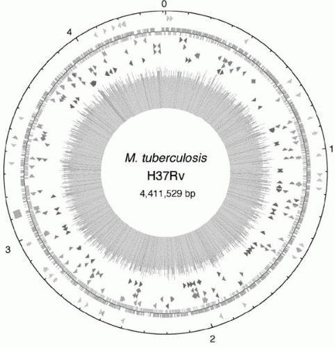
Figure 18-1 Circular map of the chromosome of M. tuberculosis H37Rv. The outer circle shows the scale in Mb, with 0 representing the origin of replication. The first ring from the exterior denotes the positions of stable RNA genes and the direct repeat region; the second ring inward shows the coding sequence by strand; the third ring depicts repetitive DNA; the fourth ring shows the positions of the PPE family members; the fifth ring shows the PE family members; and the sixth ring shows the positions of the PGR5 sequences. The figure was generated with software from DNASTAR. Reprinted by permission of Macmillan Publishers Ltd: Nature. Cole ST, et al. Deciphering the biology of mycobacterium tuberculosis from the complete genome sequence. June 11; 393(6685):538. Copyright © 1998.

Mycobacteria are slow growing and fastidious in culture. Indeed, because M. tuberculosis has a very long generation time (approximately 24 hours), culture is a slow process, often resulting in diagnostic delays and sometimes misdiagnosis.5 Traditionally mycobacteria are grown on solid, enriched media, where colonies appear 4 to 6 weeks after inoculation. They can also be grown in liquid culture, where they form characteristic strings that can be seen by light microscopy. Rapid liquid culture systems (e.g., BACTEC) have been adapted for use with mycobacteria and allow identification of organisms in as little as 9-16 days, depending on the concentration of microbes in the specimen being tested.3, 14 DNA probes speed speciation of organisms following growth. Alternatively, a number of biochemical tests can be used to speciate mycobacteria, though these approaches are time consuming.3, 15

In 1998, a consortium of scientists deciphered and published the genome map of the H37Rv strain of M. tuberculosis (Figure 18-1). The complete genome was revealed to be 4,411,529 base pairs
long and contains approximately 4000 genes.16 Since then, advances in genotyping technology have been rapidly applied to studies of the molecular epidemiology of the tubercle bacilli. Genotyping methods have been rapidly adopted, including restriction fragment-length polymorphism (RFLP), polymerase chain reaction (PCR)-based spoligotyping, and profiling of the mycobacterial interspersed repetitive units based on the number and size of the variable number tandem repeats in the genome (MIRU-VNTR). Most recently, the application of whole-genome sequencing analysis has been shown to be optimal, though this technique remains too costly for universal use. These methods vary in sensitivity and specificity, so some caution is needed when interpreting results.18

Genotyping studies are providing some interesting insights into the epidemiology of tuberculosis. For example, 15 years ago it was dogma that M. tuberculosis was a mutated form of M. bovis; the assumption being that around 7000-4000 BCE, when humans began domesticating animals, they were exposed to M. bovis, which, over time, mutated into a human pathogen.19 In a recent fingerprinting study, however, the genomes of 100 strains of M. tuberculosis complex were mapped and compared. The genetic lineage developed from these analyses provides evidence that human M. tuberculosis is not a mutation of M. bovis; rather, both bacteria diverged from a common ancestor long before either infected humans2, 13 (Figure 18-2).


Figure 18-2 Scheme of the proposed evolutionary pathway of the tubercle bacilli illustrating successive loss of DNA in certain lineages (gray boxes). The scheme is based on the presence or absence of conserved deleted regions and on sequence polymorphisms in five selected genes. Note that the distances between certain branches may not correspond to actual phylogenetic differences calculated by other methods. The top five dark arrows indicate that strains are characterized by katG463. CTG (Leu), gyr A95 ACC (Thr), typical for group 1 organisms. The bottom seven arrows indicate the strains belong to group 2 characterized by katG463 CGG (Arg), gyrA95 ACC (Thr). The light gray arrow indicates that strains belong to group 3, characterized by katG463 CGG (Arg), gyrA95 AGC(Ser), as defined by Sreevatsan et al. Reprinted from Brosch, R, AS Pym, SV Gordon, ST Cole. The evolution of mycobacterial pathogenicity: clues from comparative genomics. Trends Microbiol. Sep;9(9):452-8. Copyright © 2001, with permission from Elsevier.

DNA fingerprinting is also being used with traditional field epidemiology to help link index and secondary cases and to distinguish active disease resulting from reactivation versus recent transmission. When distinguishing between reactivation and recent transmission, the presumption is that the genotype of cases due to reactivation will not match the genotype of other cases in the community because the infection was acquired at some distant point in the past. Conversely, in cases involving recent infection, the genotype of the organism should be shared with at least the index case and probably other cases in the community resulting from the same index case. Note, however, that the “orphan” isolates that do not share a genotype with any other organism from the community could still be related to other cases
in the community, but the link cannot be identified with traditional epidemiology; this can happen when the index case is sputum negative, or when the DNA fingerprint of the index is obtained for other reasons. Nevertheless, cluster analysis is fairly reliable and has provided new and interesting epidemiologic information about tuberculosis.

Before fingerprinting was commonly available, it was believed that most infections in immunosuppressed people in low-prevalence areas represented reactivation of latent infection. The populations at risk were often transient, and linking cases epidemiologically was difficult. More recently, DNA fingerprinting studies of community cohorts have identified some surprising clusters and helped guide the shoe-leather epidemiology needed to confirm the linkages suggested by fingerprint evidence.20 Fingerprinting studies have also helped characterize the global distribution of genotypes, clarify patterns of transmission in communities, detect laboratory transmission, and prioritize control activities.21, 22, 23, 24, 25 and 26 A U.S. study helped dispel the dogma that TB can be transmitted only through prolonged contact, and that casual transmission frequently occurs.27 DNA fingerprinting is also being used to investigate the origins of susceptibility to the tuberculosis bacilli and innate host immunity to it.18, 28, 29


HISTORY


Evolution of the Tubercle Bacilli

Mycobacteria are ancient organisms that probably first appeared more than 1 million years ago in soil and water and gradually adapted to animal hosts during the Paleolithic period*.19, 30 Until recently, it was hypothesized that when humans began domesticating cattle, they began to be exposed to M. bovis, which eventually mutated into the human pathogen M. tuberculosis. A recent comparison of the genotype of 100 tubercle bacilli showed, however, that M. bovis, M. tuberculosis, and the other members of the M. tuberculosis complex all diverged from a common ancestor through successive loss of DNA (Figure 18-2).2

Regardless of the evolutionary path of the tubercle bacilli, at some point in early prehistory tuberculosis became pathogenic for humans. Skeletal remains of Neolithic humans with deformities suggestive of tuberculosis have been found in Germany, France, Italy, Denmark, and Jordan and are dated from 8000 to 5000 BCE.1, 31 It cannot be conclusively proved that these deformities are the result of infection with the tubercle bacillus, as the organism cannot be cultured from the bone. Nevertheless, the deformities are strongly suggestive of spinal tuberculosis or Pott’s disease.1, 19

Traces of tuberculosis-like disease have also been identified in Egyptian mummies dating from 3500 to 400 BCE.3, 30 One particularly well-preserved mummy of a 5-year-old boy from about 1000 BCE was discovered with the lungs intact. Again, the causative organism could not be conclusively identified, but a diagnosis of tuberculosis was made based on the observation of pleural adhesion, blood in the trachea, and the presence of acid-fast bacilli in the lung tissue.3 Egyptian artifacts from this period also begin to show people with the spinal deformities characteristic of Pott’s disease.3

Based on available evidence, tuberculosis appears to have initially been a sporadic disease in humans. This is difficult to verify, however, because relatively few large collections of skeletal remains of prehistoric human are available for examination. What is known with certainty is that as civilization developed and people began to gather together in ever increasing numbers, reports of tuberculosis became more numerous, leading to the establishment of tuberculosis as an endemic disease of humans by the beginning of the first millennium.1, 19


Global Evolution of the Epidemic

By CE 100, tuberculosis was well established in the Mediterranean states and Western Europe. It remained relatively sporadic for centuries, until people began settling in larger communities.19 The advent of the Industrial Revolution and the great migrations to the cities that followed created an ideal environment for the spread of tuberculosis.30, 32 In 1662, 1 of every 6 northern Europeans had tuberculosis; 100 years later, this figure had doubled.3 Tuberculosis was so common that most of the population became infected, and 25% of all deaths were attributed to tuberculosis.19 Thomas Sydenham was quoted in 1682 as saying, “Two thirds of those who die of chronic diseases … [are] … killed by phthisis.”32 The epidemic peaked in England in 1780s and in Western Europe by 1800.30

Although sporadic, M. bovis infections were not uncommon in the largely agrarian societies of the New World. However, it was the English and European colonists who are mostly likely responsible for spreading M. tuberculosis throughout the world.


Tuberculosis came to North America on the Mayflower and was well established in the colonies by the early 1700s.1, 30 The epidemic passed through the United States in a wave, pushing south and west with the spread of industrialization, and by the early 1900s it was endemic in North America.30 Tuberculosis was also spread to South America by colonists, but because the Spanish quarantined consumptives in the 1600s and 1700s, its introduction in that region was slowed.1 Tuberculosis also spread into Eastern Europe, Asia, and Africa from Western Europe. The epidemics in Russia came in the late 1800s and those in Asia in the early 1900s. Tuberculosis was still largely unknown in Africa at the beginning of the 1900s and spread slowly through the interior with the colonizing Europeans.1, 19

Herd immunity to tuberculosis develops slowly because of the long incubation period of the TB organism. Differences in the epidemiology in different parts of the world may be related to when tuberculosis was introduced to the population, with the most serious disease present in Africa where tuberculosis was introduced latest, leaving the least amount of time for herd immunity to develop.19, 33


CLINICAL MANIFESTATIONS

As discussed in more detail later, tuberculosis begins with latent infection that can progress to active disease. Latent tuberculosis infection (LTBI) causes no symptoms, and most latently infected people are unaware that they harbor tubercle bacilli. Tuberculosis disease generally affects the lungs and respiratory tract, but can strike nearly any organ system in the body. Both primary and reactivation tuberculosis disease can result in pulmonary or extrapulmonary manifestations. In immunocompetent people, approximately 80% of tuberculosis is pulmonary, whereas extrapulmonary disease is less common. Extrapulmonary disease is much more common in immunodeficient individuals and children.34, 35 A small percentage of immunocompetent patients and many more immunodeficient ones develop both pulmonary and extrapulmonary tuberculosis.35

The onset of active tuberculosis is insidious, and the symptoms can be nonspecific. Pulmonary disease causes symptoms ranging from very mild to severe and can present with productive cough with or without bloody sputum, fatigue, anorexia, weight loss, fever, sweating and/or chills, and chest pain.13, 34, 35 Extrapulmonary tuberculosis also causes fatigue and night sweats, but generally other symptoms specifically related to the affected organ system will be prominent.13, 34, 35 and 36 The most frequent sites of extrapulmonary infection include the pleura, pericardium, larynx, lymph nodes, skeleton (particularly the spine), genitourinary tract, eyes, meninges, gastrointestinal tract, adrenal glands, and skin.3, 37, 38 and 39 Systemic infection with tubercle bacilli occurs when hematogenous or lymphatic dissemination spreads the organism throughout the body, producing small nodules of infection in essentially every organ. Early researchers named disseminated disease miliary tuberculosis because they thought the tiny nodules resembled grains of millet, particularly when seen by chest radiography.13, 35, 40 Miliary tuberculosis is especially common in children and in people with immunosuppression.7, 34, 41


THERAPY


History of Therapy

The history of tuberculosis therapy is divided into three eras: the pre-sanatorium, sanatorium, and chemotherapeutic eras. From earliest recognition of tuberculosis as a disease until the middle of the 1800s, therapy for tuberculosis was based on the prevailing medical dogma.65 When ill airs were thought to cause tuberculosis, patients were told to move to mild, mountain, or seaside climates. When imbalance of bodily humors was thought to be the cause of all disease, bloodletting was recommended for tuberculosis. Rest or mild exercise and different variations in diet were also recommended at various times. Although most of these treatments did the patient no harm, they also did little to deter the progress of the infection.

In the 1850s, a number of physicians observed that a prolonged rest in quiet, mountainous, rural areas had cured their patients of tuberculosis, and the sanatorium movement was born. The underlying premise of this approach was that clean air combined with rest or mild exercise and good food would stimulate the body to heal itself. Consequently, patients were isolated in rural institutions built solely for the treatment of tuberculosis.42, 65 The first sanatorium was established by Brehmer in 1854 in the mountains of Germany; as the idea took hold, additional sanatoria were built throughout Europe, the United States, and England.42, 65 Isolation of tuberculosis cases in sanatoria, although perhaps no more beneficial to patients than extended rest at home, decreased the spread of tuberculosis in the community, contributing to the large decline in tuberculosis incidence seen in the late 1800s and early 1900s in Europe and the United States.65 Developments in the basic sciences during the sanatoria movement also contributed to this decline. During this time Koch discovered the causative agent of tuberculosis, and radiographic technology and surgical techniques were developed that greatly enhanced physicians’ ability to diagnose and treat tuberculosis.

Unfortunately, sanatorium care had its limitations, and in the early 1900s tuberculosis was still a major cause of death. In 1938, Rich and Follis showed that sulfanilamide inhibited the growth of M. tuberculosis in guinea pigs, and the search for effective chemotherapeutic agents for tuberculosis began. Dapsone was tested against tuberculosis in 1940, and in 1943 streptomycin was found to have anti-tuberculosis action. The identification of other anti-tuberculosis drugs, including para-aminosalicylic acid (PAS) and isoniazid (INH), soon followed.65

The tradition of randomized clinical trials has a prominent place in the history of tuberculosis research. The scarcity of streptomycin in the early 1940s led the British Medical Research Council to perform the first multicenter, randomized, controlled clinical trial to estimate the efficacy of streptomycin against a placebo.66 This elegant trial showed the profound efficacy of streptomycin against the tubercle bacilli but also the limitations of single-drug therapy in the treatment of tuberculosis. More trials followed the first rapidly as new drugs were identified, each building on the information provided by earlier work. A series of trials over several decades proved the value of combination therapy for curing tuberculosis and preventing drug resistance: the efficacy of dual therapy with streptomycin and PAS67, 68; the efficacy of combined therapy using isoniazid67; the utility of multidrug therapy in shortening the duration of tuberculosis treatment68; the minimum treatment time needed for effective cure of tuberculosis65, 66 and 67; the optimal drug combination for therapy67, 68, 69, 70, 71, 72 and 73; the efficacy of intermittent (twice or thrice weekly) treatment71, 74, 75 and 76; and the efficacy of treatment for tuberculosis in HIV-infected people.77, 78, 79 and 80


Current Therapy

The drugs most commonly used in treatment of tuberculosis today and their mode of action are shown in Table 18-3. Because the bacillary population in an infected person consists of actively growing, semi-dormant, and dormant mycobacteria,81 effective chemotherapy is complex. Some drugs that kill actively growing bacilli cannot kill those in the latent, resting phase. Drug treatment must, therefore,
continue for a minimum of six months to allow the majority of latent organisms to be exposed to the drugs during periods of metabolic activity and to be killed. Unfortunately, this long period of treatment also allows sufficient time for mutant bacilli to emerge that are resistant to the drug being used for treatment. When a single drug is used for treatment of tuberculosis, mutants resistant to that drug rapidly emerge and eventually become the predominant bacilli, and therapy fails. Use of at least two drugs to which the organisms are susceptible reduces the probability of developing drug-resistant microbes to essentially zero.


Table 18-3 First-Line Anti-Tuberculosis Drugs and Their Modes of Action


Agent


Activity


Toxicity


Isoniazid


Bactericidal


Liver, peripheral nerve, hypersensitivity


Rifampin


Bactericidal and sterilizing


Liver, gastrointestinal, discoloration of body fluids, nausea, hematological


Rifapentine


Bactericidal and sterilizing


Liver, gastrointestinal, discoloration of body fluids, nausea, hematological


Pyrazinamide


Sterilizing


Liver, hyperuricemia, gout, malaise, gastrointestinal


Ethambutol


Bacteriostatic (dose dependent)


Liver, optic neuritis, skin


Streptomycin


Bactericidal


Ototoxicity, kidneys


Data from AD Harris and AD Maher. TB/HIV: A Clinical Manual. Copyright 1996, World Health Organization; and RH Alford, Antimycobacterial Agents. Principles and Practice of Infectious Diseases, 3rd Ed., pp. 350-360 , GL Mandell et al. eds., © 1990, Churchill Livingstone.

Only gold members can continue reading. Log In or Register to continue

Stay updated, free articles. Join our Telegram channel

Jul 8, 2016 | Posted by in INFECTIOUS DISEASE | Comments Off on Tuberculosis

Full access? Get Clinical Tree

Get Clinical Tree app for offline access