Thrombocytosis and Essential Thrombocythemia


Thrombocytosis and Essential Thrombocythemia


George M. Rodgers

Robert T. Means, Jr.


The normal reference interval for platelet counts in adults approximates 150,000 to 450,000/µl; patients with platelet counts higher than the upper limit of normal have thrombocytosis, which can be broadly classified as reactive (secondary), essential (clonal or primary), or inherited (Table 51.1). Routine screening of healthy individuals has identified persons with asymptomatic thrombocytosis; some of these people will have essential thrombocythemia (ET).1 In contrast, hospitalized patients with elevated platelet counts usually have reactive thrombocytosis (RT).2 At least 90% of all patients with thrombocytosis have RT due to an underlying clinical disorder.3 It is important to distinguish ET from RT or inherited thrombocytosis because patients with the former disorder may be at risk of ischemic or bleeding complications, or evolution to myelofibrosis or acute leukemia, whereas patients with the latter disorders usually require only treatment of their underlying medical condition, with minimal or no treatment in the case of inherited thrombocytosis.


REACTIVE (SECONDARY) THROMBOCYTOSIS


Inherited Thrombocytosis

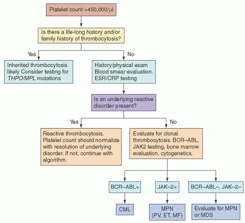
Inherited thrombocytosis is suspected in patients with a lifelong history of asymptomatic thrombocytosis, especially if other family members are also affected. The genetic mutations in inherited thrombocytosis are heterogeneous and may or may not involve mutations in the thrombopoietin gene (THPO) or its receptor (MPL).3 At least four different mutations in THPO associated with increased TPO production and inherited thrombocytosis have been described in European and Japanese kindreds.13 Thrombocytosis was inherited in an autosomal dominant manner in all four families. The TPO molecule in these patients is normal, and increased translation appears to explain elevated levels of TPO in these patients.13 At least three different MPL mutations have been linked to inherited thrombocytosis; affected kindreds are of European, African-American, Arabic, and Japanese ancestry.13 Approximately 7% of African-Americans are heterozygous for the MPL-Baltimore mutation.14 Thrombocytosis in patients with MPL mutations appears to result from either constitutive receptor activation or reduced receptor binding affinity for TPO.13

Clinical findings in patients with inherited thrombocytosis can be classified by whether the patients have THPO or MPL mutations. In general, those with THPO mutations have elevated TPO levels and mild splenomegaly, but a low incidence of vascular complications and low risk of evolution to acute leukemia and myelofibrosis,13 although development of bone marrow disease was described in two family members with inherited thrombocytosis due to THPO mutations.15 Some patients with MPL mutations and inherited thrombocytosis have been reported to experience thrombosis, splenomegaly, and myelofibrosis, but these complications have not been reported in African-American patients with MPL-Baltimore.13


Clonal Thrombocytosis Other Than Essential Thrompocythemia

Clonal or primary thrombocytosis includes not only ET (discussed later) but also a prominent feature of the other myeloproliferative neoplasms and certain myelodysplastic syndromes. Between 35% and 50% of patients with chronic myeloid leukemia (CML), polycythemia vera, or myelofibrosis may have elevated platelet counts.5, 16 The diagnosis, clinical aspects, and management of these disorders are discussed in Chapters 81, 82 and 83.

Clonal thrombocytosis is also seen in myelodysplastic syndromes associated with certain cytogenetic features (trisomy 8,17 5q- syndrome,18 and chromosome 3 abnormalities19), as well as the presence of ringed sideroblasts.20 However, the frequency of thrombocytosis in myelodysplasia is lower than in the myeloproliferative neoplasms. Myelodysplastic syndromes are discussed in Chapter 79.


DIFFERENTIAL DIAGNOSIS AND CLINICAL APPROACH TO THROMBOCYTOSIS

When evaluating a patient with thrombocytosis, the key consideration is to exclude ET and CML21 from reactive or inherited thrombocytosis and other disorders. Thrombocytosis may be a presenting feature of CML.21 Inasmuch as CML patients are at risk of conversion to acute leukemia, and there is targeted therapy for CML, BCR-ABL, and cytogenetic testing is important in patients with thrombocytosis who do not have inherited or reactive etiologies. Patients with RT usually will have major symptoms of an underlying inflammatory, infectious, or malignant disorder. However, patients with occult malignancy may initially present without obvious cancer symptoms. Post-splenectomy thrombocytosis will usually be obvious, but patients with asplenia or hyposplenia will be identified by observing Howell-Jolly bodies on the blood smear and by abdominal imaging showing a vestigial spleen or no splenic tissue. The World Health Organization (WHO) criteria for ET are discussed in the next section, and focus on distinguishing essential thrombocytosis from the other myeloproliferative neoplasms. These criteria provide no direct definitions of RT. Thus, the physician is left with practical clinical criteria to suggest an RT if a patient does not have an obvious underlying disorder. Such criteria might include the presence of fever, an elevated C-reactive protein or erythrocyte sedimentation rate, a blood smear showing reactive or toxic changes in the leukocytes, the presence of iron deficiency, or the post-splenectomy state.3, 5


FIGURE 51.1. An approach to evaluating thrombocytosis. A life-long personal history and family history of thrombocytosis would suggest a diagnosis of inherited thrombocytosis. If that diagnosis is excluded, reactive etiologies should next be considered. If an underlying cause is identified and is treatable, thrombocytosis should resolve with appropriate therapy. If neither inherited nor reactive thrombocytosis is likely, testing for clonal thrombocytosis should be performed; this should include BCR-ABL and JAK2 testing. The appropriate tests include bone marrow evaluation and cytogenetics for myeloproliferative neoplasms and myelodysplasia. CML, chronic myeloid leukemia; CRP, C-reactive protein; ESR, erythrocyte sedimentation rate; MF, myelofibrosis; ET, essential thrombocytosis; MDS, myelodysplasia; MPL, thrombopoietin receptor gene; MPN, myeloproliferative neoplasms; PV, polycythemia vera; THPO, thrombopoietin gene.

One approach to the evaluation of thrombocytosis is shown in Figure 51.1. Inherited thrombocytosis can be rapidly excluded if there is no life-long history of thrombocytosis and the family history is negative. Patients who may possibly have inherited thrombocytosis can be further evaluated with testing for THPO and MPL mutations. Because >90% of patients with thrombocytosis will have a reactive etiology,3, 4, 5 significant effort should be made in excluding secondary thrombocytosis before considering testing for myeloproliferative neoplasms, including ET.


ESSENTIAL THROMBOCYTHEMIA


Epidemiology

Evolving diagnostic criteria (discussed below) make it difficult to characterize the incidence and prevalence of ET. In studies published between 2001 and 2008, an incidence of 1.5/100,000 was reported.23, 24 Prevalence based on insurance claims data in the northeastern United States has been reported to be 24 cases/100,000 population.25 However, these reports antedate the 2008 revision of the WHO diagnostic criteria for ET, which includes a lower platelet threshold and could potentially alter the reported prevalence and incidence.

The median age at diagnosis is in the mid-50s in large reported series, and females represent 60% to 70% of cases.26, 27 Cases have been reported in children but are uncommon.28, 29, 30 First-degree relatives of ET patients have an increased risk of ET.31

Risk for developing ET appears to be associated with environmental factors linked to other myeloid neoplasms, such as radon and gamma irradiation.32, 33 Hairdressers, electricians, and farmers appear to be at increased risk to develop ET compared to other occupations, although it should be remembered that these risks are still quite low.32, 33


Diagnostic Criteria

For many years, the diagnosis of ET was based on the demonstration of significant sustained thrombocytosis (typically greater than 600 × 109/L) in the absence of a reactive etiology.34 However, the observation that a specific gain-of-function mutation in Janusassociated kinase 2 (JAK2V617F) is found in virtually all cases of polycythemia vera and in most cases of ET,35, 36 provided an additional positive criteria for the diagnosis of ET.

The 2008 revision of the WHO criteria for the diagnosis of ET is shown in Table 51.2.34 The first criterion represents a reduction in the threshold for thrombocytosis from 600 × 109/L to what is in essence the upper limit of the normal platelet count and addresses concerns that the higher threshold prevented the detection of early disease.37, 38, 39 The second criterion requires demonstration of the characteristic bone marrow morphology of ET.40

As discussed earlier in this chapter, a number of other myeloid neoplasms may be associated with thrombocytosis. This is particular true when employing the lower platelet threshold of the 2008 criteria. Polycythemia vera is excluded by the absence of an elevated hematocrit or hemoglobin concentration with adequate iron stores demonstrated on a Prussian blue-stained marrow specimen or a normal serum ferritin: if iron stores are absent, a trial of iron replacement that does not elevate the hematocrit or hemoglobin above normal is required.34 Primary myelofibrosis is excluded by the absence of excessive reticulin deposition on the marrow biopsy. Chronic myelogenous leukemia is excluded by the absence of the BCR/ABL mutation. Myelodysplastic syndromes and other myeloid neoplasms are excluded by absence of characteristic marrow abnormalities, such as ringed sideroblasts.41


TABLE 51.2 WORLD HEALTH ORGANIZATION DIAGNOSTIC CRITERIA FOR ESSENTIAL THROMBOCYTHEMIA


Diagnosis requires meeting all four criteria


  1. Sustained platelet count ≥450 × 109/L


  2. Bone marrow biopsy specimen showing proliferation mainly of enlarged mature megakaryocytes; no significant increase or left shift of neutrophil granulopoiesis or erythropoiesis


  3. Not meeting criteria for polycythemia vera, primary myelofibrosis, chronic myelogenous leukemia, myelodysplastic syndrome, or other myeloid neoplasm (details discussed in text)


  4. Demonstration of JAK2V617F or other clonal marker; or, in the absence of a clonal marker, no evidence for reactive thrombocytosis. However, the presence of a condition associated with reactive thrombocytosis does not exclude essential thrombocytosis if first three criteria are met


Modified from Tefferi et al., Proposals and rationale for revision of the World Health Organization diagnostic criteria for polycythemia vera, essential thrombocythemia, and primary myelofibrosis: recommendations from an ad hoc international expert panel. Blood 2007;110:1092-1097.


In the absence of the JAK2V617F mutation, exclusion of nonclonal reactive processes remains a key element of the diagnosis of ET. Etiologies of RT specifically cited in the criteria that should be excluded by history or laboratory studies include iron deficiency, previous splenectomy, recent surgery, active inflammation, connective tissue disorders, metastatic cancer, and lymphoproliferative diseases.34 In polycythemia vera, mutations of JAK2 in exons other than the site of the V617F mutation have been found in a significant number of JAK2V617F-negative cases.42 Similar findings have not been reported for ET.

Only gold members can continue reading. Log In or Register to continue

Stay updated, free articles. Join our Telegram channel

Oct 21, 2016 | Posted by in HEMATOLOGY | Comments Off on Thrombocytosis and Essential Thrombocythemia

Full access? Get Clinical Tree

Get Clinical Tree app for offline access