Thrombocytopenia Caused by Immunologic Platelet Destruction


Thrombocytopenia Caused by Immunologic Platelet Destruction


Meghan S. Liel

Michael Recht

David C. Calverley


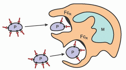
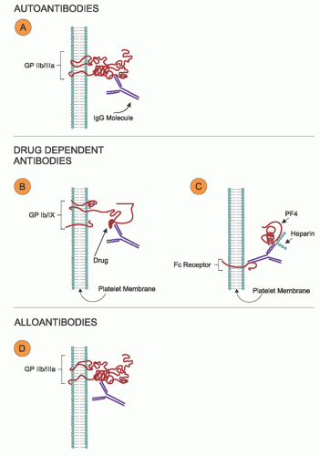
Immune thrombocytopenia (ITP) occurs when platelets undergo premature destruction as a result of autoantibody or immune complex deposition on their membranes. Although this disorder was previously known as idiopathic thrombocytopenic purpura, it is now correctly termed ITP because this nomenclature more clearly reflects the immune-mediated mechanism of the disease.1 In this chapter, both primary and secondary types of ITP are discussed (Table 47.1). Human immunodeficiencyvirus-related autoimmune thrombocytopenia, which is also in major part a result of the deposition of autoantibody or immune complexes, or both, on the platelet surface, is discussed in Chapter 64.

The diagnosis of ITP is primarily a diagnosis of exclusion, because currently available clinical assays for platelet-associated antibodies or serum antiplatelet antibodies/immune complexes are neither specific nor sensitive enough for routine clinical use. These disorders are characterized by peripheral thrombocytopenia (confirmed by examination of the peripheral smear), with a normal or increased number of megakaryocytes present on bone marrow examination, and absence of splenomegaly. Those patients who have no identifiable underlying cause, which might include infections, collagen vascular diseases, lymphoproliferative disorders (chronic lymphocytic leukemia or lymphoma), or drugs, are diagnosed as primary ITP. In some instances, ITP may be the presenting manifestation of an underlying disease, and additional manifestations appear weeks to months later.


PRIMARY IMMUNE THROMBOCYTOPENIA

Primary ITP refers to thrombocytopenia in which apparent exogenous etiologic factors are lacking and in which diseases known to be associated with secondary thrombocytopenia have been excluded. This syndrome has been recently reviewed.2, 3, 4, 5, 6

Acute ITP and chronic ITP differ in incidence, prognosis, and therapy (Table 47.2). These differences illustrate the wide spectrum of disorders that by definition are included in the syndrome, but many clinicians have long believed that acute ITP and chronic ITP are fundamentally different disorders.


TABLE 47.1 IMMUNE THROMBOCYTOPENIA


Primary


Secondary


Infections


Collagen vascular diseases


Lymphoproliferative disorders


Solid tumors


Drugs


Miscellaneous


Incidence

The annual incidence of ITP in the United States is estimated to be 1.6/10,000.7 Acute ITP, defined as thrombocytopenia occurring for <6 months and usually resolving spontaneously, most often affects children and young adults. The incidence peaks in the winter and spring, following the incidence of viral infections.8, 9 Acute ITP is most common between 2 and 6 years of age. Approximately 7% to 28% of children with acute ITP develop the chronic variety.9, 10, 11 Chronic ITP, lasting >6 months and requiring therapy to improve the thrombocytopenia, occurs most commonly in adults. In chronic ITP in adults, the median age is usually 40 to 45 years,12, 13 although in one large series of patients, 74% of 934 cases were younger than age 40 (range, 16 to 87 years of age).14 The ratio of females to males is nearly 1:1 in acute ITP9, 10, 11, 15 and 2 to 3:1 in chronic ITP.13, 14


Clinical Picture


Immune Thrombocytopenia in Children

In children with acute ITP, the onset of the disorder usually is sudden (Table 47.2). A history of infection preceding the onset of bleeding has been documented repeatedly.97, 98 In one series, such infections were noted within 3 weeks of the onset of ITP in 84% of cases.8, 9 Varicella zoster virus and Epstein-Barr virus are the most frequently identifiable viruses, although nonspecific unidentified viral infections predominate.99 Acute ITP may also occur after vaccination.100 Although it is most common between 2 and 5 years of age, ITP occurs in all pediatric age groups.101 The severity of ITP in infants is similar to that in older children, but compared to older children, a relatively small percentage of infants develop chronic ITP. A male preponderance was observed in the infant population in two studies.101, 102 Even though thrombocytopenia is likely to be severe, the bleeding manifestations of acute ITP in children usually are mild,103 and intracranial hemorrhage occurs in <1% of patients. The rare adult with the acute form of the disorder, however, may suffer hemorrhage and a more fulminant course. Acute ITP in children usually is self-limited; spontaneous remissions occur in as many as 90% of patients.8, 9 The duration of the disease ranges from a few days to a few months, with an average of 4 to 6 weeks.104 The favorable prognosis of ITP in children reflects the preponderance of the acute form of the disease in this age group. Children with thrombocytopenia of >6 months duration are classified as chronic ITP, although spontaneous remissions may still occur in an occasional child after 6 months.105, 106 Fever of mild degree has been reported, and the spleen tip may be palpable in up to 10% of patients, but this is believed to be the same incidence as is seen in normal children.107 The spleen is usually of normal weight in those patients who proceed to splenectomy.97


Immune Thrombocytopenia in Adults

In adults, the onset of the chronic form of the disorder usually is insidious (Table 47.2). A long history of hemorrhagic symptoms of mild to moderate severity is often described by the patient, but antecedent infections or fever are uncommon. Patients with chronic ITP usually have a fluctuating clinical course. Episodes of bleeding may last days or weeks and may be intermittent or even cyclic. Spontaneous remissions are very uncommon in adults, with an estimated occurrence of <5%.13, 14, 108, 109, 110 Most spontaneous remissions occur early; however, remissions have been described after 6 months in a small number of patients.13 Relapses in some cases are associated with vaccination.111 Often the clinical course is surprisingly benign.


Bleeding Manifestations

The hemorrhagic manifestations of ITP are of the purpuric type. Patients with only ecchymoses and petechiae have “dry” purpura; those with mucous membrane bleeding in addition to skin manifestations have “wet” purpura.112 Platelet counts are usually lower and the complication rates higher in those with wet purpura. In a series of 712 patients reported by the Israeli ITP study group, 82% of all patients had bleeding limited to the skin, although 43% of adult women reported menometrorrhagia.98

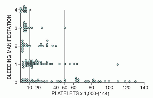
In general, the severity and frequency of hemorrhagic manifestations correlate with the platelet count (Fig. 47.3).113 Bleeding after trauma without spontaneous hemorrhage is usual in mildly affected patients with platelet counts >50,000/µl. Thrombocytopenia associated with counts between 10,000 and 50,000/µl results in spontaneous hemorrhagic manifestations of varying severity, such as ecchymoses and petechiae. Patients with platelet counts <10,000/µl are at risk for serious morbidity and mortality from bleeding, although the mortality rate is actually quite low.114 Patients who have an increased risk of bleeding include those with a history of bleeding, those with additional bleeding diatheses, and patients >60 years of age.12, 114 Older patients have also been reported to have an increased incidence of major, life-threatening bleeding.12, 115, 116


Skin and Mucous Membranes

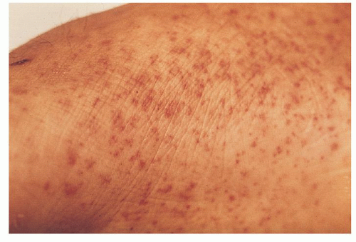
Spontaneous bleeding into the skin in the form of petechiae is characteristic. These lesions are minute, red to purple hemorrhages that range in size from that of a pinpoint to that of a pinhead (Fig. 47.4). They are flat, do not blanch with pressure, and appear and regress, often in crops, over a period of days. They are most conspicuous in areas of vascular stasis, such as the areas below tourniquet sites, the dependent portions of the body (especially around the ankles), and areas subjected to constriction from belts or stockings, as well as on skin surfaces over bony prominences. The presence of petechiae on the face and neck is unusual, except as the result of coughing.

Ecchymoses may develop on any skin surface. In ITP, they are seldom associated with subcutaneous hematomas and infrequently spread or dissect into deeper or adjacent structures. Large, purple, superficial ecchymoses may be seen, particularly on the back and thighs. Circular ecchymoses often surround even atraumatic venipuncture sites, but external bleeding from such sites is uncommon. Hemorrhagic vesicles or bullae may be seen inside the mouth and on other mucous surfaces. The bullae probably are the result of severe acute thrombocytopenia rather than a specific feature of any particular pathogenetic form.


FIGURE 47.3. Bleeding manifestations in relation to platelet count in patients with primary immune thrombocytopenia. Bleeding manifestations (or duration) are graded from 0 to 4, as follows: 0, no bleeding; 1, minimal, resulting from trauma; 2, spontaneous, but self-limited; 3, spontaneous, requiring special attention (e.g., nasal packs); and 4, massive uncontrolled or poorly controlled. (From Lacey and Penner, reference 113.)


FIGURE 47.4. Petechiae. Pinpoint, nonblanching erythematous capillary bleeding sites are most common in dependent body areas or pressure points.

Gingival bleeding and epistaxis are common. The latter usually responds for a time to conservative measures, such as nasal packing or tamponade, often to recur intermittently. Epistaxis may originate from lesions resembling petechiae in the nasal mucosa. Such lesions also may be found in the mucous membranes of the throat and mouth, sometimes in the absence of cutaneous hemorrhage. In many patients, discrete bleeding points cannot be identified.

The genitourinary tract is a frequent site of bleeding. Menorrhagia may be the only symptom of ITP and may appear for the first time at puberty. Hematuria also is a common symptom, the blood coming from the kidneys, the bladder, or the urethra, although bleeding into the kidney parenchyma is rare. Gastrointestinal bleeding is usually manifested by melena or, less often, by hematemesis.


Central Nervous System

Intracranial hemorrhage is the most serious complication of ITP. Fortunately, it is rare, affecting 1% to 2% or less of patients with severe thrombocytopenia.9, 117 The hemorrhages usually are subarachnoid, often are multiple, and vary in size from petechiae to large extravasations of blood. Numerous small hemorrhages often are seen in the retina; subconjunctival hemorrhage may also occur.


Bleeding after Trauma

Excessive bleeding often follows tooth extractions, tonsillectomy, or other operations or injuries and may first suggest the diagnosis of ITP. In contrast to the hereditary coagulation disorders, such traumatic bleeding is seldom voluminous or rapid. Slow persistent oozing may occur after trivial cuts, razor nicks, and scratches. Delayed bleeding and spontaneous hemarthrosis, which are characteristic of the hereditary coagulation disorders, are extremely rare in ITP.


Laboratory Findings


Blood

The mean platelet count of patients at the time of diagnosis of ITP is 25 to 30,000 and severe thrombocytopenia (<10,000) is frequently seen.117, 118 Abnormalities in platelet size and morphologic appearance are common. The platelets often are abnormally large (3 to 4 µm in diameter) and reveal more than normal variation in size and shape. Abnormally small platelets and platelet fragments (“microparticles”) also are evident and may represent the equivalent of microspherocytes and schistocytes.119, 120, 121, 122 Although megakaryocyte fragments may be apparent in routine blood smears, quantitative studies reveal subnormal numbers of these fragments.123

Estimates of mean platelet volume (MPV) and the extent of platelet size heterogeneity (platelet distribution width) by means of automated particle counters may, if present, provide useful information in the evaluation of patients with ITP.124 The presence of numerous megathrombocytes results in high MPV values.125 Platelet distribution width also is increased, presumably reflecting an abnormal degree of platelet anisocytosis.126 The exact mechanism underlying such megathrombocytosis is still uncertain, but it may be the result of accelerated platelet production in response to platelet destruction. When MPV is increased in patients with ITP, it is typically inversely correlated in a nonlinear manner with the platelet count. In contrast, low MPV values have been reported in association with big spleen syndromes126 and some myeloproliferative disorders, after chemotherapy with cytotoxic drugs, and in patients with septic thrombocytopenia.127

Significant abnormalities in the other blood counts should prompt a thorough evaluation for other causes of thrombocytopenia as these findings are unusual in ITP. Anemia, if present, is proportional to the extent of blood loss and is usually normocytic. If bleeding has been severe and long-standing, iron deficiency anemia may occur. Occasionally, recent severe hemorrhage may produce reticulocytosis and moderate macrocytosis. Antiplatelet antibodies in patients with ITP do not usually cross-react with erythrocytes, although erythrocyte fragmentation, presumably the result of weak complement activation, may occur.121 Patients may also have a positive direct Coombs test and autoimmune hemolytic anemia; the combination is known as Evans syndrome.128, 129

The total leukocyte count and the differential count usually are normal. Eosinophilia has been noted, particularly in children, but this finding is by no means consistent. Lymphocytosis with abnormal cells resembling those characteristic of infectious mononucleosis also has been reported.130, 131

Tests of hemostasis and blood coagulation reveal only changes attributable to thrombocytopenia, such as a prolonged bleeding time. The results of tests of blood coagulation, including prothrombin time, partial thromboplastin time, and fibrinogen, are normal in patients with uncomplicated thrombocytopenia. Slight increases in the levels of fibrinogen degradation products have been demonstrated in the plasma of some patients with ITP.132 Plasma levels of glycocalicin, a portion of platelet membrane gpIb, may be high in patients with ITP and other forms of platelet destruction. As noted previously, concentrations of thrombopoietin are not significantly increased in ITP.


Bone Marrow

ITP causes no characteristic bone marrow changes; therefore, bone marrow examination should not be routine. The American Society of Hematology (ASH) guidelines for management of ITP recommend against bone marrow biopsy in both children and adults with history, physical exam, CBC, and peripheral smear typical of ITP.5 However, bone marrow aspiration may be helpful in the differential diagnosis of ITP in patients who have atypical findings that may suggest some of these other etiologies.

In patients with ITP, alterations in the bone marrow are usually limited to the megakaryocytes, although normoblastic hyperplasia may develop as a result of blood loss. The leukocytes are essentially normal with the exception of occasional eosinophilia.133

Megakaryocytes usually are increased in size134 and are increased or normal in number,135, 136 the numbers correlating roughly with the MPV. Morphologic abnormalities of these giant cells are present in most patients with ITP. “Smooth” forms with single nuclei, scanty cytoplasm, and relatively few granules are common. Presumably, they represent the results of markedly
accelerated platelet production and the presence of many young forms.137, 138 The changes just summarized are similar to those found in most forms of thrombocytopenia caused by accelerated platelet destruction and are not characteristic or diagnostic of ITP.


Antiplatelet Antibodies

Primary ITP is a diagnosis of exclusion and relies on clinical impression. A number of different types of antiplatelet antibody tests have been developed and reported through the years.36, 139, 140, 141, 142, 143, 144, 145, 146 Most of these tests were quite cumbersome and therefore never became available for routine testing. These tests measured different types of Ig, including serum antiplatelet antibodies, platelet-associated surface Ig, or total platelet Ig and are now generally regarded as unreliable.147 The platelet Ig is released, along with other α-granule proteins, such as platelet factor 4 and β-thromboglobulin, during platelet activation and secretion. It is presumed that some of these released proteins bind to the platelet surface. These observations make it difficult to use either the platelet-associated IgG assays or the total platelet Ig assays for the diagnosis of ITP.148

In a more recent generation of antiplatelet antibody assays, monoclonal antibodies for the specific platelet membrane glycoproteins that are implicated in ITP are used in antigen-capture-type assays (also called glycoprotein immobilization assays).149 Studies have identified a specificity of 78% to 93%.150, 151 However, the sensitivity (49% to 66%) is not high enough to exclude the presence of ITP if the test is negative.68, 151, 152 None of these tests is in routine clinical use, and experts disagree on their role in the diagnosis of ITP. Future direction might include the use of flow cytometry in the diagnosis and follow-up of autoimmune thrombocytopenia.153


Treatment of Primary Immune Thrombocytopenia

Many of the treatment recommendations for ITP are based on expert opinion rather than high-level evidence from randomized controlled trials. The ASH has published a practice guideline on ITP.5, 164, 165 The reader is referred to this practice guideline for specific questions regarding the treatment of patients with ITP.


Children

Childhood ITP is usually benign and self-limited. Therefore, treatment is not required for most patients. Treatment is reserved for patients with severe thrombocytopenia (<20,000/µl) and bleeding, or patients who remain thrombocytopenic for >6 months, that is, those with chronic ITP (Table 47.4).166 Treatment guidelines recommend observation for all children with no or mild bleeding, regardless of platelet count.5 Although the greatest
fear in the acute form is intracranial hemorrhage, several large studies show that even with low platelet counts (<30,000/µl) life-threatening bleeding and intracranial hemorrhage are rare (<0.5%).166, 167


TABLE 47.4 RECOMMENDATIONS FOR INITIAL TREATMENT OF IMMUNE THROMBOCYTOPENIA (IVIG) PATIENTS WITH PLATELET COUNTS <20,000-30,000/µla


Children


Adults


Asymptomatic


None


Steroids (preferred) or IVIG


Minor purpura


Mucosal membrane bleeding that may require clinical intervention


None


IVIG or steroids


Steroids (preferred) or IVIG)


IVIG and/or steroids


Severe, life-threatening bleeding


Steroids and IVIG


Hospitalization


Consider platelet transfusion and other measures


Steroids and IVIG


Hospitalization


Consider platelet transfusion and other measures


aThe current ASH guidelines recommend treatment for adults with platelet counts of <30,000/µl.


Treatment guidelines for pediatric ITP recommend first-line treatment with either a single dose of intravenous immunoglobulin (IVIG) (0.8 to 1 g/kg) or a short course of steroids.5 Several multicenter randomized trials have been performed in high-risk patients with acute ITP to define whether treatment is associated with a prompt increase in platelet counts. These clinical trials demonstrated that treatment with either oral prednisone or IVIG was associated with a more rapid rise in platelet count to >20,000/µl than either no therapy or treatment with anti-D.168, 169 Only IVIG shortened the time to reach a platelet count >50,000/µl. More recently, children with platelet counts <10,000/µl or counts of 10,000 to 29,000/µl and mucosal bleeding were studied in a prospective randomized clinical trial, and IVIG raised platelet counts faster than three corticosteroid regimens.170 Therefore, IVIG should be preferentially used if a rapid rise in platelet count is desired.

Treatment of children who develop persistent ITP or are unresponsive to initial treatment is evolving. Splenectomy remains a standard given high response rates; however, many experts recommend delaying splenectomy for at least 12 months given the frequency of spontaneous remission.5 Rituximab treatment has been used in children and adolescents in a small study comprised of 36 patients with chronic ITP.171 In contrast to observations in adults, the median time to response was 1 week (range 1 to 7 weeks), and the overall response rate was 31%. Outcome was not associated with age, prior pharmacologic response, prior splenectomy, duration of disease, screening platelet count, refractoriness, or IgM reduction. Therefore, rituximab may be considered as an alternative to splenectomy in children with chronic ITP or in patients with persistent bleeding despite first-line treatments.5 High-dose dexamethasone (0.6 mg/kg/day) may also be considered in patients who are unresponsive to initial therapy based on small studies showing a 25% response rate in refractory patients.172


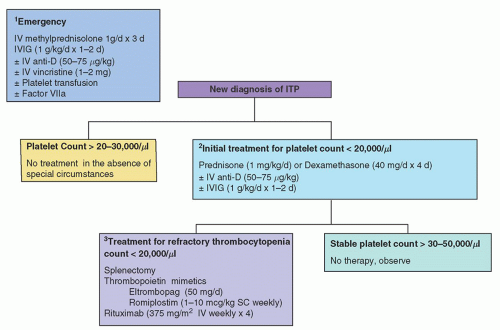
FIGURE 47.5. Therapy of adult immune thrombocytopenia (ITP). (1) Minimal emergency therapy includes intravenous (IV) methylprednisolone and intravenous immunoglobulin (IVIG). Intravenous anti-D and platelet transfusions may be given as needed. All three modalities given prior to transfusions may help preserve platelet longevity in the circulation and repeated or continuous platelet transfusions may be required in urgent situations. (2) Initial treatment of ITP typically consists of steroids (prednisone or dexamethasone) with the goal of attaining a platelet count of >30,000/µl and cessation of bleeding. IVIG or anti-D may be used if steroids are contraindicated or the patient has persistent thrombocytopenia despite steroids. (3) Thrombocytopenia recurs in most adults as corticosteroids are tapered. Treatment options for refractory ITP include splenectomy, thrombopoietin mimetics, and rituximab. (Modified from Cines and Bussel, reference 459.)


Adults

Patients with chronic ITP may have mild thrombocytopenia that can be followed without treatment. The incidence of bleeding is correlated with the platelet count; therefore, patients with platelet counts >50,000/µl rarely have spontaneous bleeding and may require treatment only if extensive operative procedures are planned. Patients with platelet counts <20,000 to 30,000/µl or significant mucosal membrane bleeding with platelet counts <50,000/µl are usually treated5 (Table 47.4 and Fig. 47.5).

No prospective studies on long-term prognosis of ITP after treatment are reported. However, it has been reported that most adult patients have a good response to treatment (without necessarily returning to normal platelet counts) and have no excess mortality when compared to the general population.173 A small group of patients who had severe thrombocytopenia after 2 years of primary and secondary therapies had a mortality risk of 4.2 (95% confidence interval, 1.7 to 10.0) resulting from both bleeding and infectious complications related to therapy.

ITP is uncommon in elderly patients; only 30% of patients in reported series are >45 years of age.133, 174 However, these patients may be more refractory to therapy115 and appear to have a higher incidence of hemorrhagic complications than younger patients.12 Guthrie and colleagues reported a 52% incidence of life-threatening or fatal bleeding in their series of 40 patients
older than age 45 years.115 This risk of fatal bleeding in patients with platelet counts that are chronically <30,000/µl is estimated at 0.4% per year for patients <40 years of age and 13.0% per year for patients >60 years of age.175

Oct 21, 2016 | Posted by in HEMATOLOGY | Comments Off on Thrombocytopenia Caused by Immunologic Platelet Destruction

Full access? Get Clinical Tree

Get Clinical Tree app for offline access