Chapter 5
The Most Common Types of Invasive Breast Carcinoma
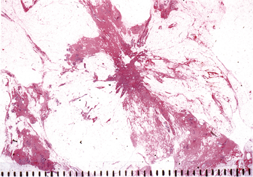
Fig. 5.1
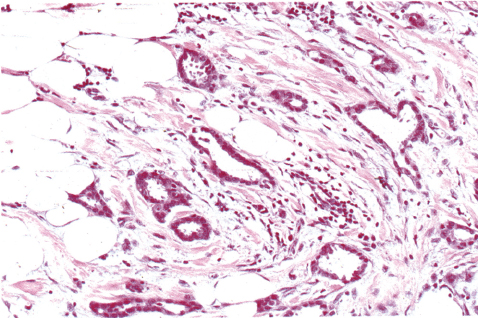
Fig. 5.3
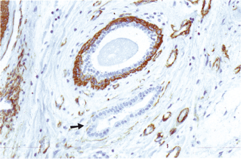
Fig. 5.4
Tubular carcinoma is usually a slow-growing, stellate tumor with stromal desmoplasia (Fig. 5.1). The typical mammographic appearance is a small stellate density with a central tumor mass (“white star”) (Fig. 5.2).
Diagnostic Criteria
More than 90% of the tumor has tubular structures, which are
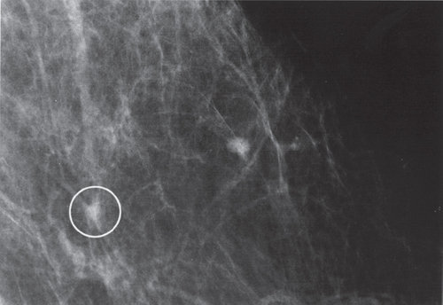
Fig. 5.2
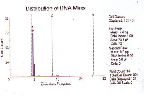
Fig. 5.5
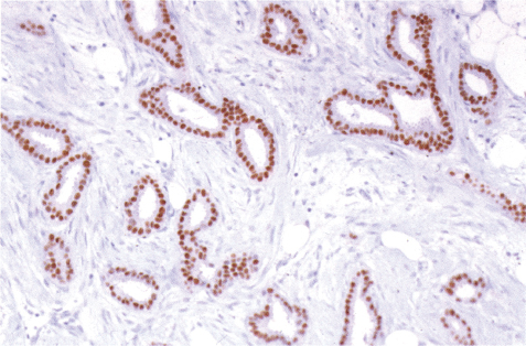
Fig. 5.6
Tubular carcinoma is usually small and of limited extent, but it may be multifocal. Structures of ductal carcinoma in situ (DCIS) grade I are regularly present in the tumor. Tubular carcinoma is regularly diploid (Fig. 5.5), estrogen-receptor positive (Fig. 5.6), and has an excellent prognosis.
Differental Diagnosis
1. Radial scar (see also Chapter 6):
– not palpable
– typical mammographic picture of a “black star”
– presence of myoepithelium
2. Microglandular adenosis:
– regular, round glands (absence of lobulocentricity and myoepithelium)
3. Invasive ductal carcinoma not otherwise specified (NOS):
– contains less than 90% tubular structures
Mucinous carcinoma is a mucin-producing, slow-growing, tumor with a favorable prognosis. Mammographically, it usually appears as a circular – oval, ill-defined, low-density tumor mass (Fig. 5.7).
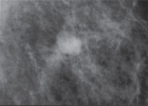
Fig. 5.7
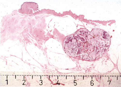
Fig. 5.8
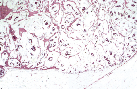
Fig. 5.9
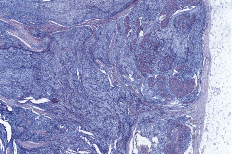
Fig. 5.10
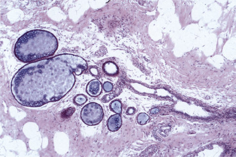
Fig. 5.11
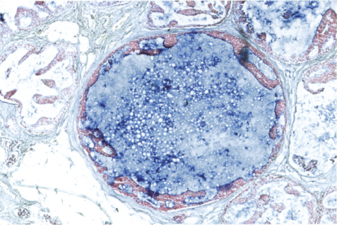
Fig. 5.12
Diagnostic Criteria
– Macroscopically well-circumscribed and gelatinous tumor (Fig. 5.8), and
– at least 90% of the tumor is composed of mucin, containing small groups of well-differentiated tumor cells. (Figs. 5.9 and 5.10).
A tumor fulfilling these criteria has an excellent prognosis. However, mucinous carcinoma often exhibits obvious intratumoral heterogeneity (see Chapter 10, case 1), which may have prognostic implications.
Differential Diagnosis
1. Mucin-containing lobules, dilated ducts, mucocele (Fig. 5.11), and mucinous DCIS (Fig. 5.12).
2. Ductal carcinoma with mucinous component:
– poorly circumscribed, often stellate
– more cellular
– often less well-differentiated tumor cells
Ductal carcinomas with a mucinous component do not share the favorable prognosis of purely mucinous carcinomas.
3. Invasive micropapillary carcinoma (see Chapter 6).
Medullary carcinoma is usually a rapidly growing, round or oval, well-circumscribed tumor appearing in younger patients. Mammographically or ultrasonographically (Fig. 5.13), a circumscribed, solid, round or oval density is observed.
Macroscopically and histologically the tumor is well-circumscribed, relatively soft, and often lobulated (Fig. 5.14).
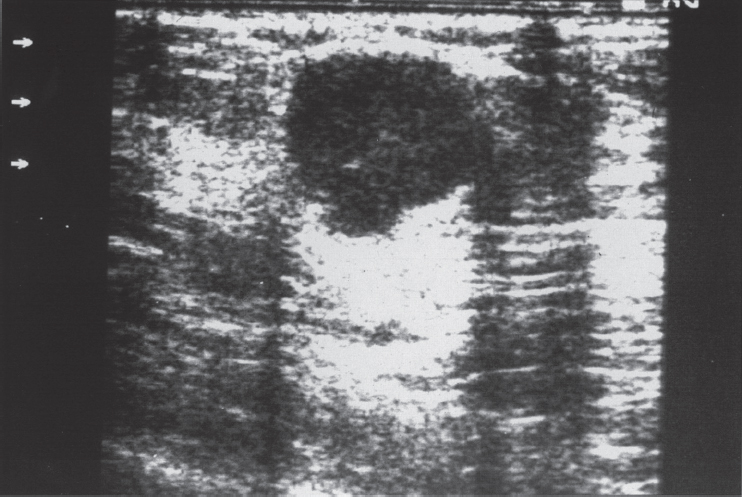
Fig. 5.13


