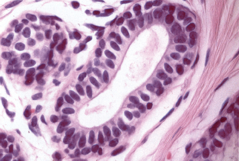
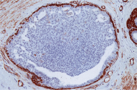
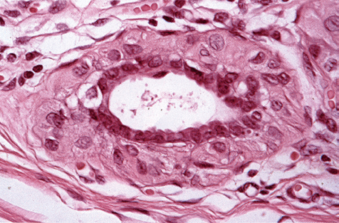
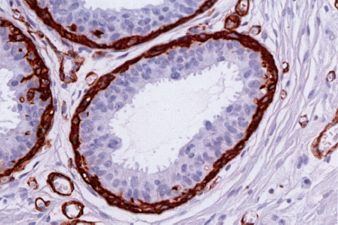
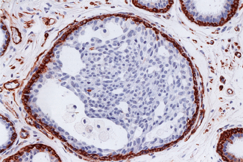
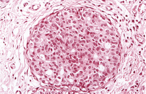
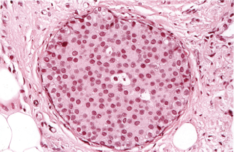
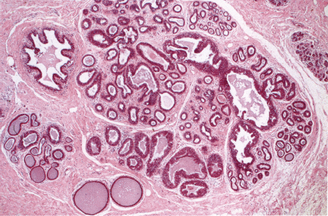
Chapter 3 Hyperplastic Changes with and without Atypia The normal ducts and lobules in the breast exhibit a single layer each of epithelial and myoepithelial cells (Fig. 3.1). Fig. 3.1 In epithelial hyperplasia, more than one layer of epithelial cells is present (Fig. 3.2). Fig. 3.2 In myoepithelial hyperplasia, more than one layer of myoepithelial cells is seen (Fig. 3.3). Fig. 3.3 Hyperplasia may be a focal or a diffuse phenomenon involving a portion of the terminal ductal-lobular unit (TDLU), the entire TDLU, or many TDLUs and ducts. Epithelial hyperplasia may result in the formation of only two to three layers of epithelial cells (Fig. 3.4), but often many layers of epithelial cells are present and form glandlike spaces or small papilloma-like structures (“florid” epithelial hyperplasia, Fig. 3.5). Fig. 3.4 Fig. 3.5 Neoplasia may also result in several layers of epithelial cells within the ducts and acini. Hyperplasia and neoplasia can be differentiated on the basis of their cellular and architectural characteristics. Cellular Characteristics Hyperplasia is a benign proliferation of several cell clones resulting in a polymorphous population of small cells (Fig. 3.6). Fig. 3.6 Fig. 3.7 Low-grade malignant epithelial cells appear as a monoclonal, monomorphous population of small cells (Fig. 3.7). These cells also give rise to several layers of cells within the ducts and lobules, resembling hyperplasia, and they may even appear within epithelial hyperplasia, partly taking over the structures. If only a portion of the TDLU is replaced by this monomorphous cell population, the lesion is called atypical ductal hyperplasia (ADH) (Fig. 3.8). If the entire TDLU is filled by the monomorphous small cells, the lesion is considered to be ductal carcinoma in situ (DCIS) grade I. ADH differs from DCIS grade I through extent and distribution, not through cellular features. Fig. 3.8 During the evolution of DCIS, different cell clones may appear within the initially monoclonal tumor cell population leading to a multiclonal malignant cell population. These lesions are grade II or grade III DCIS and consist of a population of large polymorphous cells (Fig. 3.9). Fig. 3.9
Hyperplastic Changes with and without Atypia
Only gold members can continue reading. Log In or Register to continue


Full access? Get Clinical Tree


