Chapter 1
Normal Breast Tissue or Fibrocystic Change?
The mammary gland, like all glandular organs, consists of parenchyma and stroma. The parenchyma contains ducts (Fig. 1.1) and lobules (Fig. 1.2), which are separated from the stroma by a continuous basement membrane (Fig. 1.3). The entire parenchyma (with the exception of the terminal parts of the lactiferous ducts) consists of a single inner layer of epithelial cells and an outer layer of myoepithelium (Fig. 1.4). Only the epithelial cells contain estrogen and progesterone receptors in their nuclei (Fig. 1.5).
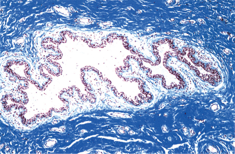
Fig. 1.1
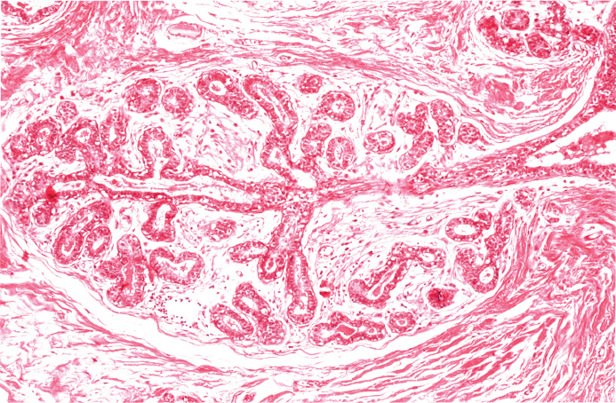
Fig. 1.2
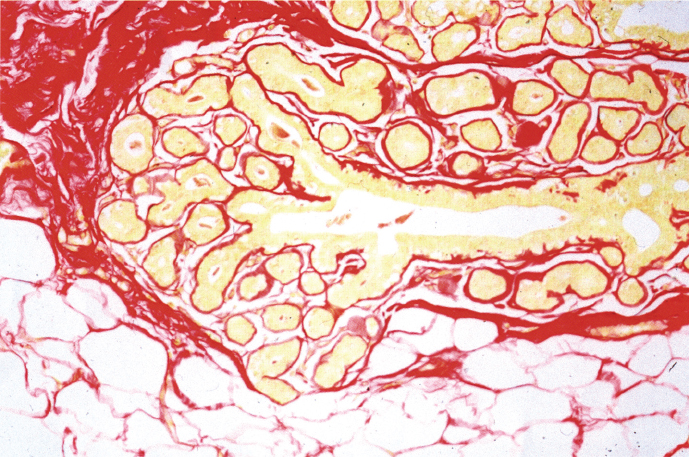
Fig. 1.3
The stroma consists of fibrous tissue and adipose tissue containing lymph vessels, blood vessels, and nerves. More specialized within the lobules and surrounding the ducts, the stroma can be divided into intralobular (mucin-rich, “active”) and interlobular stroma (see Fig. 1.16).
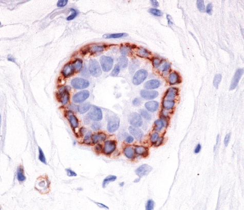
Fig. 1.4
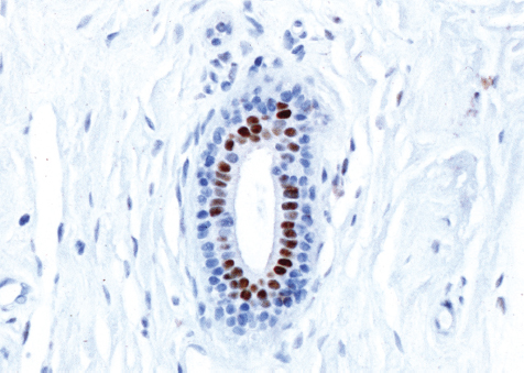
Fig. 1.5
The nipple is the origin of 15 to 25 lactiferous ducts, which branch into segmental, subsegmental, and terminal ducts that with the associated lobules comprise 15 to 25 lobes. The terminal duct and the associated lobule are collectively referred to as the Terminal Ductal-Lobular Unit (TDLU), which is the most important functional unit and the place of origin of most pathologic processes in the breast (Figs. 1.6 and 1.7).
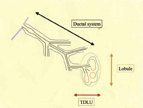
Fig. 1.6
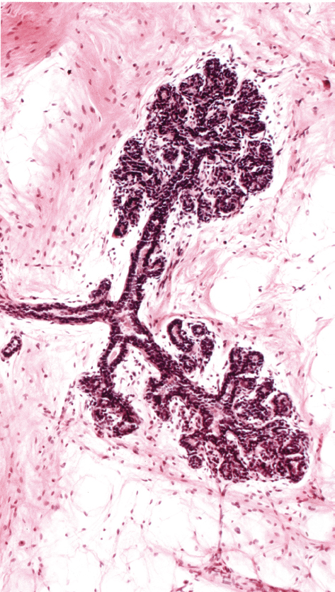
Fig. 1.7
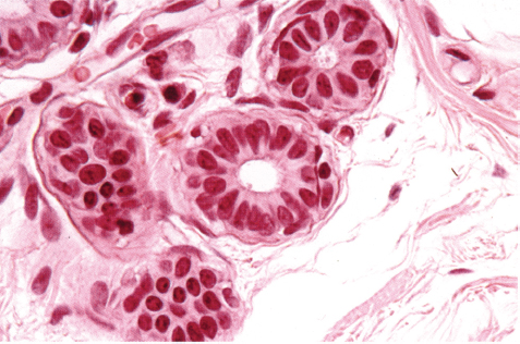
Fig. 1.8
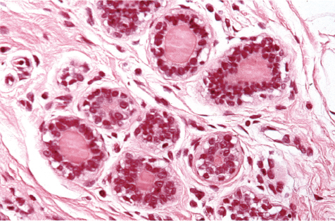
Fig. 1.9
At the beginning of the menstrual cycle, the lobules are relatively small and contain only a minimal amount of secretion, if any, in the lumina of the acini (Figs. 1.8 and 1.11). During the secretory phase of the cycle, the acini produce eosinophilic secretions and the intralobular stroma becomes edematous (Fig. 1.9). Around the time of menstruation, the myoepithelium becomes vacuolated and undergoes apoptosis (Fig. 1.10).
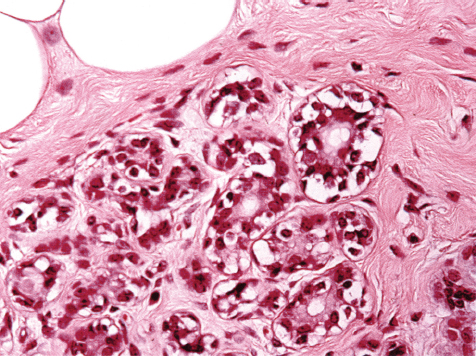
Fig. 1.10
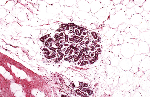
Fig. 1.11
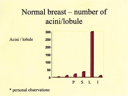
Fig. 1.12
The most obvious changes are seen during the last trimester of pregnancy and during lactation when the TDLUs are enlarged, the number of acini per lobule increases greatly (Fig. 1.12), the cytoplasm in the epithelial cells becomes vacuolated, and rich secretions are produced in large quantities (Fig. 1.13).
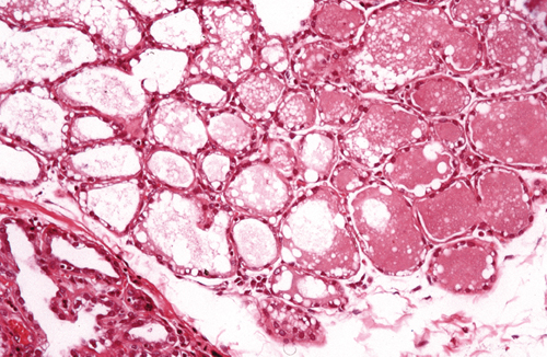
Fig. 1.13
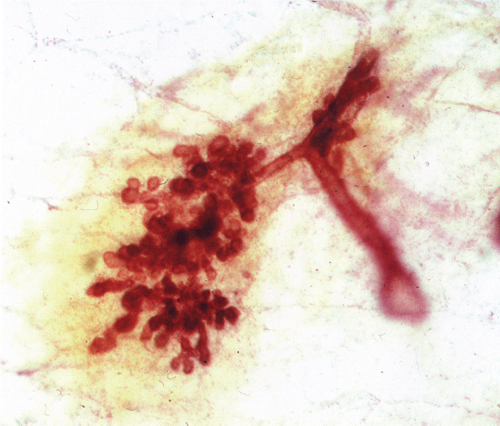
Fig. 1.14
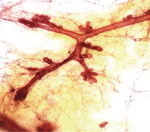
Fig. 1.15
Around the time of menopause, but often earlier, involution of the breast tissue occurs.
Involution of the parenchyma results in a diminished number of acini/lobules, lobules, and ducts (compare Fig. 1.14 showing a functioning lobule with Fig. 1.15 showing an involuted lobule, thick-section images).
Figure 1.16 shows a TDLU with normal mucin-rich intralobular stroma. Involution of the intralobular stroma is accompanied either by infiltration of fatty tissue (Fig. 1.17) or by fibrosis (Fig. 1.18). The same is true for the interlobular stroma.
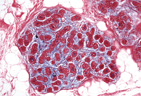
Fig. 1.16
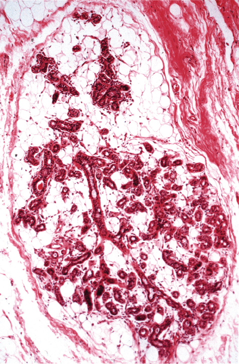
Fig. 1.17
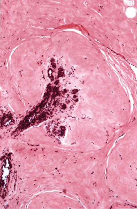
Fig. 1.18
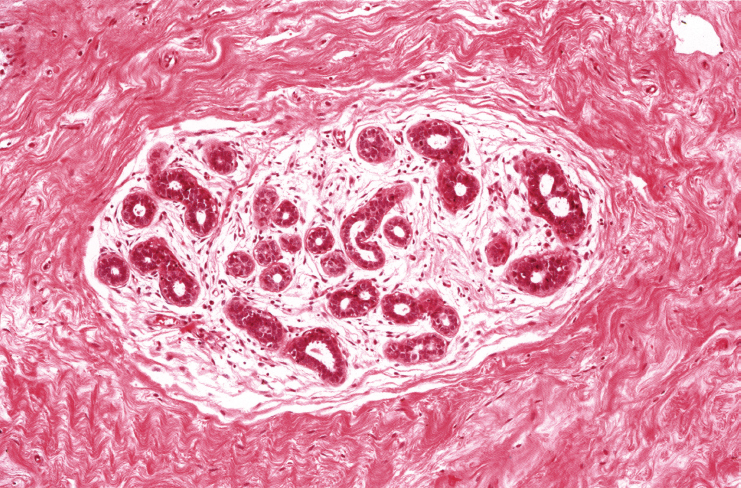
Fig. 1.19
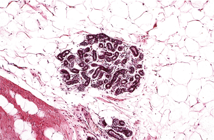
Fig. 1.20
The involution of the parenchyma and the interlobular and intralobular stroma is not necessarily a synchronized process. Functioning lobules can be seen in the presence of interlobular stroma that has undergone fibrous involution (Fig. 1.19) or has been replaced by fat (Fig. 1.20).
The intralobular and the interlobular stroma may undergo either fibrous or fatty involution in varying combinations (Figs. 1.21, 1.22, and 1.23).
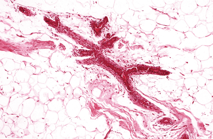
Fig. 1.21


