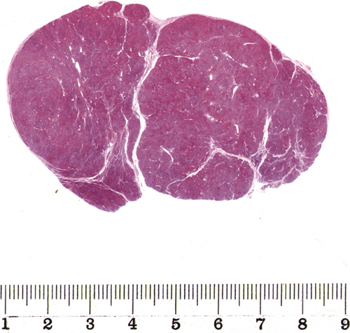
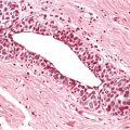
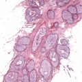
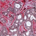
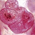
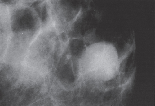
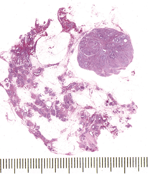
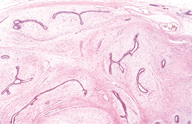
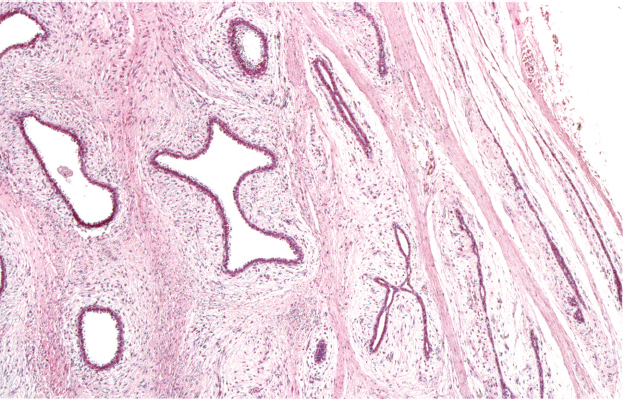
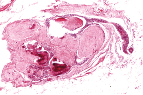
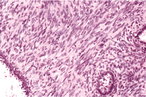
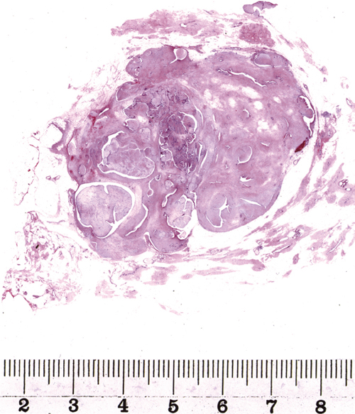
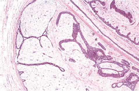
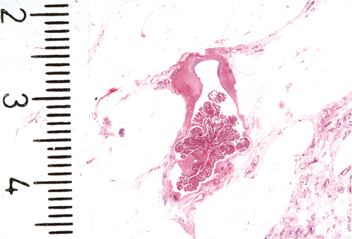
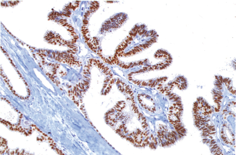
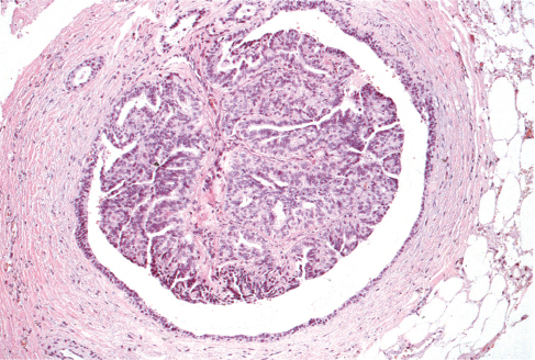
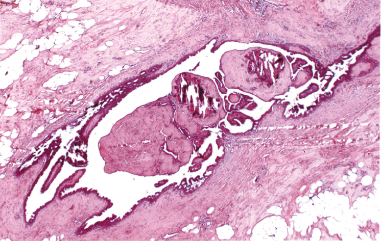
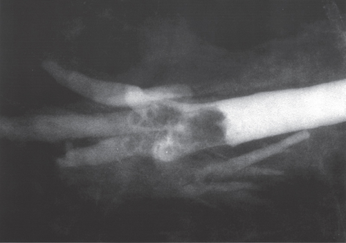
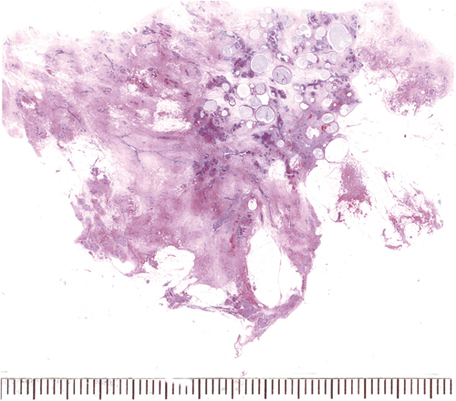
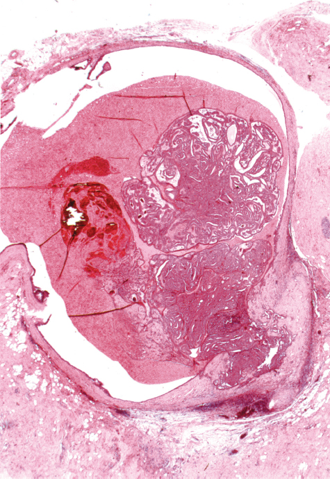
Chapter 6 The Most Common Benign Breast Lesions and Their Borderline and Malignant Counterparts Fig. 6.1 Fig. 6.2 Fig. 6.3 Fig. 6.4 Fig. 6.5 Fibroadenoma (Figs. 6.1 and 6.2) is a very common benign epithelial-stromal tumor mainly containing proliferated elements of the intralobular active stroma, but also featuring proliferated ducts and acini. The epithelial component is distorted in the so-called intracanalicular variant (Fig. 6.3) and more regular in “pericanalicular” fibroadenoma (Fig. 6.4). The epithelial component may exhibit hyperplastic or metaplastic changes. The stroma of the younger lesion is mucin-rich and becomes more fibrous over time. Fibroadenoma is usually a palpable lesion, but in about 10% of the cases, it is detected on the basis of amorphous stromal microcalcifications (Fig. 6.5) seen on the mammogram. Fibroadenoma represents a clinically detectable phase of fibroadenomatoid change (see Chapter 1). Fig. 6.7 The stroma in a fibroadenoma is poorly cellular and mitotically almost inactive. The “juvenile” variant of fibroadenoma is a more rapidly growing round or oval lesion that has a more cellular stroma with some mitotic figures (Fig. 6.6). The term “phylloides tumor” designates a group of epithelial-stromal neoplasms with dominating stroma and leaf-like structures (Figs. 6.7 and 6.8). The stroma in these lesions is more cellular and mitotically active, even in benign cases. An occurrence of more than five mitoses per ten high power fields is considered as an indicator of low malignant potential (“phylloides tumor of borderline malignancy”) with a tendency to recur. Lesions with obvious stromal cellular atypia and high mitotic activity are rare and malignant. They may contain heterologeous stromal components (e.g., liposarcoma, rhabdomyosarcoma, or osteosarcoma). Fig. 6.8 Fig. 6.9 The epithelial component of these tumors may contain foci of lobular carcinoma in situ (LCIS), ductal carcinoma in situ (DCIS) (Fig. 6.9), or invasive carcinoma. The most important issue in this group of lesions is the potential risk of overdiagnosis. In the presence of a growing palpable mass, usually in younger women, a cellular fine-needle aspirate may lead to an erroneous preoperative diagnosis of malignancy. Core biopsy usually rules out the possibility of carcinoma and detects the rare borderline and malignant variants of these tumors. Fig. 6.10 Fig. 6.11 Fig. 6.12 One of the few lesions that originate outside the terminal ductal-lobular unit (TDLU) is a papilloma. It usually develops in larger ducts in the retroareolar area and often causes serous or bloody nipple discharge. Papillomas are exophytic lesions that fill the lumen of the dilated duct (Fig. 6.10) and contain a branching fibrous central core (Fig. 6.11 and 6.12). Fig. 6.13 The stroma of elderly papillomas may calcify (Fig. 6.13) and appear as a cluster of calcifications on the mammogram. However, the radiologic method of choice in detecting papillary lesions is galactography (Fig. 6.14). Fig. 6.14 Fig. 6.15 Papillomas may be solitary or multiple. Sometimes in young women multiple papillomas appear in a fibrocystic area of the breast tissue. This is referred to as “juvenile papillomatosis” (Fig. 6.15). The epithelial component as well as the myoepithelium in papillomas may exhibit a spectrum of metaplastic, hyperplastic and neoplastic changes resulting in benign, borderline, and malignant categories of the papillary lesions. On the benign end of the spectrum are intraductal papillomas with a single layer of epithelium and a single layer of myoepithelium, but metaplasia or hyperplasia of the epithelium and the myoepithelium frequently occurs (Fig. 6.16). Fig. 6.16
The Most Common Benign Lesions and Their Borderline and Malignant Counterparts
Only gold members can continue reading. Log In or Register to continue

Full access? Get Clinical Tree


