James H. Diaz
Myiasis and Tungiasis
Flies and fleas are mostly bothersome biting nuisances of humans and animals that can also transmit infectious diseases and deeply invade living tissues, causing amputation, disfigurement, and, rarely, death. Flies can serve as mechanical vectors of shigellosis, and rat fleas can transmit bubonic plague and murine typhus. Flies may lay their eggs on human flesh, and their developing larvae, or maggots, can invade subcutaneous tissues and penetrate external body cavities, such as the orbits, ears, and nares. Flea larvae can also burrow into subcutaneous tissues to feed, complete their developmental stages or instars, and promote secondary infections with incapacitating sequelae, including autoamputation of toes and fingers, especially in impoverished tropical communities plagued by endemic jigger fleas (Tunga penetrans).
Myiasis
Myiasis is an ectoparasitic infestation of viable or necrotic tissues by the dipterous larvae of higher flies and may be broadly classified as obligatory or facultative myiasis. In obligatory myiasis, maggots must live and feed on human or animal hosts as part of their life cycle. In facultative myiasis, normally free-living maggots that preferentially feed on carrion and decaying matter attack and feed on the necrotic sores and wounds of living human and animal hosts. Maggot therapy with blowfly larvae is still used today to débride necrotic wounds.
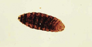
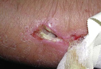
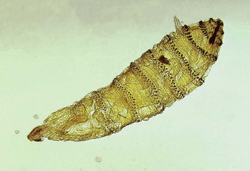
Myiasis may be further stratified clinically as furuncular (subcutaneous) myiasis, wound (superficial cutaneous) myiasis, cavitary (atrial or invasive) myiasis, intestinal myiasis, urinary myiasis, and vaginal myiasis. Furuncular myiasis is the most common clinical manifestation of myiasis and occurs when one or more larvae penetrate the skin, causing pustular lesions that resemble boils or furuncles. Larval maggots can also infest external orifices, sores, or open wounds, causing cavitary and wound myiasis. Cavitary myiasis is usually caused by screwworm larvae that can penetrate festering wounds or invade the orbits, nostrils, or external ear canals (Figs. 296-1 and 296-2). Intestinal myiasis is uncommon, usually caused by the accidental ingestion of maggot-contaminated food, and characterized by self-limited nausea, vomiting, and diarrhea. Genitourinary myiasis is also uncommon and may present as dysuria, hematuria, and pyuria, following larval invasion of the urethra (urinary myiasis) or vagina (vaginal myiasis).

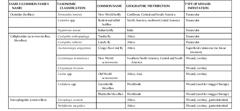
Although there are many families of dipterous flies (order Diptera), flies from three families cause most human and animal myiasis: Oestridae, or botflies; Calliphoridae, screwworms and blowflies; and Sarcophagidae, carrion-feeding flies. The most common myiasis-causing fly species are classified taxonomically and stratified by clinical type of myiasis infestation in Table 296-1.
Epidemiology
In a retrospective epidemiologic study in Rio de Janeiro, Marquez and colleagues described 71 patients with furuncular and cavitary myiasis during the period 1999 to 2003.1 Myiasis was more prevalent among adults older than 51 years (42%) and children younger than 10 years (34%). Most of the population was male (61%) and impoverished (62%). The predominant causative agent of furuncular myiasis was Dermatobia hominis, the New World human botfly, and the predominant causative agent of cavitary myiasis was Cochliomyia macellaria, an indigenous species of New World screwworm. The authors concluded that myiasis is an opportunistic infestation of disadvantaged vulnerable populations living in nonhygienic conditions. In a similar retrospective collective analysis, Jiang described 54 cases of human myiasis in China from 1995 to 2001.2 Although the Chinese cases were equally distributed between genders, most cases occurred in infants and children (72%) and were described as either hypodermic-invasive (n = 31) or ocular (n = 12). In another collective review, Schwartz and Gur reported 12 cases of furuncular myiasis caused by D. hominis, the human botfly, in 12 Israeli travelers returning from four South American countries in the Amazon Basin.3 Clyti and co-workers described an epidemic of human botfly furuncular myiasis in 30 patients living in coastal urban communities of French Guiana between January and March 2000 after heavy rainfall that accelerated soil maturation of pupae into adults.4
Tamir and associates reported two cases of furuncular myiasis caused by Cordylobia rodhaini, Lund’s fly, in Israeli travelers returning from Ghana.5 In addition to D. hominis and Cordylobia spp., Cuterebra species of botflies can also cause furuncular myiasis in North America, as well as throughout Africa and Asia (Fig. 296-3). Shorter and co-workers reported two cases of Cuterebra spp. botfly-induced furuncular myiasis in children in New England (see Fig. 296-3).6 Cuterebra or rabbit botflies are endemic in the northeastern United States and southeastern Canada, where they have established a zoonotic reservoir in lagomorphs (rabbits and hares) and cause periodic cluster outbreaks of furuncular myiasis in domestic animals and humans.7


