Inherited Coagulation Disorders


Inherited Coagulation Disorders


Jerry S. Powell

George M. Rodgers


Inherited disorders of coagulation result from the deficiency or functional abnormality of one of the plasma proteins involved in providing normal coagulation.1 There are other proteins that regulate the kinetics of coagulation, and abnormalities in one of these proteins usually result in increased risk of thrombosis; these proteins will not be covered in this chapter. If von Willebrand disease (vWD) is included, the inherited coagulation abnormalities (Table 53.1) are common; for example, vWD may affect up to 1% of the population. The other inherited coagulation disorders occur less often.

Since the purpose of the coagulation cascade is to rapidly produce fibrin to form a clot, the clinical manifestation of any deficiency or functional abnormality in any coagulation protein will be similar, i.e., clinical bleeding, with greater bleeding associated with more severe deficiency. Therefore, the manifestations and complications of clinical bleeding will be described in this chapter in the section on hemophilia A. A recent review of treatment of hemophilia A has been published with proposed algorithms for therapy.2


TABLE 53.1 INHERITED DISORDERS OF COAGULATION


X-linked recessive traits


Hemophilia A


Hemophilia B (i.e., CRM+ and CRM variants; hemophilia Bm, B Leyden, etc.)


Autosomal recessive traits


Factor XI deficiency


Prothrombin deficiency


Factor V deficiency


Factor VII deficiency


Factor X deficiency (i.e., Prower variant, Stuart variant, Friuli variant, others)


Afibrinogenemia


Hypofibrinogenemia


Factor XII deficiency


Factor XIII deficiency


Autosomal dominant traits


von Willebrand disease


Dysfibrinogenemias


Combined abnormalities


Associated with factor VIII deficiency (i.e., factor V deficiency, hemophilia B, factor XI deficiency, factor VII deficiency, von Willebrand disease, dysfibrinogenemias, platelet dysfunction)


Involving vitamin K-dependent factors (i.e., factors II, VII, IX, and X; factors IX and XII; others)


Miscellaneous


Prekallikrein deficiency


High-molecular-weight kininogen deficiency


Deficiency of physiologic inhibitors (i.e., α2-antiplasmin, abnormal α1-antitrypsin [antithrombin Pittsburgh])


CRM, cross-reacting material.


NOMENCLATURE

Historically, the best known inherited coagulation disorder is hemophilia, which dramatically manifested itself in the royal families of Europe in the 20th century, following a spontaneous mutation that apparently arose in Queen Victoria in England. The international Roman numeral designations for these disorders are summarized in Table 18.1. Like abnormal hemoglobins, qualitatively abnormal fibrinogens are designated by the name of the city in which they were first discovered, as in fibrinogen Paris.


HEMOPHILIA A

Hemophilia A refers to the inherited coagulation disorder characterized by deficiency of function of the coagulation protein called factor VIII. Except for vWD, it is the most common inherited coagulation disorder, and is X-linked. As the most common bleeding disorder, historians assume that it is the severe and often fatal hemorrhagic diathesis that affected the male children of certain families and that was well recognized in antiquity, as noted in the writings of Rabbi Simon ben Gamaliel (2nd century A.D.) in the Talmud, and those of Maimonides, the Hebrew physician and philosopher, and Albucasis, the Arab (twelfth century).4 Complete monographs have reviewed the early literature.5 However, the bleeding disorder that occurred in males throughout the royal families of Europe due to a mutation that arose with Queen Victoria, long assumed to be the more common hemophilia A, turned out, after extensive medical forensic investigation, to be hemophilia B.6


FIGURE 53.1. The relationship of factor VIII procoagulant activity and factor VIII-related antigen (von Willebrand factor [vWF]) in the plasma of normal subjects and patients with hemophilia or von Willebrand disease. (From Hoyer LW. The factor VIII complex: structure and function. Blood 1981;58:1-13, with permission.)


Incidence

Hemophilia A has been recognized in all areas of the world and in all ethnic groups. Estimates of its incidence approximate 1 in 5,000 males, or 1 in 10,000 persons.9, 10 A population-based study of the southeastern United States by the Centers for Disease Control and Prevention found that 11 cases per 100,000 persons had hemophilia A and that the prevalence was similar among different racial groups.11 This same survey estimated that in 1994, there were 13,320 cases of hemophilia A in the United States.11


Genetics

Hemophilia A is a classic example of an X-linked recessive trait. In such a disorder, the defective gene is located on the X chromosome12; the factor VIII gene maps to Xq28, on the distal end of the long arm of the X chromosome. In males who lack a normal allele, the defect is manifested by clinical hemophilia (Fig. 53.2; generation I, number 1). The affected male does not transmit the disorder to his sons (generation II, numbers 4 and 5) because
his Y chromosome is normal. However, all of his daughters are obligate carriers of the trait because they inherit his X chromosome (generation II, numbers 2 and 3). Most of these women are unaffected clinically because of the presence of a normal allele from the mother. The female carrier transmits the disorder to half of her sons (generation III, numbers 6 and 7) and the carrier state to half of her daughters (generation III, numbers 8 and 9). Due to Lyonization of the X chromosome, if the normal factor VIII allele is inactivated more often by chance, then some of the carrier females may have clinical bleeding. The severity of their bleeding depends on their factor VIII activity level; and, rarely, a woman can have very low factor VIII activity and present with symptoms of moderate or even severe hemophilia.


FIGURE 53.2. The inheritance of hemophilia A and hemophilia B. The pedigree is hypothetical. Squares indicate male; circles indicate female; fully shaded squares or circles indicate affected members; half-shaded circles indicate carriers. X, normal X chromosome; x, abnormal X chromosome.

The severity of bleeding varies in different kindreds, and closely depends on the particular genetic defect. Since the same gene defect is present in the kindred, each of the affected males will have very similar clinical phenotypes.13 As expected, hemophilia A has been observed several times in twins.14 Occasionally, however, the coinheritance of other genetic defects can influence the clinical symptoms of hemophilia A patients. An example is the coinheritance of the factor V Leiden mutation (discussed in Chapter 55) with hemophilia A. These patients have a milder clinical phenotype than expected for the same molecular defect in factor VIII.15 The coinheritance of the factor V Leiden mutation or other prothrombotic risk factors in children with severe hemophilia A may delay the first symptomatic bleeding event.16

Intriguing, and yet to be fully explained, is the fact that in over a third of families with hemophilia A, there is no evidence or history of abnormal bleeding in other members of the family.17, 18 This percentage is consistent with the Haldane hypothesis, which predicted that maintenance of a consistent frequency of a genetic disorder in the population would require that approximately onethird of cases result from spontaneous mutation. In other instances, neonatal deaths or the passage of the trait through a succession of female carriers may explain the negative family history. For practical purposes, therefore, a negative family history is of little value in excluding the possibility of hemophilia A. Mutations that cause hemophilia A originate in males at least threefold more often than in females.19 The large size of the factor VIII gene (186 kb), the presence of hot spots (e.g., CpG dinucleotides), and the fact that the X chromosome is unpaired in males may predispose for the factor VIII gene to undergo spontaneous mutation.20, 21, 22, 23

Nearly 3,000 individuals with hemophilia have had their factor VIII genes analyzed; this genetic information is collected in the Hemophilia A Mutation Database (HADB, formerly the Hemophilia A Mutation, Structure, Test and Resource Site[HAMSTeRS] database).21 The genetic defects of hemophilia A encompass deletions, insertions, and mutations throughout the factor VIII gene.24 Point mutations involving CpG dinucleotides are especially common. Approximately 5% of patients with hemophilia A have large (>50 nucleotides) deletions in the factor VIII gene.25

Approximately 45% of severe hemophilia A cases result from a major inversion of a section of the tip of the long arm of the X chromosome, one breakpoint of which is situated within intron 22 of the factor VIII gene.26 This common inversion is associated with severe hemophilia A, and a higher incidence of inhibitor formation. It is presumed that in the absence of homologous X chromosome pairing during male meiosis, an intrachromosomal recombination event occurs on the single X chromosome, resulting in this inversion. This event typically occurs in the father of the mother of a child with severe hemophilia A, and often leads to relief of guilt feelings in the mother, as the genetic mutation is not “her fault.” Another common inversion (intron 1) accounts for 5% of patients with severe factor VIII deficiency.27 Thus, two inversions of the factor VIII gene are seen in nearly one-half of all severely affected patients with hemophilia A. In contrast, point mutations are most likely found in patients with mild to moderate hemophilia A.28 The first case of hemophilia A caused by unequal homologous Alu/Alu recombination has been reported.29 Because there are ˜50 Alu repeats in the factor VIII gene, it is possible that many cases of hemophilia may result from this mechanism.29 More than 95% of hemophilia A patients have mutations detected.22, 30, 31 Approximately 2% of hemophilia A patients have no detectable mutations in the coding region of the factor VIII gene.32 These results indicate that a substantial number of families with severe hemophilia A can undergo accurate gene tracking and carrier analysis. The intron 1 and 22 inversions responsible for 50% of severe hemophilia A cases can be sought using long and inverse polymerase chain reaction (PCR) techniques. Various algorithmic approaches to rapid laboratory genetic testing have been described,30, 33, 34 and a recent method of performing linkage analysis of hemophilia A in families in whom mutations could not be identified has been reported.35

There is a strong association between genetic mutations that lead to absence of large portions of the factor VIII protein and the development of antibodies to therapeutic factor VIII infusions. These antibodies generally develop within the first 50 infusions of factor VIII, as expected when the immune system detects a protein to which it has not been exposed previously.


Variants

Hemophilia A with autosomal dominant transmission has been reported; however, it is important to distinguish these “variant hemophilia A patients” with negative X-linked transmission from patients with variant vWD (type 2N, vWD Normandy), an autosomal vWD subtype caused by defective factor VIII binding to vWF and with a clinical picture similar to hemophilia A.36


Carrier Detection

Despite the high rate of spontaneous de novo mutations in hemophilia A, detection of females who are carriers is important. When the family history of a patient suggests risk for being a carrier, then coagulation-based assays are performed, followed by DNA testing. The daughter of a man with hemophilia is an obligate carrier, whether she has symptomatic bleeding or not. Similarly, if a female has two sons with hemophilia, then she is likely a carrier; if she has one son with hemophilia and a family history of hemophilia, then she is likely a carrier; but if she has one son with hemophilia and a negative family history, then the odds are approximately 67% that she is a carrier. A number of specialized laboratories perform molecular testing for the factor VIII gene. It is most efficient and less expensive if the DNA of the relative with hemophilia is available to identify the specific genetic mutation in that kindred; then only that specific mutation is evaluated in the potential carrier.37


Coagulation-based Assays

Coagulation-based assays may be useful in confirming or excluding the carrier state, although they have limitations. The regularity with which the abnormal factor VIII gene is suppressed by the normal allele in female carriers of hemophilia varies because of the phenomenon of random X chromosome inactivation (the Lyon hypothesis). Thus, although the mean concentration of factor VIIIc in the plasma of heterozygous female carriers is ˜50% of that in normal women,13, 38 observed values scatter widely around this mean and often overlap with those found in the normal population. This is a result, in part, of the large error of assay methods and the wide range of factor VIIIc levels in normal subjects.

Although the demonstration of low levels of factor VIIIc by means of the usual assay methods strongly suggests the presence of the carrier state, the converse statement cannot be made with equal certainty—that is, the presence of normal levels of factor VIIIc does not reliably exclude the carrier state.39 Furthermore, pregnancy and hormonal medications may increase the levels of factor VIIIc in female carriers.40 Normal women and carriers with blood types A, B, or AB have higher levels of factor VIIIc and vWFAg than those with blood type O.41

The use of immunoassays for vWF has improved carrier detection in hemophilia A.42 These methods allow measurement of levels of vWF, which are normal43 or increased in carriers of the disorder, despite mild but variable deficiencies of factor VIIIc. Results obtained when a bioassay and an immunoassay are performed on the same sample, and the ratio of VIIIc to vWF is computed, differentiate between the carrier population and the normal population with minimal overlap.44 This ratio normally ranges from 0.74 to 2.20 and was found to be from 0.18 to 0.90 in obligatory carriers. The overall detection rate ranged from 72% to 94% in such obligatory carriers46 and from 48% to 51% in women without hemophilic sons or fathers.42 In the latter group (possible carriers), 50% would be predicted to be carriers. Abnormally low ratios of VIIIc:vWF (false positives) have been encountered in an occasional normal subject,43, 45 which may be attributable to nonspecific variables such as stress.46 Pregnancy and the use of oral contraceptives, blood type,41 or contamination of plasma samples with thrombin46 or other proteolytic enzymes may produce falsely high ratios (false negatives) in documented obligatory carriers. Linear discriminant analysis47 has been recommended for the expression and analysis of such laboratory data. The carrier detection rates using standardized assays with discriminant analysis exceed 90%.48


DNA-based Assays

Molecular analyses of the factor VIII gene identify mutations in 97% of hemophilia A patients. Based on the frequency of the intron 22 inversion, severe hemophilia A patients should be initially screened for this defect. Inversion-negative patients and those with mild or moderate hemophilia A should have systematic sequencing of the factor VIII promoter, exons, and splice junctions performed. Such an approach has been reported to identify mutations in 97% of hemophilia A patients.30

DNA-based assays have been adapted for prenatal diagnosis,49 and for pre-implantation genetic diagnosis in association with in vitro fertilization. One commonly used method is to obtain chorionic villi samples during the eleventh to twelfth gestational week and perform direct genotype testing.50 Alternatively, genetic linkage analysis of polymorphisms can be performed. The subject of genetic counseling in hemophilia has been reviewed.51


Hemophilia in the Female

Hemophilia has been well documented in human females.44, 52 The most common form is that seen in a minority of heterozygous carriers, discussed previously, in whom X chromosome inactivation may occur at an unusually early stage of embryogenesis, resulting in unusually low levels of factor VIII.

A second cause of female hemophilia is a mating between an affected male and a carrier female (Fig. 53.2; generation IV, number 10).53 One-half of the female offspring of such a match would inherit two abnormal X chromosomes, one from the father and one from the mother. Such homozygous female hemophilia was once thought to be lethal and to inhibit the development of the embryo. That this is not true was first suggested by the successful experimental production of hemophilia in female dogs. Homozygous hemophilia is now well authenticated in several women53, 54 and resembles the disorder seen in affected males in all respects.


Clinical Manifestations of Hemophilia

Prior to effective replacement therapy, the life expectancy for a child born with severe hemophilia was less than 20 years. These boys would die from exsanguinating hemorrhage after a trivial traumatic injury, from spontaneous internal bleeding or from spontaneous intracranial hemorrhage. It is dramatic that a boy born today with severe hemophilia A can expect to have a normal life expectancy.

However, the most characteristic bleeding manifestations in hemophilia are spontaneous bleeding in weight-bearing joints, leading to severe hemarthrosis. The frequency and severity of these joint bleeds are related to the functional activity level of factor VIII in plasma8, 55 (Table 53.3). Three categories of severity have been defined by a consensus committee on the basis of FVIII activity levels (Table 53.3).56 Severe hemophilia (factor VIII level <1 IU/dl) is manifested clinically by repeated and severe hemarthroses, resulting almost invariably in crippling arthropathy in the absence of replacement therapy; such severe cases often are called classic hemophilia. Moderate hemophilia (factor VIII level of 1 to 5 IU/dl) is associated with less frequent and less severe hemarthroses and seldom results in serious orthopedic disability. In mild hemophilia (factor VIII level of 6 to 40 IU/dl), hemarthroses and other spontaneous bleeding manifestations may be absent altogether, although serious bleeding may follow surgical procedures or traumatic injury.57 As indicated in Table 53.3, most patients with hemophilia A have severe disease. However, one epidemiologic survey reported variable differences in the incidence of severity between individual states within the United States; for example, 13% of hemophilia cases were of moderate
severity in Massachusetts, whereas 35% of cases in Georgia were classified as moderate severity.11


TABLE 53.3 PREVALENCE AND SEVERITY OF HEMOPHILIA A AND HEMOPHILIA B IN THE UNITED STATES


Incidence (%)b


Factor VIII or IX Level (IU/dl)


Clinical Picturea


Hemophilia A


Hemophilia B


<1


Severe, spontaneous bleeding


70


50


1-5


Moderate bleeding with minimal trauma or surgery


15


30


6-40


Mild bleeding with major trauma or surgery


15


20


a The criteria for classifying hemophilia severity are taken from White GC, Rosendaal F, Aledort M, et al. Definitions in hemophilia. Thromb Haemost 2001;85:560.

b Incidence figures are for the United States in 1989 and were provided by the National Hemophilia Foundation (Courtesy of Kathleen F. Cortes, PhD).


From Rodgers GM. Common clinical bleeding disorders. In: Boldt DH, ed. Update on hemostasis: contemporary management in internal medicine. New York: Churchill Livingstone, 1990:75-120, with permission.


Hemarthrosis

Hemarthrosis is the most common manifestation of the inherited coagulation disorders (Fig. 53.3). Joint bleeding and the consequent damage to weight-bearing joints remain the most common debilitating symptom in severe hemophilia.


Clinical Presentation

Most persons with severe hemophilia report a characteristic warm, tingling sensation before the onset of symptoms of joint bleeding and hemarthrosis; this is called the aura. The earliest definite symptom is pain, which in the acute form may be excruciating. Physical examination reveals muscle spasm and limited motion of the affected joint. If therapy is not initiated immediately, typically within approximately 30 minutes, then the joint may progress so that it is warm and grossly distended and discolored,
but external evidence of bleeding may be minimal or absent in chronically damaged large joints because of thickening of the articular capsule. Generally, only one joint is involved at a time, although bleeding may develop simultaneously in two or more joints. The weight-bearing joints are most commonly affected and the knees are the joints most often severely affected. However, because of the success of total knee and total hip replacement surgeries, the ankles are the most commonly affected joints that lead to chronic problems that interfere with quality of life for persons with severe hemophilia.


FIGURE 53.4. Elbow and knee joints in a patient with hemophilia A. Thickening of synovium with deposition of calcium is shown in (A) and (A1); increased intercondylar notch is shown in (B); increased density and decreased interarticular space are shown in (A), (B), and (C); and lipping along the borders of the joint surfaces is shown in (C).


TABLE 53.4 SCORING SYSTEM FOR HEMOPHILIC ARTHROPATHY USING MAGNETIC RESONANCE IMAGING


Score


Abnormalities on Imaging


O


None


I


Minimal hemosiderin


II


Large amount of hemosiderin and cartilaginous erosion


III


Cartilage destruction, bone erosion, subchondral cysts


IV


Osteoarthritis with or without ankylosis


Subcutaneous and Intramuscular Hematomas

Large ecchymoses and subcutaneous and intramuscular hematomas were common in hemophilia A prior to the use of regular infusion therapy, reflecting the substantial amounts of time patients had factor levels less than 1% of normal. With modern treatment protocols designed to keep plasma levels above 1% of normal, and immediate home infusion when a bleeding episode occurs, the frequency of large soft tissue bleeds has decreased dramatically. Such hemorrhages, when not treated promptly, characteristically spread within fascial spaces and dissect deeper structures (see Fig. 45.2). Subcutaneous bleeding may be extensive. When not treated promptly with factor infusion, the bleeding continues; and at the site of origin, the tissue is hard, indurated, raised, and purplish black. From this center, the hemorrhage extends in all directions, with each successive concentric extension less deeply colored. The point of origin of the hemorrhage may be absorbed entirely while the margin is still progressing. Intramuscular and subcutaneous hematomas may produce leukocytosis, fever, and severe pain in the absence of significant discoloration of the overlying skin.

Hematomas may produce serious consequences from the compression of vital structures. Bleeding into the tongue, throat, or neck may develop spontaneously and is especially dangerous because it may compromise the airway with surprising rapidity.63 Gangrene may result from pressure on arteries; and the development of compartment syndrome and, if not promptly treated, ischemic contractures are common sequelae of hemorrhage into the calves or forearms. Peripheral nerve lesions of varying severity are common complications of untreated hemorrhage into joints or muscles. Hemophilic cysts are discussed in the section Special Aspects of Treatment.


Psoas and Retroperitoneal Hematomas

Spontaneous hemorrhage into internal fascial spaces and muscles of the abdomen is common in severe hemophilia A,64 reflecting plasma factor levels less than 1% of normal. Bleeding into or around the iliopsoas muscle produces pain of progressively increasing severity and tenderness; when it occurs on the right side, it may closely simulate acute appendicitis. Femoral nerve involvement may be partial or complete, with the development of pain on the anterior surface of the thigh. The psoas sign is positive, and the hip is held in partial flexion. Paresthesias, partial or complete anesthesia, and, ultimately, weakness or paralysis of the thigh extensors with eventual muscular atrophy may ensue. Retroperitoneal hemorrhage and intraperitoneal hemorrhage also are common. Computed tomography may be helpful in the diagnosis of these hematomas.64 Prompt factor infusion is critical to normalize coagulation and should be continued until the hematoma has resolved completely, as any residual hematoma is at higher risk for rebleeding and pseudocyst formation.


Gastrointestinal and Genitourinary Bleeding

Hemorrhage from the mouth, gums, lips, frenulum, and tongue is common and often serious. The eruption and shedding of deciduous teeth usually occur without abnormal bleeding, but may occasionally be accompanied by hemorrhage that lasts for days or weeks if not treated. Epistaxis occurs in many patients and may be of exsanguinating proportions.

Hematemesis, melena, or both are not uncommon. The source of the bleeding is usually the upper gastrointestinal tract. In most patients in whom bleeding is persistent or recurrent, it originates from a structural lesion, most commonly a peptic ulcer or gastritis. Hemorrhage may be accompanied by abdominal pain, distention, increased peristalsis, fever, and leukocytosis. Intramural bleeding into the intestinal wall may result in intussusception or obstruction.

Hematuria is more common than gastrointestinal bleeding, but it is less often the result of a demonstrable pathologic condition in the genitourinary tract. The bleeding may arise in the bladder or in one or both kidneys and may persist for days or weeks.65 When clots form, ureteral colic may develop. Typically, the hematuria resolves after factor infusion, but if persistent, then a short course of prednisone may prove helpful to shorten the course of hematuria.66


Traumatic Bleeding

Patients with coagulation disorders seldom bleed abnormally from small cuts such as razor nicks, reflecting the normal function of platelets, as measured by platelet function tests. All laboratory assays of platelet function are normal in patients with severe hemophilia A. After larger injuries, however, hemorrhage out of proportion to the extent of the injury is characteristic. The bleeding reflects both increased acute bleeding rates beyond what is expected after the trauma, and persistent bleeding as slow continuous oozing occurs for days, weeks, or months. Such traumatic bleeding may be massive and life-threatening unless coagulation replacement therapy is provided by immediate factor infusion.

Delayed bleeding is common. Thus, although hemostasis after an injury or a minor surgical procedure may appear to be adequate, hemorrhage, often of sudden onset and serious proportions, may develop several hours or even days later. This phenomenon apparently occurs because the processes of primary hemostasis are only temporarily effective. Delayed bleeding may occur in patients with mild hemophilia and is a significant hazard after minor surgical procedures, particularly those performed on an outpatient basis, such as tooth extraction and tonsillectomy.

Venipuncture, if skillfully performed, is without danger for the person with severe hemophilia, primarily because of the elasticity of the venous walls. If venipuncture is traumatic, digital pressure on the puncture site or a pressure dressing may prevent further complications. Subcutaneous, intracutaneous, and small intramuscular injections seldom produce hematomas if firm finger pressure is maintained for at least 5 minutes. Large intramuscular injections should be avoided. Vaccination using intramuscular injections is acceptable with minimal increase in bleeding risks.


Other Clinical Aspects

Infants usually are asymptomatic because they are insulated from trauma.67 However, trauma during birth or afterwards may trigger
life-threatening bleeding. Infants of women who are known carriers may be at risk during delivery, and instrumentation during vaginal delivery should be avoided. Currently, the usual standard of care is to avoid testing the infant in utero due to the risks inherent in current techniques available to test the fetus, and to allow normal vaginal delivery with back-up Caesarean section available if any difficulties arise during birth. Typically, hematomas are seen first when children become active, and hemarthroses seldom develop until they begin to walk. Occasionally, evidence of the disorder is not seen until patients reach teenage years or young adult life. Spontaneous hemorrhage may be cyclic in nature. Petechiae, which are characteristic of disorders of platelets and blood vessels, are rare in patients with hemophilia but have been noted in severely affected patients during an exacerbation of bleeding. Hemorrhage from the umbilical cord or stump is unusual, but prolonged bleeding after circumcision is common and brought hemophilia to the attention of the ancient Hebrews. Pulmonary and pleural bleeding are uncommon, although mediastinal and pleural shadows have been noted radiographically and presumably originate from fresh or old hematomas. Intraocular hemorrhage is uncommon, but bleeding into the orbit and conjunctiva occurs often. Spontaneous rupture of the spleen has been reported. Intracranial bleeding is discussed in the section Special Aspects of Treatment. The website for the National Hemophilia Foundation is www.hemophilia.org and contains useful information and links to more detailed information on this bleeding disorder.


Course and Prognosis

In recent years, the prognosis in severe hemophilia has improved dramatically. With modern therapy using regular factor infusions to maintain adequate plasma factor concentrations to prevent nearly all spontaneous bleeding episodes, a person born with severe hemophilia A can expect to live an essentially normal lifespan. No other genetic disease has made such dramatic progress in the past 50 years. It is not yet clear whether hemophilia protects older hemophilia patients from thromboembolic disorders,68 atherosclerosis,69 or cardiovascular diseases. Despite the lack of stringent prospective clinical studies for this emerging problem of aging patients with severe hemophilia and other medical problems, reviews of current practice and expert opinion on how to treat aging patients with severe hemophilia have been published.70, 71, 72, 73


TABLE 53.5 LABORATORY FINDINGS IN COMMON INHERITED COAGULATION DISORDERS


Disorder


Partial Thromboplastin Time (PTT)


Prothrombin Time


Thrombin Time


Ancillary Tests


Hemophilia A


A


N


N


vWF antigen and activities are normal or increased, ratio VIIIc:vWF is low.


Hemophilia Ba


A


Na


N


von Willebrand diseasea,b


A or N


N


N


vWF antigen and VIIIc are usually low, ratio VIIIc:vWF is variable; ristocetin-induced platelet aggregation and ristocetin cofactor activity are usually diminished.


Afibrinogenemia


A


A


A


Platelet function may be abnormal.


Dysfibrinogenemiaa


vA


vA


Ac,d


Hypofibrinogenemia,e reptilase time is prolonged,a fibrin(ogen) degradation products levels are increased.a


Hypoprothrombinemiaa


A


A


N


Two-stage assay is abnormal.f


Factor V deficiency


A


A


N


Factor VII deficiencya


N


A


N


Stypven (Russell viper venom) time is normal.


Factor X deficiencya


A


A


N


Stypven time is abnormal.


Factor XI deficiency


A


N


N


Factor XII deficiency


A


N


N


Factor XIII deficiency


N


N


N


Clot solubility tests are abnormal.


A, abnormal; N, normal; v, variable; vWF, von Willebrand factor.


Certain PTT reagents may not detect mild deficiency of factors VIII, IX, and XI.


a Findings are significantly different in some variants.

b Coagulation abnormalities are caused by deficiency of factor VIIIc.


c Patient’s plasma may inhibit normal coagulation.


d Abnormality may be corrected by increasing calcium concentration and may be magnified by diluting the thrombin solution.


e Abnormality varies depending on technique.


f Results of one-stage techniques may be uninterpretable.


HEMOPHILIA B

Hemophilia B was recognized as a separate disorder from hemophilia A in 1947.94 Hemophilia B (Christmas disease, factor IX deficiency) was established as different from hemophilia A by Aggeler et al. in 1952.95, 96 Hemophilia A is approximately 5 times more common than hemophilia B.18 A 1994 survey estimated that there were 3,640 cases of hemophilia B in the United States.11 As expected, it appears that the severity of hemophilia B is a consequence of the plasma factor IX concentration, and that for any particular plasma concentration, the clinical severity is similar for hemophilia A and B. However, in contrast with hemophilia A, most of the genetic causes of hemophilia B are not large deletions or inversions; therefore in many patients there is some antigenic protein, and often some low level of function of the mutated protein. Consequently, the clinical phenotype is often less severe. As with hemophilia A, hemophilia B is classified as severe, moderate, or mild based on the percentage of functional factor IX in coagulation activity assays. In addition, because of the presence of low levels of factor IX protein in more patients with hemophilia B, there are fewer persons with hemophilia B that develop neutralizing inhibitors (approximately 3% versus more than 25% in hemophilia A).

Historically, when plasma from patients with hemophilia B was tested against autologous antibodies, three distinct groups were defined: A cross-reactive material (CRM)-positive variant, the most common form; a CRM-negative variant; and a third form of the disorder in which antibody neutralization was variable in extent and was approximately proportional to coagulant factor IX activity (the CRM-reduced variant).97 These distinctions are only of historical interest now.

In a disorder called hemophilia B Leyden,98 the clinical manifestations tend to diminish during puberty in association with a rise in the factor IX level from as low as 1 IU/dl in childhood to levels of 20 IU/dl or more in adult life. The Leyden variant is characterized as CRM-negative at birth but becomes CRM-positive or CRM-reduced with advancing age.98 The genetic basis for factor IX Leyden is that mutations occur in the factor IX gene promoter region; this region contains an androgen response element that, with age, stimulates factor IX gene transcription and protein synthesis.99

In another variant of hemophilia B, the PT is prolonged when performed with ox brain thromboplastin.100 This disorder has been called hemophilia Bm and is characterized by the presence in the plasma of CRM that neutralizes both autologous and heterologous antibodies to factor IX.101 The degree of abnormality of the ox brain PT is proportional to the plasma level of CRM in both affected males and carrier females. This variant results from mutations in the carboxy terminus of the factor IX molecule, resulting in a factor IX molecule that interacts abnormally with ox brain thromboplastin.

Numerous factor IX mutations have been recognized.22, 102 Factor IX purified from the plasma of affected members of one kindred did not fragment normally when activated in vitro (hemophilia B Chapel Hill).101 The molecular defect was found to be a substitution of histidine for arginine at position 145, a defect that inhibits cleavage by factor XIa.103 Studies of factor IX obtained from another family104 revealed biochemical abnormalities identical to those characteristic of the descarboxy analog of this factor that is found in vitamin K deficiency or produced by coumarin drugs. Such molecules lack Ca2+-binding sites and do not undergo conformational changes induced by Ca2+. Another report concerns a unique variant with deficient Ca2+ binding and an abnormally high-molecular-weight (factor IX Zutphen).105


Genetics

Hemophilia B is inherited as an X-linked recessive trait, but the locus on the X chromosome of the gene controlling factor IX production is remote from that involved with factor VIII biosynthesis.106 Factor IX levels <10% have been documented in a few women, including some with chromosomal abnormalities.107, 108, 109 Ten useful polymorphisms have been described that are associated with the factor IX gene.49 An updated listing of mutations is available at www.kcl.ac.uk/ip/petergreen/haemBdatabase.html. Unlike hemophilia A, the spontaneous mutation rate is low,17 and most patients with hemophilia B have positive family histories.

Factor IX defects may be severe, moderate, or mild. Severe defects result from large gene deletions, nonsense mutations, and inversions; these defects are associated with absence of factor IX protein. Milder gene defects such as splice-site or missense mutations result in a dysfunctional protein with some residual activity. Missense mutations account for ˜80% of mutations in severe hemophilia B patients.22, 110, 111, 112


Detection of Carriers

Detection of heterozygous carriers of hemophilia B involves the same principles and limitations as described for hemophilia A. Carrier detection based on coagulation assay alone usually is more reliable than is the case with hemophilia A.113 Thus, in one series of 45 obligatory carriers, the mean factor IX level in the plasma was 33 IU/dl; 40 from the group had levels <50 IU/dl, and 10 had levels <25 IU/dl. As a consequence of these low levels of factor IX, abnormal hemorrhage is not uncommon in carriers of hemophilia B.

Results of immunoassays of factor IX and the ratio of factor IX-related antigen to coagulant factor IX levels in the carrier population overlap with the normal population to a considerable degree, particularly in the CRM-positive variants.114, 115 Such studies provide a lower overall detection rate among obligatory carriers of hemophilia B than is the case with hemophilia A.114, 115 Levels of factor IX-related antigen obtained by immunoassay
are significantly increased by the use of oral contraceptives. The use of DNA probes116 and monoclonal immunoassays117 provide highly accurate methods for determining carrier status. Rapid carrier testing using allele-specific microarray methods has been described. This method permits detection of a majority of common factor IX mutations in a single assay.118 Prenatal diagnosis of hemophilia B has been successful in the CRM-negative variants.119 Modern methods for prenatal diagnosis use genetic testing and are quite reliable.120, 121 Determination of carrier status is based on several factors, including pedigree analysis, factor IX assay results, and genotype.122


Clinical Features

Severely affected patients (those with factor IX activity levels <1 IU/dl) are less common than in hemophilia A (Table 53.3), but the clinical manifestations of the two disorders are identical for the same activity levels of the coagulation factors. Specific factor assays are necessary to distinguish between hemophilia A and hemophilia B. Mild factor IX deficiency should always be considered in the differential diagnosis of patients with coagulation-type bleeding and normal routine coagulation test results (PT, PTT).123 Many PTT reagents do not detect mild factor IX deficiency (factor IX levels of 20% to 30%).74


VON WILLEBRAND DISEASE

The historical confusion that has surrounded the pathogenesis of vWD is apparent from the many names that have been applied to this disorder. These designations include angiohemophilia, vascular hemophilia, pseudohemophilia, constitutional thrombopathy, and idiopathic prolonged bleeding time. von Willebrand first recognized the disorder in a 1926 study of the inhabitants of the Åland Islands.
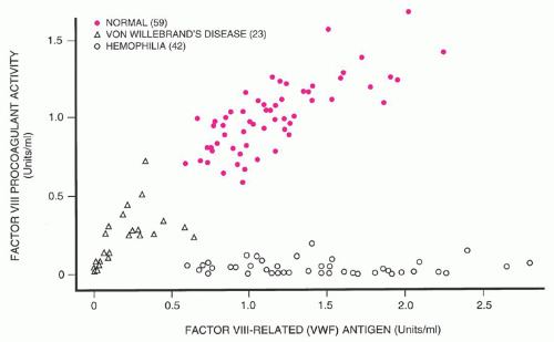
125 Three cardinal manifestations of this bleeding disorder are mucocutaneous hemorrhage rather than hemarthrosis and deep-muscle bleeds; autosomal dominant inheritance, rather than sex-linked as seen in hemophilia A; and prolonged bleeding time. Evaluation of one large kindred revealed variable severity of bleeding symptoms, with some obligate heterozygote carriers being asymptomatic. von Willebrand thought that the hemostatic defect resulted from combined defects in platelet function and vascular endothelium. The discovery that plasma from normal volunteers or patients with hemophilia A corrected the hemostatic defect of vWD suggested that a plasma protein distinct from factor VIII caused vWD. In 1972, von Willebrand factor (vWF) was purified. Genetic variants were correlated with various vWF structural abnormalities, and demonstration of the heterogeneous nature of vWD was furthered by development of vWF multimer analysis by gel electrophoresis.126 In its most common form (type 1), the disorder is characterized by mild mucocutaneous hemorrhage, which is attributable to deficiency of both vWF and factor VIIIc. The disorder is not homogeneous, however, in part because of the multiple physiologic functions played by vWF. The recognition of a number of variants, together with the demonstration of multiple genetic patterns, suggested that vWD is very heterogeneous. However, all forms of vWD can be traced to insufficient amounts of vWF or defective function of this protein. Recent reviews of this area have been published,127, 128 and a consensus document on vWD sponsored by the National Institutes of Health reviewed all aspects of the disorder.129, 130


Incidence

Epidemiologic studies indicate that vWD is the most common bleeding disorder, affecting about 1% of the population.131 The high incidence of the disease is not limited to certain ethnic groups 131, 132; however, only a fraction of people come to medical attention because of bleeding symptoms. This may be because of either the relatively mild nature of the disease in many affected individuals, or a lack of recognition by patients of excessive bleeding in response to either physiologic challenge (e.g., heavy menstrual bleeding) or trauma.


Nomenclature

The identification of numerous variants of vWD has led to attempts to simplify classification of this disorder. An updated classification system has been proposed (Table 53.6).128 Quantitative defects are divided into partial deficiency (type 1) and severe deficiency with virtually complete absence of vWF (type 3). The qualitative defects (type 2) are divided into four categories according to the nature of the defect of vWF function. Type 2A refers to variants with impaired interaction between vWF and platelets, resulting from a deficiency of intermediate- and high-molecular-weight multimers of vWF. Type 2B refers to variants in which vWF exhibits increased affinity for its receptor, platelet GPIb. Paradoxically, bleeding in these patients develops as a result of clearance of larger vWF multimers and platelets from the circulation. Type 2M refers to variants with defective interaction between vWF and the platelet GPIb receptor that is not due to deficiency of high-molecular-weight multimers of vWF from plasma, but rather results from defects within the GPIb-binding domain of vWF. Finally, variants of vWD in which decreased affinity of vWF for factor VIII results in depressed plasma factor VIII levels are classified as type 2N.128 Thus, six major categories of vWD are defined, each having distinct pathophysiology.


TABLE 53.6 REVISED CLASSIFICATION OF VON WILLEBRAND DISEASE


Revised Type


Features


1


Partial deficiency of vWF


2


Qualitative vWF defects


2A


vWF variants with loss of high-molecular-weight multimers and decreased vWF-dependent platelet adhesion


2B


vWF variants with loss of high-molecular-weight multimers caused by increased affinity for platelet glycoprotein Ib


2M


vWF variants with decreased vWF-dependent platelet adhesion not associated with the loss of high-molecular-weight multimers


2N


vWF variants with decreased binding affinity for factor VIII


3


Severe deficiency of vWF


vWF, von Willebrand factor.


Data from Sadler JE, Budde U, Eikenboom JC, et al. Update on the pathophysiology and classification of von Willebrand disease: a report of the Subcommittee on von Willebrand Factor. J Thromb Haemost 2006;4:2103-2114.


Genetics

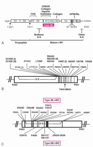
A gene on chromosome 12 codes for the synthesis of the vWF macromolecule. The genetic message is composed of 52 exons covering a span of 158 kb.132, 133 The vWF gene encodes for a protein with multiple copies of homologous motifs, including three “A,” three “B,” two “C,” and four “D” motifs (see Fig. 53.5). These motifs in turn encode protein domains that subserve the various functions of vWF. The A1 domain contains binding sites for platelet GPIb, ristocetin, and collagen,134, 135 the A2 domain contains a protease-sensitive domain that may have a role in regulating vWF function,136 the A3 domain contains a second collagen-binding domain,135 the C1 domain has an RGD sequence capable of interacting with platelet glycoprotein IIb/IIIa, and the D’ and D3 domains contain a factor VIII-binding sequence.135

vWD appears to be inherited by multiple genetic mechanisms (Table 53.7). The most common form of the disorder (type 1 vWD) accounts for ˜70% of all cases of vWD. Diagnostic criteria for type 1 vWD have been proposed,128 including a history of bleeding symptoms, a positive family history of bleeding symptoms, and bleeding that is attributable to quantitatively low vWF levels. Population screening suggests that the prevalence of vWD may be as high as 1% of the population, but only a minority of these patients subsequently present with clinically significant bleeding.
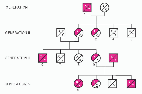
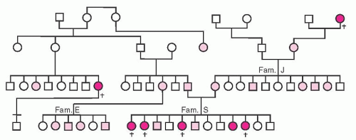
137 vWD is inherited as an incompletely dominant autosomal trait with variable penetrance, even among members within a single kindred. Because of variable penetrance and expression of vWD, only ˜33% of children may be affected. Figure 53.6 illustrates one of the original pedigrees described by von Willebrand.

Twin studies demonstrated that ˜60% of the variation in vWF level and ˜50% of the variation in factor VIII level is attributable to genetic factors.138 Genetic loci outside of the vWF gene contribute to variation of vWF level. Individuals with blood group O have vWF levels that are on average 30% lower than those of people with blood group A, B, or AB,139 and it is likely that carbohydrate groups attached to vWF play a subtle role in clearance of vWF from plasma.140, 141 In addition, variation in expression of levels of other hemostatically active proteins, such as variation in the level of platelet adhesion receptors, may modulate the severity of symptoms conferred by vWF deficiency.142 vWF levels are under hormonal and other controls, which further complicates disease expression. Variation in levels of as much as 20% has been reported with menstrual cycle, with levels lowest in the early follicular phase (before day 7 of the cycle); and levels increase with age, rising ˜15% for each decade increase in age.143 As will be discussed later, the criteria for diagnosing quantitative vWD deficiency, and the specificity of historic bleeding, have introduced controversy in what constitutes type 1 vWD and have led to the potential for the new diagnostic entity of “low vWF”.137, 144

Mild to moderate quantitative deficiency of vWF characterizes patients with type 1 vWD, whereas virtual absence of vWF characterizes type 3 disease. Two large studies of families with a diagnosis of type 1 vWD and genetic evaluation of patients with type 3 disease have begun to shed light on the genetics of quantitative vWF deficiency.

Very low vWF levels in the range of 5 to 20 IU/dl tend to be highly inheritable and are frequently associated with “dominant negative”-acting mutations. Mutations in these patients with type 1 vWD appear to be different than those with type 3 disease, such that type 1 disease is not simply explained by being heterozygous for a type 3 allele. Genetic defects associated with these more severe type 1 phenotypes encode amino acid changes that in turn lead to defects in protein expression. Mechanisms responsible for decreased plasma vWF level include reduced secretion related to impaired intracellular transport of vWF subunits, increased clearance from the circulation, or accelerated catabolism as a result of increased susceptibility to degradation by ADAMTS13. Changes that were most likely to cause a “dominant negative” mechanism
are often missense mutations that change the number of cysteine residues in the vWF protein. Although such changes probably affect vWF synthesis and trafficking in the cell, at least one common mutation (Tyr1584Cys) also results in enhanced degradation by ADAMTS13.145, 146 One non-cysteine-related mutation is seen in vWD Vicenza. In these patients, a mutation encoding for Arg1205His results in accelerated clearance of plasma vWF, with levels of vWF in the range of 15 IU/dl. With DDAVP stimulation, timed plasma sampling revealed that the released vWF had a circulation half-life that was approximately one-sixth of normal.147 Several other similar mutations have recently also been described.148


FIGURE 53.5. A: Structure and functional domains of von Willebrand factor (vWF). The open rectangles indicate domains of pre-pro-vWF. The first 22 amino acids of pre-pro-vWF are the signal sequence. The left arrow identifies the site of cleavage of the signal peptide, resulting in pre-vWF monomers. Pro-vWF monomers form dimers, and the vWF propeptide (vWAgII) is cleaved from mature vWF (right arrow indicates cleavage site). Dimer S-S and multimer S-S indicate locations of disulfide bonds that covalently link monomeric vWF into dimers and dimeric vWF into multimers, respectively. The red rectangle identifies exon 28, which encodes the A1 and A2 domains of mature vWF. Functional domains are identified above the open rectangles. The factor VIII binding site is located in the D’and D3 domains in the amino-terminal 272 amino acids of mature vWF. The A domains of vWF contain the binding site for the platelet glycoprotein (gp) Ib/IX complex, as well as binding sites for heparin, collagen, and sulfatides. The gpIIb/IIIa complex binding site of activated platelets is located at the carboxy-terminal portion of the C1 domain and contains the Arg-Gly-Asp sequence. The numbers below the open rectangles indicate amino acid numbers: Signal peptide, 22 amino acids; propeptide (vWA-gII), 741 amino acids; mature vWF, 2,050 amino acids. B: Mutations associated with type 2A von Willebrand disease (vWD). The open rectangle represents vWF exon 28 encoding domains A1 and A2. The arrow indicates the site of proteolysis seen in type 2A vWD. Mutations are identified using the single-letter code for amino acids. I and II identify group I and group II missense mutations that result in impaired synthesis of vWF or increased sensitivity to proteolysis in plasma, respectively. The disulfide bridge shown represents the boundary of the Cys 509-Cys 695 loop in the A1 domain. Type 2A vWD mutations associated with defective vWF multimerization are not shown in this figure; these latter mutations occur in the propeptide (vWAgII) D1 and D2 domains. C: Mutations associated with types 2B and 2M vWD. The large open rectangle represents exon 28, which encodes domains A1 and A2 of vWF. The mutations responsible for type 2B vWD are shown above the rectangle. Most type 2B mutations lie between residues 540 and 578 of mature vWF. The mutations responsible for type 2M vWD are shown below the rectangle. Asterisks identify mutations that reduce the platelet-dependent function of vWF and vWF multimeric size. δ indicates deletion of an amino acid sequence. [Modified from the Symposium Proceedings of the National Hemophilia Foundation’s 1995 Annual Meeting. Diagnosis and management of severe von Willebrand disease (types 2 and 3). Kroner PA, Montgomery RR. The molecular basis of von Willebrand disease, 1996:15-25. Published with permission of Advanstar Communications.]


TABLE 53.7 FEATURES OF COMMON VARIANTS OF VON WILLEBRAND DISEASE


Features


Type 1


Type 2A


Type 2B


Type 3


Platelet Type


Inheritance


Autosomal dominant


Autosomal dominant


Autosomal dominant


Autosomal recessive


Autosomal dominant


Factor VIIIc in plasma


Normal or reduced


Normal or reduced


Normal or reduced


Markedly reduced


Normal or reduced


vWF antigen


Normal or reduced


Normal or reduced


Normal or reduced; increased affinity for platelets


Markedly reduced


Normal or reduced; increased affinity for platelets


Ristocetin cofactor activity


Normal or reduced


Reduced


Normal or reduced


Markedly reduced


Reduced or normal


vWF multimeric analysis


Normal (plasma and platelets)


Absence of large and intermediate-sized multimers in plasma


Absence of large multimers from plasma; normal in platelets


Small multimers or absent multimers in plasma and platelets


Reduction in large multimers caused by “consumption” by platelets


Ristocetin-induced platelet aggregation


Normal or diminished


Diminished


Increased aggregation at low ristocetin concentrations


Markedly diminished


Hyperaggregation with patient’s platelets, normal plasma, and low concentration of ristocetin


vWF in platelets


Normal or reduced


Normal or absence of large and intermediatesized multimers


Normal


Absent


Normal


Ancillary findings


DDAVP usually produces significant increase in plasma VIIIc and vWF


DDAVP produces rise in factor VIIIc, but functional vWF increase is variable and may be of short duration


Variable response to DDAVP, with intravascular platelet aggregation and thrombocytopenia in some cases; ristocetin-induced platelet aggregation enhanced in presence of patient’s plasma; cryoprecipitate does not aggregate platelets in vitro unless ristocetin is added


Response to DDAVP lacking; endothelial vWF absent


Transfusion of vWF or DDAVP may produce intravascular platelet aggregation and thrombocytopenia; cryoprecipitate produces in vitro platelet aggregation


DDAVP, 1-deamino-8-d-arginine vasopressin; vWF, von Willebrand factor.


At higher levels of vWF, linkage of the vWF level to the vWF gene is found less often. The recent European type 1 vWF cohort study demonstrated this observation. Almost all families with vWF levels <30 IU/dl showed linkage to the vWF gene, but the proportion fell to only 51% with vWF levels >30 IU/dl.98 These data were corroborated in the Canadian study.146


FIGURE 53.6. Pedigree of patients described by von Willebrand. Three families are emphasized: S, E, and J. Clinical details of bleeding events in these families have been reported. Open circles indicate female nonbleeders, open squares indicate male nonbleeders. Shaded circles and squares indicate female and male bleeders, respectively. Solid symbols indicate family members who experienced severe bleeding, and crosses indicate family members who experienced hemorrhagic deaths. (Modified from von Willebrand EA. Über hereditäre pseudohämophilie. Acta Med Scand 1931;76:521-550, with permission of the publisher.)

Complete absence of vWF is responsible for the most extreme form of vWD, and this is classified as type 3 vWD.128 As one might predict, a large variety of genetic abnormalities scattered throughout the vWF gene have been reported in families with type 3 vWD. These have included large gene deletions, small gene deletions, frame-shift mutations, splice-site mutations, nonsense mutations, and point mutations.149

Type 2 vWD is characterized by production of a qualitatively defective protein, and the genetics of these variant forms is more straightforward than that of type 1 vWD. In families affected by types 2B and 2M, as well as in the majority of families with type 2A vWD, inheritance is autosomal dominant. Rare cases of type 2A disease are transmitted in an autosomal recessive fashion, as are most cases of type 2N disease. vWF gene sequencing studies combined with vWF mutation-expression analysis has revealed that single missense point mutations underlie the majority of type 2 disorders.128 Type 3 vWD is also inherited as a recessive trait and occurs when both copies of the vWF gene are defective. In type 3 disease, gene abnormalities include large or partial gene deletions, disruption of orderly messenger RNA transcription (frame-shift mutation, splicesite, or nonsense mutation), and missense mutation. For patients with recessively inherited vWD phenotypes (type 2N, type 3, or some rare type 2A), genetic analysis may reveal either homozygous or compound heterozygous gene abnormalities. Members of the International Society of Thrombosis and Hemostasis maintain a database of vWD mutations (www.vwf.group.shef.ac.uk).


Response to Transfusion

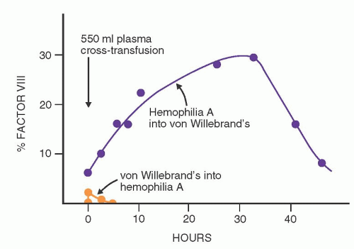
When blood products containing the vWF-factor VIII complex are infused into patients with hemophilia A, peak levels of factor VIIIc are present immediately after the infusion; this activity then declines rapidly, with an overall half-life of 8 to 10 hours (Fig. 53.7). Twenty-four hours later, factor VIIIc activity is minimal. This response is highly predictable and is discussed in a later section concerning replacement therapy for hemophilia A. In most patients with vWD, the infusion of normal vWF-factor VIII complex produces an initial rise in factor VIIIc that is predicted from the preinfusion level and the amount of factor infused. This is followed by a sustained but variable rise in factor VIIIc activity that reaches a plateau about 24 hours later and may persist for 48 to 72 hours. This phenomenon is highly variable and irregular; in some patients, a rapid initial fall of factor VIIIc is followed by a secondary rise in activity. This disproportionate response to transfused factor VIII has been called the secondary transfusion response, a feature that is apparently unique to vWD. Various hypotheses have been advanced to explain the disproportionate response to transfusion described above. The leading hypothesis is that the infused vWF results in stimulation of increased factor VIII release into the plasma with the attachment of the newly released factor VIII onto infused vWF.161


FIGURE 53.7. The phenomenon of new factor VIII synthesis in patients with von Willebrand disease (vWD) who receive plasma transfusion. Open circles indicate changes in the plasma factor VIII levels of a patient with vWD after infusion of plasma from a patient with severe hemophilia A. A significant and sustained increase in the factor VIII levels of the recipient was observed even though no active factor VIII was present in the infused plasma. Solid circles indicate effects of infusing plasma from a patient with vWD into a patient with severe hemophilia A. The factor VIII level in the infused plasma was 15% of normal. Note the slight and transitory effects. (From Shulman NR, Cowan DH, Libre EP, et al. The physiologic basis for therapy of classic hemophilia (factor VIII deficiency) and related disorders. Ann Intern Med 1967;67:856-882, with permission.)


Transfusions of blood products containing vWF shorten the bleeding time to a variable degree in patients with vWD. This corrective effect seldom persists for more than a few hours, even after massive transfusions that raise vWF to high levels. The correction of the bleeding time apparently requires the large molecular forms of vWF that are present in cryoprecipitate and select intermediate-purity factor VIII concentrates,162 but that are completely absent from monoclonally purified or recombinant concentrates of factor VIII.163 Failure of the skin template bleeding time of patients with vWD to show sustained correction with appropriate replacement therapy has been noted, and does not necessarily predict defective surgical hemostasis. This apparent paradox may be a result of sensitivity of the bleeding time test to platelet α-granule vWF, which is not replenished during vWF replacement therapies.164


Clinical Manifestations

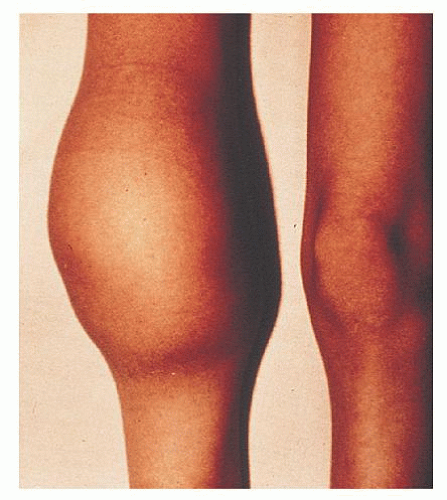
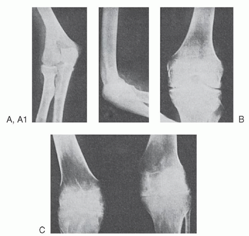
The bleeding manifestations in vWD are heterogeneous but consistent with the dual roles of vWF in supporting both primary and secondary hemostasis. Thus, in the mild forms of vWD, the clinical picture is dominated by cutaneous and mucosal bleeding, which appears to be mainly the result of disordered primary hemostasis. In the most severe forms of the disorder, in which factor VIII levels are low, hemarthroses and dissecting intramuscular hematomas may develop. As in mild classic hemophilia, serious hemorrhage resulting from traumatic injuries or after surgical procedures is a significant hazard in severe vWD. Petechiae are rare, but hematoma formation and the extent of bruising are excessive compared with the inciting trauma.

The bleeding manifestations in the usual patient with type 1 vWD are mild, however, and many patients are virtually asymptomatic. The disorder may be symptomatic at any age, but it is not always recognized. Easy bruising is common in vWD, but it is not specific. Mucosal bleeding is particularly common. Childhood epistaxis, a life-long history of easy bruising, bleeding with dental extraction, heavy menstrual bleeding or anemia attributed to excessive menstrual blood losses, or postpartum hemorrhage are all included in the spectrum of vWD symptomatology. Systemic bleeding disorders are often not considered in women with menorrhagia,165 but clinical studies reveal that the prevalence if vWD is significant in individuals who present with this complaint.166 Angiodysplasia of various vascular beds, particularly those in the gut, has been demonstrated in some patients with vWD and may be an important contributory factor in chronic gastrointestinal bleeding.167 In type 1 vWD, symptoms usually become milder during pregnancy or with estrogen therapy, when the vWF and factor VIIIc levels rise significantly.40 The disorder may also decrease in severity with advancing age. Bleeding manifestations tend to be more prominent in patients with more severe quantitative deficiency and in patients with qualitative (type 2) defects.

While eliciting the history of bleeding, one should bear in mind that women are often inaccurate assessors of whether their menstrual flow is normal or excessive. Supplemental questioning regarding the frequency of changes of menstrual protection, and questions regarding a history of iron deficiency anemia and transfusion, may be informative. Retrospective studies of women with vWD show that abnormal bleeding can often be traced to menarche.168 Family history also may provide important clues to a diagnosis. The physician should specifically seek a history of the response to hemostatic challenge, such as dental extraction, tonsillectomy, surgical procedures, menstruation, peripartum hemorrhage, and transfusion. A model questionnaire with a scoring system has been published.169 Coexistent conditions or drug effects can strongly influence the severity of bleeding symptoms. Risk of hemorrhage may increase in patients with concomitant liver disease, uremia, gastrointestinal ulcer, or angiodysplasia. Excessive bleeding in response to challenge with aspirin may also point toward a bleeding disorder, but this history is not specific for vWD. Valproic acid use can result in lower vWF activity levels, as may hypothyroidism and valvular heart disease. Conversely, oral contraception frequently ameliorates menorrhagia.127


Genetics of Type 1 and Type 3 vWD

Mild to moderate quantitative deficiency of vWF characterizes patients with type 1 vWD, whereas virtual absence of vWF characterizes type 3 disease. Genetic analysis is not generally required to make these diagnoses, but may provide useful additional information concerning pathogenesis of a patient’s condition, responsiveness to DDAVP, or risk for alloantibody formation. In distinction from type 2 vWD, genetic defects underlying quantitative vWF deficiency are distributed throughout the vWF gene, complicating evaluation. In addition, for families with patients who demonstrate normal multimer structure and higher vWF levels, the chances of finding linkage to the vWF gene are reduced,145, 146 and family members are usually asymptomatic.137 Type 3 patients with large gene deletions are particularly prone to develop anti-vWF alloantibodies after treatment with vWF-containing concentrates.


Type 1 von Willebrand Disease and “Low von Willebrand Factor”

Patients with bleeding attributed to decreased production of qualitatively normal vWF are classified as having type 1 vWD. Most patients with vWD (70% to 80%) fall into this category.134 The majority of patients have a mildly symptomatic disorder, but bleeding may increase with physical trauma, surgery, or during menstruation. vWF levels are low, with concordant reduction of vWF:Ag and vWF:RCo (and vWF:CB, if that is measured). Factor VIII levels are generally equal to or higher than vWF levels, and all vWF multimers should be present if that analysis is performed as part of the patient evaluation. The diagnosis of type 1 vWD may be simpler in an individual with mucosal bleeding symptoms, a strong family history of similar symptoms, and quantitative vWF level (vWF:Ag and/or vWF:RCo) <30 IU/dl. Although not required for a diagnosis of type 1 vWD, the probability of finding linkage of the phenotype to the vWF gene in such an individual would be high, such that both therapeutic choices and genetic counseling would be straightforward.


Unfortunately, many factors make the diagnosis of type 1 vWD difficult.144 Mild bleeding symptoms are common in hemostatically normal people (see Chapter 45). In addition, the relative risk of bleeding for individuals with only modest reductions in vWF level appears to be minimal. Finally, because of the strong influence of blood group on vWF levels, there is a high prevalence of blood group O among individuals with mildly depressed vWF levels. These observations have led to the recent proposal of the concept of “low vWF”.137,144 The term “low vWF” could be applied to patients with vWF levels below the normal reference range but above some lower limit. A recently convened “expert group” brought together by the National Heart, Lung and Blood Institute is considering this proposal.129 Studies of families of patients with a diagnosis of type 1 vWD suggest that linkage to the vWF gene is less often seen when the vWF level rises above 30 IU/dl, suggesting that this might be a candidate for the lower limit of the “low vWF” category.145, 198 Empiric therapy to raise vWF levels in patients with “low vWF” at times of hemostatic challenge would be reasonable for patients with a history of bleeding, but the genetic counseling issues would clearly be different.144

On the other hand, two population-based studies have shown that “low vWF” causes increased bleeding.199, 200 These latter studies would suggest that abnormally low ristocetin cofactor activity, and not the presence of a mutation, should primarily be used to identify bleeders.


Type 3 von Willebrand Disease

Type 3 vWD is the most severe form of vWD, because it results from complete failure of vWF synthesis. This bleeding disorder is generally diagnosed during infancy. Hematoma formation is common, epistaxis may be life-threatening, and hemarthrosis may occur as a result of the low factor VIII levels that are seen in this condition. Plasma from patients with type 3 vWD contains virtually no detectable vWF, and vWF is not present in either platelets or endothelial cells. Factor VIII level is generally in the range of 1 to 10 IU/dl, similar to that seen in moderate to mild hemophilia A. Multimeric analysis of the little vWF present yields variable results, in some cases revealing only small multimers.193 Type 3 vWD is rare, with an estimated frequency of 1 in a million people.201 Genetic analysis reveals either homozygous or compound (double) heterozygous defects of the vWF gene, with gene deletions, frame-shift mutations, missense mutations, or nonsense mutations. Careful laboratory evaluation of the parents of patients with type 3 disease may reveal mild quantitative deficiency of vWF. In a review of 117 obligate heterozygotes, the mean plasma vWF level was 45 IU/dl, with a range from 5 to 130 IU/dl.144 Parents are frequently asymptomatic, consistent with the impression that type 3 disease is inherited as a recessive trait. Consanguinity is common in kindreds with this variant. In some patients with type 3 vWD, large gene deletions have been identified on one or both vWF alleles.128,202 Such patients are at increased risk of developing inhibitory alloantibodies to vWF after transfusion therapy.202

Because these patients lack vWF in their endothelial cells and platelets, there is no rise in vWF level in response to DDAVP. For replacement therapy, these patients require vWF-containing concentrates. This is usually provided in the form of specific intermediate-purity factor VIII concentrates that have been documented to contain intact vWF.155 Because these patients possess a normal factor VIII gene but are missing the vWF chaperone, posttransfusion factor VIII recovery and survival may be longer in a patient with type 3 vWD than in a patient with hemophilia A, owing to endogenous factor VIII production. Although life-threatening bleeds require immediate replacement of both vWF and factor VIII, replacement with vWF alone may be sufficient if therapy is begun 12 to 24 hours before elective surgery.203


Qualitative Defects of von Willebrand Factor

Several subsets of patients with vWD differ substantially from those with the common quantitative deficiency states (Table 53.6).
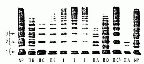
128 Classified as type 2 variants, these qualitative defects of vWF are less common, but they may represent up to 20% to 25% of cases of vWD. Type 2 forms of vWD are suspected when the severity of the patient’s symptoms seems in excess of the observed vWF and factor VIII levels, when there is discordant reduction between vWF antigen and vWF functional activity or factor VIII assay, or when there is concomitant vWF deficiency and thrombocytopenia. Discrimination between qualitatively normal and abnormal vWF can be difficult; however, when the ratio of vWF:RCo to vWF:Ag or vWF:CB to vWF:Ag is found to be <0.6, dysfunctional vWF should be considered. The sensitivity and specificity of such ratio screening depends to a large extent on the expertise and precision of the laboratory performing the assays, but the predictive value of ratios decreases when the level of vWF antigen is <20 IU/dl. Examining the structure of patient vWF may provide further definition of the nature of the qualitative defect. This can be demonstrated by vWF multimer analysis. If required, further information is provided by supplemental studies of vWF function, or via genetic analysis of the vWF gene (Fig. 53.5).126,128,193 Type 2 defects were initially classified using a Roman numeral system, but further understanding of the genetics of vWD led to the consolidation and reclassification into types 2A, 2B, 2M, and 2N,128 based on the nature of the vWF functional defect. In addition to the well-defined variants described in this chapter, other forms of vWD continue to be described.


Type 2A von Willebrand Disease

vWD with defective interaction of vWF with platelets due to deficiency of intermediate- and high-molecular-weight forms of vWF is classified as type 2A vWD.128 This form of the disorder is generally inherited as an autosomal dominant trait,204 and it accounts for the majority of patients with type 2 defects. Levels of factor VIIIc and vWF:Ag in the plasma may be normal or reduced. The vWF activity as assayed by either vWF:RCo or vWF:CB is significantly lower than vWF:Ag because of the absence of the larger multimers (which are more potent in their ability to interact with platelet GPIb and collagen). Analysis of vWF multimers reveals a relative reduction in intermediate- and high-molecularweight species.127 Protein studies followed by genetic analysis of the basis of type 2A vWD revealed multiple mechanisms for the generation of this disorder.205, 206, 207 In some patients, there is failure to synthesize full-length multimers (type 2A, subgroup 1). Mutations associated with impaired vWF multimerization have been localized to the vWFpp (formerly called type IIC), the region of the mature vWF protein associated with the amino terminus area of multimer formation (formerly called type IIE), and the carboxy terminus region that is involved in initial dimer formation (formerly called type IID). Subtle abnormalities uncovered by high-resolution multimer analysis or genetic evaluation can be used to distinguish these subtypes of type 2A vWD. The spectrum of multimers released into plasma after DDAVP administration would not be expected to improve significantly in these patients whose defects prevent normal multimer assembly.

A second group of type 2A patients has been described in whom there is excessive catabolism of fully multimerized vWF after vWF is released into the plasma. These patients demonstrate mutations in exon 28, encoding the A1 and A2 regions of vWF. These mutations allow increased proteolysis of multimerized vWF by ADAMTS13. Again, high-resolution multimer gel studies may be informative by demonstrating increased “satellite” band intensity, which accumulates as a result of excessive ADAMTS13 activity. Because intracellular vWF is protected from cleavage by ADAMTS13, these patients may demonstrate very transient
improvement in both vWF:RCo and multimer spectrum after DDAVP administration.

Inheritance of type 2A vWD is generally autosomal dominant, with the majority of cases caused by mutations clustered in the region of exon 28, which encodes the vWF A2 homologous repeat (Fig. 53.5).128 Rare recessive forms of type 2A vWD have been reported with some multimerization defects (formerly called types IIC and IID).

Because of the underlying structural abnormality of the vWF produced in these patients, neither stress nor pregnancy significantly increases the functional amount of vWF protein in the plasma. As noted above, the administration of DDAVP does not result in a significant rise in vWF:RCo in patients with type 2A vWD, in which the underlying mechanism is defective multimerization or transport, but a very short-lived correction may be observed in patients with mutations associated with increased vWF cleavage by ADAMTS13.


Type 2B von Willebrand Disease

Type 2B vWD is a paradoxical bleeding disorder, characterized by increased interaction of patient vWF with platelets which is demonstrated in vitro in the presence of low doses of ristocetin. Increased in vivo interaction of the larger multimers of type 2B vWF with platelets is thought to result from mutations that either allow increased access of platelets to the A1 loop of vWF or that stabilize that interaction.127 This may in turn allow increased generation of vWF-platelet complexes, which are subsequently cleared from circulation, resulting in thrombocytopenia.

This bleeding disorder is characterized by deficient vWF function attributable to mild reduction of vWF:Ag, a somewhat more marked deficiency of vWF:RCo activity (and vWF:CB, if measured). Multimeric analysis reveals a deficiency of the highestmolecular-weight vWF multimers. Measurements of factor VIIIc are variable. Many patients with the type 2B variant have mild persistent thrombocytopenia.208 The platelet count may fall further during physiologic stresses, pregnancy, in association with surgical procedures, or after the administration of DDAVP.209

Additional laboratory evaluation is required to support a diagnosis of type 2B vWD. In most cases, multimeric analysis reveals absence of higher-molecular-weight forms of plasma vWF, but distinction of type 2B from type 2A vWD is not possible by multimeric analysis alone. To show the distinct “gain in function” in type 2B disease, RIPA studies are done to reveal enhanced interaction of patient vWF and platelets with low doses of the drug. Finally, to differentiate the more common type 2B vWD from the rare platelet-type (pseudo) vWD, one should prove that the defect resides in vWF. This is done either through performing mixing studies of patient plasma with donor platelets187 or by analysis of the patient’s vWF gene.

Type 2B vWD is inherited as an autosomal dominant trait. A small cluster of mutations within the portion of vWF exon 28 that encodes the vWF A1 domain that interacts with platelet GPIb accounts for all the cases of type 2B vWD that have been reported (Fig. 53.5).128

Recent studies evaluating a large number of patients with type 2B vWD mutations revealed diversity in laboratory findings.210 Not all patients had reduced vWF:RCo activity or abnormal multimeric analysis, and not all patients had thrombocytopenia.210 Type 2B patients with normal multimers did not develop thrombocytopenia.


Type 2M von Willebrand Disease

The type 2M vWD variant is defined by diminished platelet-dependent vWF function that is not attributed to deficiency of vWF multimers.128 Thus, these patients have an initial laboratory profile that in many ways is similar to that of patients with type 2A vWD, revealing variable deficiency of vWF:Ag but disproportionately decreased interaction of vWF with platelets in the presence of ristocetin as measured by vWF:RCo assay. Factor VIII level is proportionate to the vWF:Ag, and the platelet counts are normal. What differentiates type 2M vWD from the type 2A patients is that the vWF multimeric analysis is normal (and, if measured, the vWF:CB is usually similar to the vWF:Ag). Type 2M vWD is inherited as an autosomal dominant trait, and, when investigated, mutations have been found in the region of exon 28 that encodes the A1 domain of vWF. Unlike the type 2B mutations, 2M mutations cause impairment of the binding of vWF to the GP1b receptor and have been shown to be localized to an alternative area in the A1 domain (Fig. 53.5).196

One case classified as a type 2M defect was described in which the vWF interaction with collagen was defective. In that case, a mutation in the A3 domain that contributes to the interaction of vWF with collagen was found.189


Type 2N von Willebrand Disease

Mutations affecting the association of vWF with factor VIII can result in “autosomal hemophilia” in which factor VIII levels are significantly reduced relative to vWF.128 Affected patients have factor VIII levels in the range of 5 to 30 IU/dl, attributed to mutations at the factor VIII-binding site near the amino terminus of the vWF subunit.211 Indeed, genetic studies indicate that the majority of mutations underlying type 2N vWD involve vWF exons 18, 19, and 20, which encode the bulk of the factor VIII-binding domain of vWF. The other functions of vWF in patients with type 2N vWD are qualitatively normal, and thus vWF laboratory parameters (vWF:Ag, ristocetin cofactor activity, etc.) are usually normal (unless there is coinheritance of type 1 vWD). This variant was originally named vWD Normandy but has been renamed type 2N vWD in the revised classification.128 The factor VIII-binding defect in these patients is inherited in an autosomal recessive manner, and thus affected patients must inherit 2N alleles from each of their two parents, or inherit type 1 vWD from one parent while inheriting a 2N allele from the other. Patients with factor VIII deficiency and a bleeding disorder that is not clearly transmitted as an X-linked disorder or who demonstrate an unexpectedly short in vivo survival of infused factor VIII should be evaluated for type 2N vWD.212 Diagnosis requires performance of an assay that assesses the interaction of factor VIII with vWF,191, 192 or genetic studies of the vWF gene. A survey of almost 400 unrelated patients with either hemophilia A or type 1 vWD indicated a prevalence of type 2N vWD in these patient populations of 3% and 1.5%, respectively.213 Consideration of this diagnosis is important because both therapy and genetic counseling are distinct from that of mild hemophilia A.


Platelet-type (Pseudo) von Willebrand Disease

Platelet-type (pseudo) vWD resembles type 2B vWD in most respects, except that the basis for platelet-type vWD is a structural defect in platelet GP1b214 rather than a defect of vWF (thus platelet-type [pseudo] vWD is a form of platelet dysfunction). However, as with type 2B vWD, a genetic defect results in increased interaction between GP1b and vWF. Phenotypically, platelet-type (pseudo) vWD is manifested by mild thrombocytopenia, a prolonged bleeding time, and variable deficiency of plasma vWF and factor VIIIc. The reductions in plasma vWF and factor VIIIc may be a result of the attachment of these proteins to platelets that are subsequently removed from the circulation. This “consumption” of vWF and platelets results in preferential loss of high-molecular-weight multimers from patient plasma.214 In vitro platelet aggregation studies reveal platelet agglutination at unusually low concentrations of ristocetin. Spontaneous intravascular platelet clumping may also occur. Platelet-type (pseudo) vWF is inherited as an autosomal dominant trait in most families.214, 215


Distinction of platelet-type vWD from type 2B vWD is based on showing that the enhanced interaction of platelets with vWF is either platelet-based or resides in the plasma phase (type 2B vWD).216 This can sometimes be demonstrated through low-dose ristocetin-based agglutination assays using mixtures of normal donor and patient samples, or genetic analysis can be used. To date, all cases of platelet-type vWD have been traced to genetic variations of platelet GPIbα.217 These include missense mutations that alter the amino acid sequence from Gly233 to Met 239. This region of the protein adopts a β-sheet conformation after interaction with vWF, and these mutations may function to stabilize this conformation.218 In addition, a 27-bp gene deletion that encodes for deletion of proline 421 through serine 429 has been shown to underlie platelet-type vWD in one kindred. Thus, genetic analysis of the GPIbα gene could confirm a diagnosis of platelet-type vWD in cases in which phenotypic studies were unsuccessful.


CLINICAL DISORDERS OF THE FIBRINOGEN MOLECULE

The clinical disorders of fibrinogen are complex, and depending on the types of mutation may present with bleeding or with thrombotic symptoms, or both. These disorders can be divided based on whether the defect is predominantly a quantitative or qualitative abnormality of the fibrinogen molecule.219, 220 Quantitative abnormalities are disorders associated with the complete absence of fibrinogen (afibrinogenemia) or with low levels of fibrinogen (hypofibrinogenemia). Qualitative abnormalities are disorders resulting from synthesis of an abnormal fibrinogen molecule (dysfibrinogenemia) with altered functional properties.219, 220, 221, 222 Dysfibrinogenemia may be asymptomatic and identified only through laboratory studies, or it may be associated with either hemorrhagic or thrombotic consequences.

Fibrinogen is a 340-kDa plasma protein that circulates at a concentration of 1.5 to 3.5 mg/ml. It is a symmetric disulfidelinked dimer with a central E domain linked via “coiled coil” peptide chains to outer D domains.223 Each half-molecule consists of a set of three different peptide chains, termed Aα, Bβ, and γ, which are linked at their amino termini by disulfide bonds to form the E domain. The D domains are formed by disulfide linkages near the carboxy termini of the peptides. Fibrinogen is synthesized in hepatocytes224 by coordinated expression of three separate genes on chromosome 4.225 The rate-limiting step in fibrinogen synthesis is transcription of the Bβ gene. Fibrinogen is an acute-phase reactant, and levels may rise considerably with inflammation. The circulation half-life of plasma fibrinogen is ˜4 days. Fibrinogen participates in multiple physiologic processes, including fibrin clot formation mediated by the enzymatic activities of thrombin and factor XIIIa, and cohesion of activated platelets through interaction with the GP IIb/IIIa receptor. Fibrinogen also acts as a plasma carrier for factor XIII. Fibrinogen physiology has been reviewed.223

Only gold members can continue reading. Log In or Register to continue

Stay updated, free articles. Join our Telegram channel

Oct 21, 2016 | Posted by in HEMATOLOGY | Comments Off on Inherited Coagulation Disorders

Full access? Get Clinical Tree

Get Clinical Tree app for offline access