Hereditary Hemolytic Anemias Due to Red Blood Cell Enzyme Disorders


Hereditary Hemolytic Anemias Due to Red Blood Cell Enzyme Disorders


Bertil Glader


OVERVIEW OF ERYTHROCYTE METABOLISM

Mature red blood cells (RBCs) are anucleate (thereby incapable of cell division), devoid of ribosomes (thereby incapable of protein synthesis) and lack mitochondria (thereby incapable of oxidative phosphorylation). Despite these limitations RBC survive 100 to 120 days in the circulation, and effectively deliver oxygen to peripheral tissues. Glucose is the main metabolic substrate of red blood cells and it is metabolized by two major pathways: the glycolytic or “energy producing” pathway and the hexose monophosphate (HMP) shunt or “protective” pathway. Under normal conditions approximately 90% of glucose flows through glycolysis, with a much smaller fraction being channeled through the HMP pathway. However, the fraction of glucose entering the pentose phosphate pathway can increase significantly under conditions of increased oxidative stress. The major products of glycolysis are ATP (a source of energy for numerous RBC membrane and metabolic reactions), NADH (a necessary cofactor for methemoglobin reduction by cytochrome b5 reductase), and 2,3-diphosphoglycerate ([2,3-DPG], an important intermediate that modulates hemoglobin-oxygen affinity) (Fig. 28.1). Mature RBC are incapable of de novo purine or pyrimidine synthesis, although many enzymes of nucleotide metabolism are present in erythrocytes. The latter are now known to be important for RBC preservation in vitro; and it also is recognized that abnormalities in purine and pyrimidine metabolism are associated with inherited hemolytic disease.

The consequences of red cell enzymopathies are diverse. Some enzyme variants cause hemolytic disease, with anemia being the sole expression of the enzymopathy. In other enzyme disorders hemolysis is one feature of a multisystem disease affecting many tissues. Also, in some cases erythrocyte enzyme abnormalities have no adverse effects on RBC function, and, if they occur in patients with hemolytic anemia, it is not always clear that the enzyme deficiency and hemolysis are causally related. This chapter focuses on the varied enzyme defects associated with hemolysis. These RBC enzyme disorders are due to abnormalities in the HMP shunt and glutathione metabolism, glycolytic enzyme deficiencies, and abnormalities in purine and pyrimidine metabolism.


DISORDERS OF HEXOSE MONOPHOSPHATE SHUNT AND GLUTATHIONE METABOLISM

The HMP shunt pathway metabolizes 5% to 10% of glucose utilized by red blood cells, and this is critical for protecting red cells against oxidant injury (Fig. 28.2). The HMP pathway is the only RBC source of reduced nicotinamide adenine dinucleotide phosphate (NADPH), a cofactor important in glutathione metabolism. Red blood cells contain relatively high concentrations of reduced glutathione (GSH), a sulfhydryl-containing tripeptide (glutamylcysteinylglycine) which functions as an intracellular reducing agent that protects cells against oxidant injury. Oxidants, such as superoxide anion (O2) and hydrogen peroxide (H2O2), are produced by exogenous factors (i.e., drugs, infection) and also are formed within red cells as a consequence of reactions of hemoglobin with oxygen. However, when these oxidants accumulate within red cells, hemoglobin and other proteins are oxidized, leading to loss of function and RBC death. Under normal circumstances this does not occur, since GSH, in conjunction with the enzyme glutathione peroxidase (GSH-Px), rapidly inactivates these compounds. During the oxidant detoxification process, however, GSH itself is converted to oxidized glutathione (GSSG), and GSH levels fall. In order to sustain protection against persistent oxidant injury, GSH levels must be maintained, and this is accomplished by glutathione reductase (GSSG-R), which catalyzes reduction of GSSG to GSH. This reaction requires the NADPH generated by glucose-6-phosphate dehydrogenase (G6PD), the first enzymatic reaction of the HMP shunt. Thus, it is the tight coupling of the HMP shunt and glutathione metabolism that is responsible for protecting intracellular proteins from oxidative assault. Almost all hemolytic episodes related to altered HMP shunt and glutathione metabolism are due to G6PD deficiency, and this enzyme deficiency is known to affect millions of people throughout the world.1, 2, 3, 4 Rare cases of hemolysis associated with decreased GSSG-R activity, GSH-Px deficiency, and deficiencies of GSH synthetic enzymes also have been described.


Glucose-6-Phosphate Dehydrogenase Deficiency

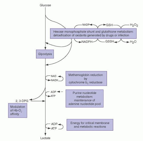
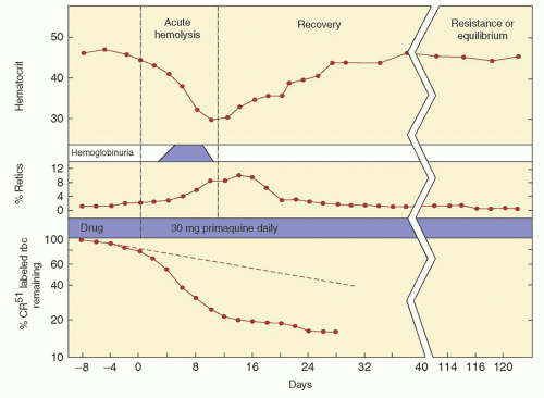
The importance of this enzyme for red cell integrity was first recognized following the observation that some African-American soldiers taking the antimalarial drug primaquine would develop acute hemolytic anemia with hemoglobinuria. Initially it was observed that GSH was decreased in the RBC of susceptible individuals during acute hemolytic episodes. Subsequently, the activity of G6PD, one of the enzymes needed to keep adequate GSH levels, was found to be deficient in affected red cells.5, 6 Soon thereafter, the worldwide distribution of G6PD deficiency became apparent, and the variation of clinical expression of enzyme deficiency was discovered. In most individuals with G6PD deficiency, there is no anemia in the steady state, reticulocyte counts are normal, but RBC survival may be slightly decreased. However, episodic exacerbations of hemolysis accompanied by anemia occur in association with the administration of certain drugs, with some infections, and with the eating of fava beans. In a minority of cases G6PD deficiency is associated with a chronic hemolytic process. To date, over 400 G6PD biochemical variants have been recognized.2, 3, 7


Genetics

The gene for G6PD is located on the X chromosome (band X q28),8, 9, 10 The fact that normal males and females have the same enzyme activity in their red cells is explained by the Lyon hypothesis.1, 11 This hypothesis maintains that one of two X chromosomes in each cell of the female embryo is inactivated and remains inactive throughout subsequent cell divisions for the duration of life. In fact, it was studies by Beutler on females with G6PD deficiency which were used in proof of the Lyon hypothesis.12 Enzyme deficiency is expressed in males carrying a variant gene, whereas heterozygous females usually are clinically normal. However, dependent upon the degree of lyonization, and the degree to which the abnormal G6PD variant is expressed, the mean red blood cell enzyme activity in females may be normal, moderately
reduced, or grossly deficient. A female with 50% normal G6PD activity has 50% normal red cells and 50% G6PD-deficient red cells. The G6PD-deficient cells in females, however, are as vulnerable to hemolysis as are enzyme-deficient red blood cells in males.


FIGURE 28.1. Summary of overall glycolysis, hexose monophosphate shunt, glutathione metabolism, and red blood cell (RBC) nucleotide metabolism. GSH, reduced glutathione; GSSG, oxidized glutathione; 2,3-DPG, 2,3-diphosphoglycerate NADPH, nicotinamide adenine dinucleotide phosphate.

The concept of X chromosome inactivation and the study of G6PD has been informative in other areas. In particular, it has facilitated our understanding of monoclonal and multiclonal disorders of cell proliferation. Most tumors, both benign and malignant, can be demonstrated to have a clonal origin, being derived from one cell.13, 14, 15 For example, analysis of the G6PD enzyme in uterine myomata of women heterozygous for G6PD A and G6PD B revealed that any given tumor had either G6PD A or enzyme B, but not both.16 The same principle has been used to demonstrate the clonal origin of the malignant transformation of acute leukemia17, 18 and chronic myelocytic leukemia,19 and also the clonal nature of polycythemia vera,20 primary thrombocythemia,21 and paroxysmal nocturnal hemoglobinuria.22


FIGURE 28.2. Hexose monophosphate (HMP) shunt, enzymes of glutathione (GSH) metabolism, protection from oxidant assault, and relationship to glycolytic metabolism. Enzyme abbreviations: GCS, γ-glutamyl-cysteine-synthetase; G-6-PD, glucose-6-P dehydrogenase; GS, glutathione synthetase; GSH-Px, glutathione peroxidase; GSSG-Red, glutathione reductase; GPI, glucosephosphate isomerase; HK, hexokinase; PFK, phosphofructokinase; 6-PGD, 6-phosphogluconate dehydrogenase. Substrate abbreviations: DHAP, dihydroxyacetone phosphate; G-3-P, glyceraldehyde-3-phosphate; GSH, reduced glutathione; GSSG, oxidized glutathione; R-5-P, ribose-5-phosphate; 6-P-G, 6-phosphogluconate.


Prevalence and Geographic Distribution

Deficiency of G6PD is the most common metabolic disorder of red blood cells and has been estimated to affect over 400 million people worldwide.2, 3, 23 Although global in its distribution, G6PD deficiency is encountered with greatest frequency in the tropical and subtropical zones of the Eastern Hemisphere. The incidence of the deficiency state is approximately 20% among African Bantu males,24, 25 12% in African-American men,26 and 8% in Brazilian blacks. As many as 20% of female African-Americans may be heterozygous for G6PD mutants,27 and as many as 1% are homozygous. In Sardinia, the incidence varies from 35% at low altitudes to 3% in areas above 600 meters.28 The deficiency state has been reported from most areas of Greece, again with greatest frequency (20% to 32%) in the lowlands.29 The condition is also prevalent among Sephardic Jews, and as many as 60% to 70% of Kurdish Jews may be affected.3, 30, 31 In the male Asian population, the incidence of G6PD deficiency is estimated to be 14% in Cambodia,32 5.5% in South China,33, 34 2.6% in India,32 and less than 0.1% in Japan.35 In China the frequency of G6PD deficiency in males ranges from 0% to 17.4%, with the highest prevalence being seen in ethnic groups geographically related to historical malaria. It is rare among Native Americans.36

Because of its high incidence among populations in which malaria was once endemic, G6PD deficiency is thought to have conferred a selective advantage against infection by falciparum malaria.37, 38, 39 Partial indirect support for this is that G6PD deficiency in Sardinia is more common at sea level compared to higher elevations, and this also parallels the endemicity of malaria. In addition, it has been observed that parasitized female heterozygotes for G6PD deficiency (who therefore have normal and G6PD-deficient RBC) have more malaria parasites in normal erythrocytes compared to their own G6PD-deficient cells.40 Moreover, it has been demonstrated that the in vitro growth of malarial parasites is inhibited in G6PD-deficient red cells.41 The
precise reasons for the observed inhibition of parasite growth in G6PD-deficient red cells are not known. One possibility is that the oxidant stress which causes GSH instability, and destroys the host RBC, also kills the parasite.2, 42, 43, 44


The Enzyme and Its Variants

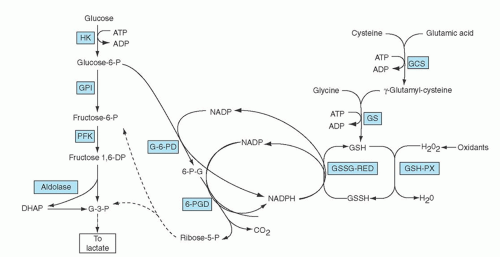
The monomeric form of G6PD contains 515 amino acids and has a molecular weight of 59 KDa.45, 46 The active form of G6PD in vivo is a dimer which requires NADP for its stability.47 The G6PD gene has been cloned and sequenced. It is known to contain 13 exons, and is over 18 kb in length.46, 48, 49

The normal or wild-type enzyme is G6PD B, although hundreds of variant enzymes now have been identified. By international agreement, standardized methods have been used to characterize these enzyme variants, which differ on the basis of biochemical properties such as kinetic activity, electrophoretic mobility, the Michaelis constant for its substrate glucose-6-phosphate and cofactor NADP, the ability to utilize different substrate analogues, heat stability, and pH optima. Throughout the years the published list of recognized G6PD biochemical variants has been periodically updated,50, 51 and over 400 biochemical variant forms of G6PD are recognized.3 However, differences between some variants are subtle, and most likely reflect minor technical differences between laboratories rather than true enzyme differences. Moreover, advances in molecular biology have revealed that many biochemical G6PD variants, in fact, have the same DNA defect (see below).

The World Health Organization (WHO) has classified G6PD variants on the magnitude of the enzyme deficiency and also the severity of hemolysis.52 Class I variants have very severe enzyme deficiency (less than 10% to.20% of normal) and have chronic hemolytic anemia. Class II variants also have severe enzyme deficiency (less than 10% of normal), but there is usually only intermittent hemolysis. Class III variants have moderate enzyme deficiency (10% to 60% of normal) with intermittent hemolysis usually associated with infection or drugs. Class IV variants have no enzyme deficiency or hemolysis. Class V variants are those in which enzyme activity is increased. Variants in the last two groups, although of much interest to biologists, geneticists, and anthropologists, are of no major clinical significance.

The normal wild-type enzyme, G6PD B, is found in most Caucasians, Asians, and a majority of blacks. It has normal catalytic activity and is not associated with hemolysis (Class IV). A commonly encountered variant is G6PD A+ which is found in 20% to 30% of blacks from Africa.53 It has normal catalytic properties and does not cause hemolysis (Class IV). It differs from G6PD B in that it has a much faster electrophoretic mobility (the letters A and B refer to relative electrophoretic mobilities). The structure of G6PD A+ differs from that of G6PD B by the substitution of a single amino acid, an asparagine for aspartate at the 126th position of the protein.54 Another common variant, G6PD A, is the enzyme responsible for primaquine sensitivity in blacks, and it is the most common variant associated with mild to moderate hemolysis (Class III). This G6PD variant is found in 10% to 15% of African-Americans, and with similar frequencies in western and central Africa.55 It has an electrophoretic mobility identical to that of G6PD A+. However, this is an unstable enzyme and its catalytic activity, although nearly normal in bone marrow cells and reticulocytes,56 decreases markedly in older RBC.57 Hence, this variant is designated G6PD A compared with G6PD A+ (the + and – denote enzyme activity). G6PD Mediterranean is a common abnormal variant found in people whose origins are in the Mediterranean area. However, this same variant also is found in the Middle East and India.2 The electrophoretic mobility of G6PD Mediterranean is identical to that of G6PD B, but its catalytic activity is markedly reduced, and hemolysis can be severe (Class II).56 G6PD Canton is a common variant enzyme seen in Asians.58 Its biochemical properties are very similar to those of G6PD Mediterranean.

Advances in molecular biology have further enhanced our understanding of G6PD deficiency, and now over 160 different gene mutations or mutation combinations have been identified.2, 59, 60, 61 These DNA changes almost all are missense mutations leading to single amino acid substitutions in the enzyme. Large deletions have not been identified, suggesting that complete absence of G6PD might be lethal.3 The mutations are located throughout the entire coding region of the gene.62 However, in Class I variants associated with chronic hemolysis, mutations are clustered around exon 10, an area that governs the formation of the active G6PD dimer.62, 63 The correlation between the different biochemical variants, the site of genetic mutation, and the extent of hemolysis is a matter of current investigation.2

An interesting example of how molecular biology has enhanced our understanding relates to G6PD A, once thought to be a single unstable variant found in blacks throughout the world. However, molecular analysis now has demonstrated that G6PD A may have more than one genotype. In all cases there is a mutation at nucleotide 376 (A→G), which also is the nucleotide substitution characteristic of G6PD A+. In addition, the G6PD A variants have a second mutation, and in the majority of cases it is at nucleotide 202 (G→A).3, 64 A smaller fraction of G6PD A subjects have the second substitution at nucleotide 680 (G→T) or at nucleotide 968 (T→C).65 Thus, the G6PD A variant, once thought to be a single homogeneous mutation in Africans, now turns out to represent at least three different genotypes.3 Also, a number of G6PD variants originally described in non-Africans are now found to have one of the known G6PD A mutations (Table 28.1). For example, G6PD Betica,66 a Spanish variant and G6PD Matera,67 an Italian variant, have demonstrated base substitutions at nucleotides 202 and 376, identical to the common G6PD A variant. They are examples, therefore, of G6PD A. One subject with G6PD Betica had base substitutions at nucleotides 376 and 968, identical to the less common G6PD A variant.66

There are several other variants that appear clinically and biochemically heterogeneous but have been found to be genetically more uniform. For example, G6PD Mediterranean involves many different ethnic groups, although most subjects have the same genetic defect, a single base substitution (C→T) at nucleotide 563.66, 67, 68 Moreover, just as in the case of G6PD A, many of the different biochemical variants have turned out to have the same molecular defect as G6PD Mediterranean (Table 28.1).

In China there are at least 21 variants causing G6PD deficiency. The three most common are G6PD Canton (G→T at nucleotide 1,376), G6PD Kaiping (G→A at nucleotide 1,388), and G6PD Gaohe (A→G at nucleotide 95).69

G6PD Canton and G6PD Gaohe are mainly regarded as WHO Class II variants, while G6PD Kaiping is considered a WHO Class III variant.

Because leukocyte and platelet G6PD is regulated by the same gene as that of red cells, documentation of decreased activity in the white blood cells70, 71, 72 and platelets71 of deficient individuals is not surprising. Because of the normally short survival of leukocytes and platelets, however, most individuals with G6PD deficiency do not manifest impairment of phagocytosis or bactericidal activity of granulocytes.71, 72 The exception to this occurs with Class I G6PD deficiency, where some affected individuals may have neutrophil dysfunction and increased susceptibility to infection.73, 74


Clinical and Hematologic Features

The clinical expression of G6PD variants encompasses a continuous spectrum of hemolytic syndromes. In most affected individuals the deficiency state goes unrecognized, while in some it causes episodic or chronic anemia. The common clinical entities encountered are acute hemolytic anemia, favism, neonatal hyperbilirubinemia, and congenital nonspherocytic hemolytic anemia.


Acute Hemolytic Anemia

With most G6PD variants, hemolysis occurs only after exposure to oxidant stresses. In the steady state there is no anemia, no evidence of increased red cell destruction, nor alteration in blood morphology. Sudden destruction of enzyme-deficient erythrocytes is triggered by drugs having a high redox potential and by selected infectious or metabolic perturbations. The clinical and laboratory features of an acute hemolytic episode are best illustrated in a figure from a classic study with primaquine-induced hemolysis in subjects with G6PD A (Fig. 28.3)5, 6 After 2 to 4 days of primaquine ingestion, all the signs, symptoms, and
laboratory results characteristic of an acute hemolytic episode are observed. Jaundice, pallor, and dark urine, with or without abdominal and back pain, are sudden in onset. An abrupt decrement of 3 to 4 g/dl in hemoglobin concentration occurs. The peripheral blood smear contains spherocytes and eccentrocytes or “blister” cells. In response to anemia, red cell production increases; an increase in reticulocytes is apparent within 5 days and is maximal by 7 to 10 days after onset of hemolysis. Despite continued drug exposure, the acute hemolytic process ends spontaneously after about 1 week, and the hemoglobin concentration thereafter returns to normal levels. The anemia is self-limited because the old susceptible population of erythrocytes is replaced by younger RBC with sufficient G6PD activity to withstand an oxidative assault. Although red cell survival remains shortened as long as use of the drug continues, compensation by the erythroid marrow effectively abolishes the anemia in subjects with G6PD A. In contrast, hemolysis occurring with the G6PD Mediterranean variant is more severe because a larger population of circulating erythrocytes is vulnerable to injury.83 Hemolytic crises occur in heterozygous female subjects, as well as in hemizygous male patients. Of interest, the incidence of clinically significant G6PD deficiency in elderly women reportedly is increased, and this is thought to reflect skewed lyonization that occurs with aging. In a fascinating study from Hong Kong, the incidence of G6PD deficiency in elderly females (80 to 107 years) was increased (1.73%) compared to newborn girls (0.27%).84 In virtually all cases acute hemolytic episodes are due to administration of drugs, or are associated with infection or fava bean exposure.


FIGURE 28.3. The course of primaquine-induced hemolysis in the glucose-6-phosphate dehydrogenase (G6PD) A variant. From Alving A, et al. Mitigation of the haemolytic effect of primaquine and enhancement of its action against exoerythrocytic forms of the Chesson strain of Plasmodium vivax by intermittent regimens of drug administration. Bull WHO 1960;22:621-631.

Drug-induced Hemolysis. Primaquine is but one of several drugs that can precipitate hemolysis. The common denominator of these drugs is their interaction with hemoglobin and oxygen, thus accelerating the intracellular formation of H2O2 and other oxidizing radicals. The published lists of suspect drugs are lengthy; however, many of the putative hemolytic agents were incriminated before it was recognized that infections often mimic the adverse effects of drugs. Consequently, many hemolytic events previously ascribed to drugs may, in fact, have resulted from infections for which drugs were given. Aspirin is such a drug, and it now is recognized that it can safely be given to individuals with Class II and III G6PD variants. Some drugs and chemicals, however, are predictably injurious for all G6PD-deficient subjects,3, 30 and these agents are listed in Table 28.2. Other drugs, although producing a modest shortening of survival of G6PD-deficient red cells, can be given safely in usual therapeutic doses to individuals with Class II and III G6PD variants (Table 28.2). Ascorbic acid is safe in usual therapeutic doses, although large amounts may pose problems.85 Similarly, acetaminophen (Tylenol), aminopyrine, sulfisoxazole (Gantrisin), sulfamethoxazole, and vitamin K can be given safely in usual therapeutic doses.30 It also should be noted that other agents that are not drugs also can cause hemolysis in G6PD-deficient individuals. Examples of these include naphthalene (moth balls), henna compounds (used for hair dyes and tattoos), and some Chinese herbs.86

Infection-induced Hemolysis. Infection is probably the most common factor inciting hemolysis,87, 88 About 20% of G6PD-deficient subjects with pneumonia experience an abrupt drop in hemoglobin concentration.87 A variety of infectious agents has been implicated: salmonella,89, 90, 91 Escherichia coli,87 β-hemolytic streptococci,92 and rickettsiae.93 Hemolysis is particularly prominent in G6PD-deficient subjects with viral hepatitis.91, 94, 95 The accelerated destruction of red cells imposes a bilirubin load on an already damaged liver, resulting in an exaggerated increase in serum bilirubin level. Despite the magnitude of bilirubin retention, however, convalescence is generally complete and uneventful. Although hemolysis triggered by infection characteristically is mild, on rare occasions acute renal failure secondary to massive intravascular hemolysis can occur.93, 94 The mechanism for destruction of G6PD-deficient red cells during infection is not known. One possible explanation for this relationship is that oxidants generated by phagocytosing macrophages may diffuse into the extracellular medium, where they pose an oxidative threat to G6PD-deficient erythrocytes.96


TABLE 28.2 DRUGS AND CHEMICALS ASSOCIATED WITH HEMOLYSIS IN G6PD DEFICIENCY


Unsafe (Class I, II, and III G6PD Variants)


Aceteanilide


Thiazolesulfone


Diaminodiphenyl sulfone (Dapsone)


Phenazopyridine (Pyridium)


Furazolidone (Furoxone)


Phenylhydrazine


Glibenclamide


Primaquine


Henna (Lawsone)


Sulfacetamide


Isobutyl nitrate


Sulfanilamide


Methylene blue


Sulfapyridine


Nalidixic acid (Neg—Gram)


Thiazolesulfone


Naphthalene (Mothballs)


Trinitrotoluene (TNT)


Niridazole (Ambilhar)


Urate oxidase (Rasburicase)


Nitrofurantoin (Furadantin)


Safe in Usual Therapeutic Doses (Class II and III G6PD Variantsa)


Acetaminophen (Tylenol)


Trimethoprim


Acetophenetidin (Phenacetin)


Phenylbutazone


Acetylsalicylic acid (Aspirin)


Phenytoin


Aminopyrine (Pyramidon)


Probenecid (Benemid)


Antazoline (Antistine)


Procainamide hydrochloride (Pronestyl)


Antipyrine


Pyrimethamine (Daraprim)


Ascorbic Acid


Quinine


Benzhexol (Artane)


Streptomycin


Chloramphenicol


Sulfacytine


Chloroguanidine (Proguanil, Paludrine)


Sulfadiazine


Chloroquine


Sulfamethoxazole (Gantanol)


Colchicine


Sulfamethoxypyridazine (Kynex)


Diphenhydramine (Benadryl)


Sulfisoxazole (Gantrisin)


Isoniazid


Tiaprofenic acid


L-Dopa


Trimethoprim


Menadione sodium bisulfate (Hykinone)


Tripelennamine (Pyribenzamine)


p-Aminobenzoic acid


Vitamin K


p-Aminosalicylic acid


a Safety for Class I G6PD variants is not known.


Data modified from Beutler, E. Glucose-6-phosphate dehydrogenase deficiency: a historical perspective. Blood 2008;111(1):16-24.


Hemolysis Associated with Diabetic Acidosis. Diabetic ketoacidosis rarely is associated with triggering destruction of G6PD-deficient red cells.97 Correction of acidosis and restoration of glucose homeostasis reverses the hemolytic process. Changes in blood pH, glucose,98 and pyruvate99 have been proposed as possible mechanisms for hemolysis. Also, occult infection may be a common trigger for inducing both acute hemolysis and diabetic acidosis.


Favism

Exposure to the fava bean (Vicia fava, broad bean) is toxic and potentially fatal for some individuals, and this has been known, allegedly, since the time of Pythagoras.1 Studies several years ago revealed that individuals made ill by the fava bean invariably are deficient in G6PD.100 Unlike other agents capable of inducing hemolysis, the fava bean is toxic for only some G6PD-deficient individuals. The variant most frequently implicated is G6PD Mediterranean and, as a result, favism is encountered commonly in Italy, Greece, and the Middle East, areas where fava beans are a dietary staple.3, 101 It also occurs in the Asian G6PD variants.32 Of particular interest, Africans and African-Americans with G6PD deficiency are much less susceptible, although hemolytic episodes have been reported.102

Most cases of favism result from ingestion of fresh beans. Consequently, the peak seasonal incidence of this disorder in the Mediterranean is in the spring and coincides with harvesting of the bean.103 Hemolysis of comparable severity can follow consumption of fried fava beans, a popular Chinese snack. Favism has been observed in nursing infants of mothers who have eaten the beans,103 as well as in a newborn infant whose mother had eaten fava beans 5 days before delivery.104 Enzyme deficiency was held responsible for fatal hydrops fetalis in the male infant of a hematologically normal Chinese woman who ingested fava beans during the final month of pregnancy.105 Moreover, inhalation of pollen from the fava plant has been incriminated.106

Favism occurs most commonly in children between the ages of 1 and 5 years. As with other clinical manifestations of G6PD deficiency syndromes, it is seen primarily in males, although it also can occur in females with severe enzyme deficiency. Symptoms of acute intravascular hemolysis occur within 5 to 24 hours of ingestion of the bean. Headache, nausea, back pain, chills, and fever are followed by hemoglobinuria, anemia, and jaundice. The drop in hemoglobin concentration is precipitous, often severe, and may require a red cell transfusion.

Favism does not occur in all susceptible G6PD individuals,107 and it is thought that additional genetic factors are involved, presumably related to how fava bean oxidants are metabolized. Furthermore, the reaction to the fava bean by the same individual at different times may not be consistent.103 Clearly, a factor other than enzyme deficiency is operative. Two pyrimidine aglycones, divicine and isouramil, have been implicated as the toxic components of fava beans.108, 109 Both compounds rapidly overwhelm the GSH-generating capacity of G6PD-deficient cells and reproduce many of the metabolic derangements noted during hemolytic episodes.110 To date, however, there are no convincing data to explain the erratic hemolytic episodes seen in favism.


Neonatal Hyperbilirubinemia

Hyperbilirubinemia and hemolysis resulting from G6PD deficiency are well documented in the newborn period, rarely present at birth but with a peak incidence of clinical onset between days 2 and 3.111, 112, 113 In most cases there is more jaundice than anemia. Close monitoring of serum bilirubin levels in infants known to be G6PD-deficient is warranted.114, 115, 116 Neonatal hyperbilirubinemia is seen with G6PD Mediterranean (Class II) variants. An increase in the incidence of neonatal hyperbilirubinemia also is seen in Southeast Asia32, 117 and China118; and a significant fraction of the latter are associated with G6PD Canton.119 African-American infants with G6PD A (Class III) once were considered to be at minimal risk, but this is no longer held; and significant hyperbilirubinemia can occur in these neonates.111, 120, 121 In Africa, untreated hyperbilirubinemia often leads to kernicterus with severe neurologic injury or death.122, 123 It is of interest that data from the USA Kernicterus Registry from 1992 to 2004 indicate that over 30% of kernicterus cases are associated with G6PD deficiency.124

The observation that the incidence of hyperbilirubinemia in G6PD-deficient infants born in Australia to Greek immigrants is lower than that noted in deficient infants in Greece suggests that local environmental variables are probably important.125 Herbs used in traditional Chinese medicine and clothing impregnated with naphthalene also are examples of covert oxidants to which susceptible infants may be exposed. Lastly, drugs (e.g., sulfonamides) and fava bean ingestion by mothers in late gestation have been implicated as the inciting stimulus of hemolysis in newborns.105, 121, 126


Although the cause of hyperbilirubinemia in G6PD-deficient infants sometimes reflects accelerated red cell breakdown,112, 123, 127 often there is no obvious RBC destruction or oxidant exposure. It has been suggested that hyperbilirubinemia may have another etiology, possibly related to impaired liver clearance of bilirubin. In support of this hypothesis are the observations that carboxyhemoglobin production, a marker of hemolysis or RBC breakdown, is the same in G6PD Mediterranean deficient neonates with and without hyperbilirubinemia.128 It is now thought that the variable degree of hyperbilirubinemia in G6PD-deficient neonates reflects the presence or absence of the variant form of uridine-diphosphoglucuronosyl-transferase responsible for Gilbert’s syndrome.116 The relative importance of the latter is underscored by the observation that most jaundiced G-6-PD neonates are not anemic and that evidence for increased bilirubin production secondary to hemolysis often is lacking.129


Congenital Nonspherocytic Hemolytic Anemia

A very small fraction of G6PD-deficient individuals have chronic lifelong hemolysis in the absence of infection or drug exposure. These rare Class I G6PD variants are extremely heterogeneous with respect to biochemical kinetics, but have in common very low in vitro activity and/or marked enzyme instability.63 Most of these variants have DNA mutations at exon 10, an area which affects monomer-dimer interactions and thereby enzyme activity.63 The hemolytic anemia associated with Class I variants is indistinguishable from the congenital nonspherocytic hemolytic syndromes related to glycolytic enzyme deficiencies.

Anemia and jaundice often are noted first in the newborn period. Hyperbilirubinemia often necessitates exchange transfusion. Typically, hemolysis occurs in the absence of a recognized triggering factor, although exposure to drugs or chemicals with oxidant potential exaggerates an already established hemolytic process. Beyond infancy, signs and symptoms of the hemolytic disorder are subtle and inconstant. Exaggeration of anemia occurs after exposure to drugs with oxidant properties, even those that are safe for individuals with Class II and III G6PD variants. Hemolysis also can be accelerated with exposure to fava beans.130

No hematologic alterations of the Class I variants are distinctive. The hemolytic process may be fully compensated, although mild to moderate anemia is the rule (hemoglobin 8 to 10 g/dl). Under basal conditions the usual reticulocyte count is 10% to 15%. Splenectomy is sometimes of benefit.131

In a few instances, leukocyte dysfunction associated with Class I variant G6PD deficiency has been described.73, 74, 132 The abnormality is characterized functionally by defective bactericidal activity (but normal chemotaxis and phagocytosis), and clinically by recurrent infections with catalase-positive organisms. Overall, however, clinical infections are not a major problem in G6PD deficiency.


Screening

Aside from the District of Columbia, there is no generalized neonatal screening program for G6PD deficiency in the United States. The approach here has been to recognize that neonatal hyperbilirubinemia may be caused by G6PD deficiency and these children should be tested and monitored closely. In some countries where the predominant G6PD variants are Class II mutations, screening programs have been instituted. Neonatal screening for G6PD deficiency has been very effective in reducing the incidence of favism later in life in Sardinia145 and other regions where this potentially fatal complication is common.112 Prenatal diagnosis utilizing molecular techniques is potentially available, but the benign course of most G6PD variants has precluded its development.146

Routine blood bank screening has been considered unwarranted and G6PD deficiency is not considered a problem in transfusion medicine. Even in areas where G6PD deficiency is endemic, screening of blood donors is not required. One careful evaluation of the recipients of G6PD-deficient blood uncovered no deleterious consequences.147 However, patients receiving G6PD Mediterranean blood may have an increased serum bilirubin and lactate dehydrogenase (LDH) concentration following transfusion, and this can be confused with a transfusion reaction.148 In premature infants, simple transfusions with G6PD-deficient red cells have been associated with hemolysis and severe hyperbilirubinemia requiring exchange transfusion.149 Also, massive intravascular hemolysis has occurred in an Indian neonate following an exchange transfusion with G6PD-deficient blood.150 In view of these occurrences, it has been recommended that in areas where G6PD deficiency (presumably Class II variants) is common, donor blood should be screened for the enzyme before transfusing premature infants149 or using the blood for a neonatal exchange transfusion.150 This recommendation currently is not standard blood banking practice.


Related Disorders of Hexose Monophosphate Shunt and Glutathione Metabolism

In addition to G6PD, other enzymes of the HMP shunt pathway (6-phosphogluconate dehydrogenase [6PGD]), the closely linked reactions of glutathione metabolism (GSSG-R, GSH-Px), and the glutathione synthetic pathway are important in protecting RBC against oxidant injury. Rare abnormalities in these enzymes have been reported, and in some cases they are associated with hemolysis.


6-Phosphogluconate Dehydrogenase Deficiency

The enzyme 6PGD catalyzes the conversion of 6-phosphogluconate to pentose-5-phosphate (Fig. 28.2), and, in the process, NADPH is generated from NADP. Although deficiency of 6PGD is well documented, it appears to have little or no significance for red cell viability. Presumably this reflects the fact that NADPH is generated by the proximal enzyme, G6PD, suggesting that the second dehydrogenase may not be necessary for cell integrity.


Glutathione Reductase Deficiency

GSSG is reduced in the presence of NADPH by GSSG-R (Fig. 28.2). The enzyme contains flavin adenine dinucleotide (FAD) as a prosthetic component, and as a result, normal enzyme activity is dependent on the dietary availability of riboflavin. Not surprisingly, partial GSSG-R deficiency is a relatively common feature of disorders that are compounded by suboptimal nutrition.151 GSSG-R levels are restored within days by the administration of physiologic quantities of riboflavin.152 The association between riboflavin induced GSSG-R deficiency and various disease states is of no hematologic consequence.152, 153

Genetically determined GSSG-R deficiency has been documented in three siblings who were offspring of a consanguineous marriage.154 Enzyme activity was not enhanced by incubation of hemolysates with FAD. Despite near absence of erythrocyte GSSG-R activity, the siblings were hematologically normal, except for episodes of hemolysis after the ingestion of fava beans. All three of the siblings acquired cataracts at an early age (24 to 32 years).155 During the past 35 years, there have been no new reports of hereditary GSSG-R deficiency associated with hemolysis.


Glutathione Peroxidase Deficiency

GSH-Px catalyzes the oxidation of GSH by peroxides, including hydrogen peroxide and organic hydroperoxides (Fig. 28.2). Rare cases of hemolysis in association with moderate deficiency of erythrocyte GSH-Px activity have been described in adults and children.156, 157, 158 Of all the reported cases suggesting a relationship between hemolysis and GSH-Px deficiency, one of the most persuasive was that of a 9-month-old Japanese girl with chronic nonspherocytic hemolytic anemia.159 This patient’s erythrocyte GSH-Px activity was 17% of control activity, while her hematologically normal parents had 51% to 66% control enzyme activity. However, it is not known whether this specific enzyme defect was responsible for the patient’s chronic hemolytic anemia. The general consensus today is that GSH-Px deficiency is probably not a cause of hemolysis or other hematologic problems. The reason for this opinion is that many healthy normal individuals, particularly those of Jewish or Mediterranean ancestry, have reduced GSH-Px activity without evidence of hemolysis.160 Moreover, low GSH-Px activity, in the absence of hemolysis, also is observed in normal people from New Zealand with selenium (Se) deficiency (Se being an integral part of GSH-Px).161, 162 In view of these observations, the role of GSH-Px deficiency as a cause of hemolysis is questioned. Some argue that GSH-Px is only one of the cellular mechanisms available to detoxify peroxides. Under physiologic conditions, catalase and nonenzymatic reduction of oxidants by GSH also may be important factors regulating the rate of H2O2 detoxification. From a clinical perspective, because of the questionable role of GSH-Px, any patient with hemolytic anemia and reduced GSH-Px activity should be extensively evaluated for other causes of hemolysis.


Defects in Glutathione Synthesis

Glutathione is actively synthesized in RBC and has an intracellular half-life of only 4 days, in part due to cellular efflux of GSSG. RBC are capable of de novo GSH synthesis, and this is
accomplished by two critical enzymes (Fig. 28.3). γ-Glutamylcysteine synthetase (GCS) catalyzes the first step in GSH synthesis, the formation of γ-glutamyl-cysteine from glutamic acid and cysteine. Glutathione synthetase (GS) catalyzes the formation of GSH from glutamyl-cysteine and glycine. In many tissues, but not RBC, these two enzymes are part of the γ-glutamyl cycle, which is involved with the synthesis and degradation of GSH and is also thought to have a role in amino acid transport across cell membranes. Hereditary hemolytic anemia, characterized by reduced GSH content, has been reported in patients with deficiencies of both GCS and GS activity. The clinical effects of these disorders depends on the severity of enzyme deficiency and whether the γ-glutamyl cycle also is affected in non-erythroid tissues.

γ-Glutamyl-cysteine synthetase deficiency is a rare hemolytic anemia which was first described in two adults who were brother and sister.163 Both of these patients had a lifelong history of mild hemolytic anemia, intermittent jaundice, cholelithiasis, and splenomegaly. They also manifested severe neurologic dysfunction and generalized aminoaciduria.164 This disorder is an autosomal recessive condition and, in the family studied, presumed carriers had reduced GCS activity, although erythrocyte GSH levels were normal. Hemolytic anemia was seen only in the homozygous state where erythrocyte GSH levels were approximately 5% of normal, and there was markedly reduced GCS activity. A third patient with GCS deficiency, unrelated to the first cases, was a 22-year-old woman with markedly reduced RBC GCS activity, severely reduced erythrocyte GSH concentration, and chronic hemolytic anemia.165 Family members of this patient had 50% reduced enzyme activity but no decrease in RBC-glutathione content or evidence of hemolysis. Of particular interest, in contrast to the first patients described with GCS deficiency, this patient had no neurologic disease. To date there have been a total of 8 probands reported with GCS deficiency and hemolysis; and 4 of these also have had severe neurologic disease.166, 167, 168 The molecular defect in these cases has been associated with different missense mutations. There is no specific therapy for GCS deficiency, although it would seem prudent to obtain periodic gallbladder ultrasound examinations since affected patients have had cholecystectomies.

Glutathione synthetase deficiency has been incriminated as the cause of chronic hemolytic anemia alone (due to isolated RBC enzyme deficiency) and as the cause of a generalized syndrome (due to enzyme deficiency in many tissues) characterized by mild hemolytic disease, severe metabolic acidosis, and mental deterioration. The first syndrome, mild hemolytic anemia and intermittent jaundice, has been described in several families.164, 169, 170, 171 Exposure to oxidant drugs and to fava beans has occasioned temporary acceleration of hemolysis. Splenomegaly has been noted in approximately one-half of the reported cases. A concurrent deficiency of glutathione-S-transferase is thought to be caused by the instability of this enzyme in the absence of adequate intracellular GSH.172 The second more generalized syndrome is characterized by mild hemolytic anemia, persistent metabolic acidosis presenting in the newborn period, and progressive cerebral and cerebellar degeneration.173, 174, 175, 176 Acidosis is caused by the accumulation of 5-oxoproline, a metabolic product of γ-glutamylcysteine. Abnormally large quantities of the dipeptide are produced because of the loss of feedback inhibition of γ-GCS by GSH. This disorder is suspected in patients with hemolytic anemia and markedly reduced red blood cell GSH content. Virtually no GS activity is detected in homozygous-deficient individuals.163, 169, 177 Rarely, therapy is required for the hematologic consequences of GS deficiency. Exposure to drugs and chemicals with oxidant potential should be avoided by those individuals with chronic hemolytic anemia. In some cases, splenectomy has been efficacious in modifying the anemia, although hemolysis may continue as manifested by persistent reticulocytosis.170, 178 In those individuals with GS deficiency and oxoprolinemia, administration of oral sodium bicarbonate or citrate is necessary to control acidosis.176

Only gold members can continue reading. Log In or Register to continue

Stay updated, free articles. Join our Telegram channel

Oct 21, 2016 | Posted by in HEMATOLOGY | Comments Off on Hereditary Hemolytic Anemias Due to Red Blood Cell Enzyme Disorders

Full access? Get Clinical Tree

Get Clinical Tree app for offline access