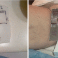
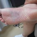
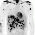
Fig. 3.1
(a) Coronal PET image showing the right groin mass. (b) Axial PET imaging showing the right groin mass
Case (2). The patient had a PET–CT scan ordered for a 10 × 7–cm mesenteric lymph node mass within the right side of the mid abdomen with an SUV max of 18.2 and several enlarged hypermetabolic mesenteric and para–aortic lymph nodes surrounding it. Additionally, the patient had bilateral pelvic, inguinal, axillary, and cervical lymph nodes present (Fig. 3.2a, b). Considering the patient had known extensive relapsed disease, a PET–CT scan was not necessary, and a CT of the chest/abdomen/pelvis would suffice in this scenario.
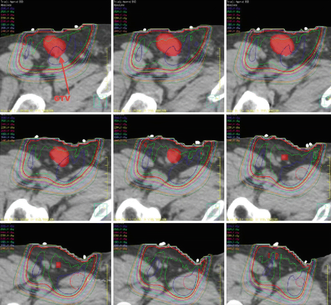

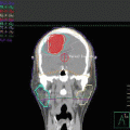
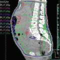
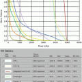
Fig. 3.2
Showing the coverage of the groin mass
Staging of FL follows that of other NHLs using the Ann Arbor system. Work-up should include a patient medical history and physical examination (focused on the lymphatic region) for evaluation of B symptoms, extent of disease, and performance status. Blood work should include a complete blood count, a lactate dehydrogenase test (LDH), a beta-2 (B2) microglobulin test, a comprehensive metabolic panel test, hepatitis B testing, and a pregnancy test for women of child-bearing age. Bone marrow biopsy should be performed in all cases. Imaging should include a diagnostic CT scan of the chest/abdomen/pelvis with consideration of a CT of the neck and/or a PET-CT scan. PET-CT scans are important if considering definitive treatment with radiation for early-stage FL. Wirth et al. demonstrated that a PET-CT scan upstaged patients who were thought to have early-stage (I/II) FL by conventional assessment to stage III/IV in 31 % of the cases [6]. Additionally, PET-CT scan showed more extensive stage I/II disease than previously thought, which would increase the radiation field in 14 % of the cases.
In a large pooled analysis that led to the development of the FLIPI, several important clinical features were observed, including an even distribution of disease between the sexes (male, 51 %, female, 49 %), 63 % of patients under 60 years old, 22 % with stage I/II disease, 19 % with B symptoms, 35 % with five or more nodal sites, 38 % with extranodal involvement (other than bone marrow), 48 % with bone marrow involvement, 22 % with splenic involvement, 41 % with elevated serum B2 microglobulin, 21 % with elevated LDH, 18 % with hemoglobin below 120 g/L, and 10 % with albumin <35 g/L [2]. The factors that were found to be most prognostic for outcomes were age ≥60 years, stage III/IV disease, hemoglobin <120 g/L, elevated LDH, and >4 involved nodal sites. Based on these factors, the following risk groups were created: low risk, 0–1 factor; intermediate risk, 2 factors; and high risk, 3 or more factors. The 5-year and 10-year overall survival rates are 91 %, and 71 % for low-risk disease were 78 and 51 % for intermediate-risk disease and 53 and 36 % for high-risk disease.
In a follow-up project that involved a more modern patient cohort treated in the era of rituximab, a FLIPI2 was developed [7]. The factors found to have the strongest prognosis were B2 microglobulin levels (above normal), longest diameter of the largest involved node (>6 cm), bone marrow involvement, hemoglobulin <12 g/dL, and age >60 years. Based on these factors, the 3-year overall survival and progression-free survival (PFS) rates were 99 and 91 % for low-risk disease (0 factor), 96 and 69 % for intermediate-risk disease (1–2 factors), and 84 and 51 % for high-risk disease (3–5 factors).
Treatment Strategies and Their Variations
Case (1). Several options exist for the management of stage II low–risk FLIPI and intermediate–risk FLIPI2. With this patient, we discussed definitive treatment with radiotherapy or management with immunotherapy. The consensus among his oncologists was definitive treatment with involved–site radiotherapy to a dose of 24 Gy in 12 fractions, which the patient agreed to pursue.
Case (2). The patient was tired of chemotherapy but had symptomatic disease. Palliative radiotherapy of 4 Gy in two fractions of 2 Gy/fraction was offered to the patient.
Early-Stage Disease
The diagnosis of stages I and II FL is rare; consequently, few prospective clinical studies have been conducted to determine the most successful approach to treatment. Historically, definitive management with radiotherapy has been the recommended standard of care based on large historical retrospective cohorts (Table 3.1). However, the National LymphoCare study, a multicenter, longitudinal observational study that included patients from both academic and community practices, demonstrated that there was great variety in how patients with early-stage FL were being managed [3, 8]. Approaches included observation alone (29 %), radiation therapy alone (23 %), rituximab alone or with chemotherapy (35 %), and rituximab followed by radiation therapy (8 %). Although similar excellent outcomes were seen during the short median follow-up of just less than 5 years, the cohort represents a heterogenous group of patients, with only 62 % staged with a PET-CT scan [3, 8].
Table 3.1
Literature review of definitive treatment of stage I/II follicular lymphoma with radiotherapy alone
Institution | No. of patients | Median follow-up (years) | Percentage of patients with stage I disease | Percentage of patients with stage II disease | 10-year progression-free survival | 10-year overall survival | Radiotherapy dose (Gy) | Radiotherapy field |
|---|---|---|---|---|---|---|---|---|
University of Florida [9] | 72 | 8.5 | 75 % | 25 % | 59 % | 46 % | 20–50 | EFRT/IFRT |
Stanford University [10] | 177 | 7.7 | 41 % | 59 % | 44 % | 64 % | 35–50 | TLI/EFRT/IFRT |
British Columbia Cancer Agency [11] | 237 | 7.3 | 76 % | 24 % | 49 % | 66 % | 20–40 | EFRT/INRT + 5 cm |
Princess Margaret [12] | 460 | 10.6 | 74 % | 26 % | 52 % | 62 % | 16–48 | IFRT |
Radiation therapy has been the standard treatment of early-stage FL for over 50 years based on large single-institution retrospective studies. Table 3.1 lists an assortment of these studies and their outcomes [9–12]. The largest studies of approximately 200 patients included those conducted by the British Columbia Cancer Agency, Stanford University (California), Princess Margaret Hospital (Toronto, Ontario), and British National Lymphoma Investigation, which all demonstrated 10-year PFS rates of 44–53 % and 10-year overall survival rates of 58–66 % using radiation doses between 20 and 50 Gy and extended-field radiation therapy (EFRT) as well as involved-field radiation therapy (IFRT) designs. Considering the significant upstaging with PET-CT scans, it is likely that some stage I/II patients treated before the use of PET-CT had stage III/IV disease. Therefore, outcomes might be improved for patients evaluated during the PET-CT era.
Radiation dose has been evaluated across several single-institution databases to understand the doses needed for definitive treatment of patients with FL. In 2011, a published randomized study from the United Kingdom compared 24 Gy in 12 fractions to 40–45 Gy in 20–23 fractions among patients with indolent NHL, including those with advanced-stage disease and those treated with chemotherapy [13]. With a median follow-up of 5.6 years, overall PFS was 55 %, and no difference was seen in freedom from local progression (79 % vs 76 %, p = 0.68). Additionally, no difference in overall survival was seen at 5 years (73 % vs 74 %, p = 0.84). Among patients treated with radiotherapy alone, no difference was seen in PFS; 54 % in the high-dose arm and 64 % in the low-dose arm were alive without a recurrence at 5 years. Furthermore, a randomized study comparing 24 Gy in 12 fractions with 4 Gy in two fractions among patients with indolent histologies demonstrated that when the intent was cure, local control improved with 24 Gy (p = 0.0051) [14].
Stay updated, free articles. Join our Telegram channel

Full access? Get Clinical Tree


