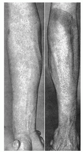
Diagnostic Approach to the Bleeding Disorders


Diagnostic Approach to the Bleeding Disorders


George M. Rodgers

Christopher M. Lehman


Except for that which occurs during menstruation, spontaneous bleeding is abnormal. Surprisingly, little blood is lost, even after large injuries, because of the efficiency with which vascular integrity is normally maintained and the rapidity with which it is restored after injury. In general, these phenomena reflect the functional effectiveness of normal hemostasis (see Chapters 17, 18, and 19). It must be recognized, however, that the adequacy of hemostasis is only relative, and despite the presence of normal vessels, platelets, and coagulation factors, bleeding can occur as the result of localized pathologic processes.

The 11 chapters in Part V deal with disorders that result from abnormalities of the hemostatic process. This chapter is a summary of the diagnostic approach to these disorders and includes a brief discussion of laboratory methods for their study. In subsequent chapters, individual disorders are considered in six categories: thrombocytopenia (Chapters 46, 47, 48, and 49), bleeding disorders caused by vascular abnormalities (Chapter 50), thrombocytosis (Chapter 51), disorders of platelet function (Chapter 52), inherited coagulation disorders (Chapter 53), and acquired coagulation disorders (Chapter 54). The pathophysiology of thrombosis and the principles of antithrombotic therapy are summarized in Chapter 55.


CLINICAL EVALUATION OF THE BLEEDING PATIENT

A careful evaluation of the patient presenting with a bleeding disorder can often provide valuable clues as to whether the abnormality resides in the vessels, platelets, or the process of blood coagulation; a carefully obtained history can usually establish whether the disorder is inherited or acquired; and the physical examination may reveal findings such as the characteristic skin lesions of hereditary hemorrhagic telangiectasia, which alone may provide the diagnosis of a previously perplexing bleeding problem. Results of the clinical evaluation should lead to a rational and efficient laboratory investigation.

It is important to ask specific questions about bleeding because people with normal hemostasis may believe they bleed excessively.1 Certain questions may discriminate between those with normal and abnormal hemostasis, including whether excessive bleeding occurs after tooth extraction or small cuts, whether spontaneous bruising or muscle bleeding occurs, or whether the patient has ever been transfused or treated with blood products.1


MANIFESTATIONS OF DISORDERED HEMOSTASIS

Certain signs and symptoms are virtually diagnostic of disordered hemostasis. They can be divided arbitrarily into two groups: those seen more often in disorders of blood coagulation and those most commonly noted in disorders of the vessels and platelets. The latter group is often called purpuric disorders because cutaneous and mucosal bleeding usually are prominent. The clinical findings that are most valuable in distinguishing between these two broad categories are summarized in Table 45.1. Although these criteria are relative, they provide valuable clues to the probable diagnosis if they are applied to the predominant clinical features in a given patient.


TABLE 45.1 CLINICAL DISTINCTION BETWEEN DISORDERS OF VESSELS OR PLATELETS AND DISORDERS OF BLOOD COAGULATION


Finding


Disorders of Coagulation


Disorders of Platelets or Vessels


Petechiae


Rare


Characteristic


Deep dissecting hematomas


Characteristic


Rare


Superficial ecchymoses


Common; usually large and solitary


Characteristic; usually small and multiple


Hemarthrosis


Characteristic


Rare


Delayed bleeding


Common


Rare


Bleeding from superficial cuts and scratches


Minimal


Persistent; often profuse


Sex of patient


80%-90% of inherited forms occur only in male patients


Relatively more common in females


Positive family history


Common


Rare (except von Willebrand disease and hereditary hemorrhagic telangiectasia)


Hemarthrosis

Hemorrhage into synovial joints is virtually diagnostic of a severe inherited coagulation disorder, most commonly hemophilia A or hemophilia B, and is rare in disorders of the vessels and platelets or in acquired coagulation disorders. This disabling problem often develops with pain and swelling as chief symptoms but without discoloration or other external evidence of bleeding (see Fig. 53.3). Subperiosteal hemorrhages in children with scurvy and swollen painful joints that may develop in some patients with allergic purpura occasionally may be confused with hemarthrosis.


FIGURE 45.1. Diffuse petechial rash induced by a tourniquet in a patient with chronic idiopathic thrombocytopenic purpura (platelet count = 40 × 109/L).


FIGURE 45.2. Large dissecting hematoma of thigh in a patient with hemophilia. A. The lesion resulted from a slight bump to the inguinal area and spread to involve the entire thigh. (Courtesy of Dr. John Lukens.)


Traumatic Bleeding

The unavoidable traumas of daily life and even minor surgical procedures are a greater challenge to hemostasis than any test yet developed in the laboratory. In contrast to “spontaneous” bleeding manifestations, bleeding after trauma in a person with a hemorrhagic diathesis differs in a quantitative way from that which would normally be expected in terms of the amount, duration, and magnitude of the inciting trauma. Such variables are extremely difficult to assess accurately by taking the patient’s history. The amount of blood lost may be exaggerated by the patient. The need for transfusions and the number administered may serve as a rough guide. The patient’s statement concerning the duration of bleeding is more reliable. Detailed inquiry as to past injuries and operations must be made because the patient is likely to forget procedures or injuries that were uncomplicated and to dwell on those in which bleeding was a problem. Whether reoperation was required for prolonged bleeding after tooth extraction or other minor surgical procedures may be helpful in identifying a patient with abnormal hemostasis.

In individuals with a coagulation disorder, the onset of bleeding after trauma often is delayed. For example, bleeding after a tooth extraction may stop completely, only to recur in a matter of hours and to persist despite the use of styptics, vasoconstrictors, and packing. The temporary hemostatic adequacy of the platelet plug despite defective blood coagulation may explain this phenomenon of delayed bleeding, as well as the fact that patients with coagulation disorders seldom bleed abnormally from small superficial cuts such as razor nicks. In contrast, posttraumatic or postoperative surgical bleeding in thrombocytopenic patients usually is immediate in onset, as a rule responds to local measures, and rarely is as rapid or voluminous as that encountered
in patients with coagulation disorders. However, it may persist for hours or days after surprisingly small injuries.

Valuable information often is obtained from a careful review of dental procedures, because most patients have had one or more teeth extracted at some time during their lives. The amount of bleeding normally encountered varies greatly, but as a rough guide, uncomplicated extraction of a single molar tooth may result in brisk bleeding for up to 1 hour and slight oozing for up to 2 days in normal persons.2 Typically, bleeding is more profuse from upper than from lower sockets and is more extensive after extraction of molar teeth, particularly impacted third molars, than after removal of other teeth. In patients with inherited coagulation disorders, the shedding of deciduous teeth often is uncomplicated.

The response to trauma is an excellent screening test for the presence of an inherited hemorrhagic disorder, and a history of surgical procedures or significant injury without abnormal bleeding is equally good evidence against the presence of such a disorder. The removal of molar teeth is a major challenge to hemostasis, as is a tonsillectomy, and it is a rare hemophiliac, however mildly affected, who can withstand these procedures without excessive bleeding.


Miscellaneous Bleeding Manifestations

Despite the fact that structural causes for bleeding (such as polyps, varices, and tumors) are commonly seen in patients with hematuria, hematemesis, and melena, bleeding from these sites may also be associated with both purpuric and coagulation disorders. Severe menorrhagia may be the sole symptom of women with von Willebrand disease (vWD), mild thrombocytopenia, or autosomally inherited coagulation disorders. Recurrent gastrointestinal bleeding or epistaxis in the absence of other bleeding manifestations is common in hereditary hemorrhagic telangiectasia. A coagulation disorder or a disorder of platelet function should be considered if protracted hematuria is the only symptom.

Bleeding into serous cavities and internal fascial spaces often occurs in patients with inherited coagulation disorders and may create serious diagnostic problems. In hemophilia, retroperitoneal hemorrhage or bleeding into the psoas sheath may mimic appendicitis, and hemorrhage into the bowel wall may be confused with intestinal obstruction. Signs and symptoms simulating a variety of acute intra-abdominal disorders also may be seen in association with allergic purpura. Bleeding into the central nervous system may complicate thrombocytopenia and may occur after minor trauma in patients with coagulation disorders. Multiple small retinal hemorrhages are common in patients with thrombocytopenia and other purpuric disorders but are uncommon in those with inherited coagulation disorders; large hematomas of the orbit may be seen in the latter group. The coexistence of bleeding and thromboembolic phenomena or bleeding from previously intact venipuncture sites is suggestive of diffuse intravascular coagulation (DIC). Protracted wound healing, wound dehiscence, and abnormal scar formation have been described in inherited afibrinogenemia, the dysfibrinogenemias, and in factor XIII deficiency.3 Hemoptysis rarely is associated with hemorrhagic disorders.


CLINICAL FEATURES OF INHERITED BLEEDING DISORDERS

An inherited bleeding disorder is suggested by the onset of bleeding symptoms in infancy and childhood, a positive family history (particularly if it reveals a consistent genetic pattern), and laboratory evidence of a single or isolated abnormality, most commonly the deficiency of a single coagulation factor.


Age at Onset: Bleeding in the Neonate

Birth and the neonatal period provide unique challenges to the hemostatic mechanism,4 and bleeding during the first month of life often is the first evidence of an inherited disorder of hemostasis. Small cephalohematomas and petechiae are common in the newborn as a result of the trauma of delivery. Large cephalohematomas that progressively increase in size may result from hemophilia but are more common in association with acquired bleeding disorders such as hemorrhagic disease of the newborn (see Chapter 54). Bleeding from the umbilical stump and after circumcision is common in the acquired coagulation disorders, and it also occurs in the inherited coagulation disorders,5 with the exception of hypofibrinogenemia, dysfibrinogenemia, and factor XIII deficiency. The onset of bleeding from the umbilical cord may be delayed in these latter disorders. In the evaluation of bleeding in the neonate, the clinician should remember that hematochezia and hematemesis may originate from swallowed blood of maternal origin. Simple tests to distinguish such maternal blood from fetal blood have been described.5

Many infants with inherited coagulation disorders do not bleed significantly in the neonatal period. Less than one-third of patients with hemophilia A and B and only 10% of those with other inherited coagulation disorders have hemorrhagic symptoms during the first week of life. In such patients, the disorder may become clinically silent for a time. Hematomas may first be seen only when the child becomes active. Hemarthrosis commonly does not develop until a child is 3 or 4 years of age.

A mild inherited hemorrhagic disorder may be difficult to distinguish from the insidious onset of an acquired defect. Patients with mild inherited coagulation disorders may enter adult life before characteristic bleeding manifestations occur. These patients and those with some forms of inherited thrombocytopenia and disordered platelet function often describe a history of posttraumatic bruising and hematoma formation that they have come to accept as normal. In hereditary hemorrhagic telangiectasia, the lesions become more prominent with advancing age and may not be symptomatic until middle age. Similarly, in patients with Ehlers-Danlos syndrome, bleeding may not be a problem until adult life.


Family History

The family history is of great importance in the evaluation of bleeding disorders. In disorders inherited as autosomal dominant traits with characteristic symptoms and high penetrance, such as hereditary hemorrhagic telangiectasia, an accurate pedigree spanning several generations can often be obtained. The presence of typical bleeding manifestations in male siblings and maternal uncles is virtually diagnostic of X-linked recessive inheritance, which characterizes hemophilia A and hemophilia B. In such X-linked traits, the family history also may be helpful in a negative sense—that is, it may clearly exclude the disorder in certain offspring, such as the sons of a known hemophiliac. Details of the various genetic patterns that may be encountered are discussed in the chapters that deal with these conditions.

The limitations of the family history, however, are greater than is commonly realized. Hearsay history is difficult to evaluate, and it is often impossible to assess the significance of easy bruising or to differentiate between manifestations of a generalized bleeding disorder and more common localized lesions, such as peptic ulcer and uterine leiomyomas. A negative family history is of no value in excluding an inherited coagulation disorder in an individual patient. As many as 30% to 40% of patients with hemophilia A have a negative family history.6 The family history usually is negative in the autosomal recessive traits, and consanguinity, which is commonly present in these kindreds, is notoriously difficult to document or exclude.


CLINICAL FEATURES OF ACQUIRED BLEEDING DISORDERS

Generalized bleeding may be a prominent feature of a wide variety of acquired disorders that encompass virtually the entire field of medicine. Bleeding manifestations usually are less severe than in the inherited forms, and the clinical picture often is dominated by evidence of the underlying disorder rather than by bleeding alone. In the neonate, for example, DIC usually is associated with significant complications such as sepsis, hypoxia, acidosis, or problems related to prematurity. The physician should suspect sepsis or occult thrombosis in any sick neonate with unexplained thrombocytopenia.5 Multiple hemostatic defects commonly are present in patients with acquired hemorrhagic diseases, which often include thrombocytopenia and significant coagulation abnormalities. In contrast, a single abnormality usually is found in patients with inherited hemorrhagic disorders.

In general, the emphasis of the study of the acquired bleeding disorders should be on the patient, not on the laboratory. A thorough history and the physical examination often reveal the cause of thrombocytopenia, such as a drug or acute leukemia. In most vascular disorders, including senile purpura, allergic purpura, scurvy, and amyloidosis, the history and physical examination are of primary diagnostic importance, and the laboratory has little to offer.


Drug History

The importance of exhaustive interrogation regarding drug use and chemical exposure cannot be overemphasized. The list of drugs associated with thrombocytopenia (see Table 47.6) or vascular purpura grows longer each year. Less common but more serious is drug-induced aplastic anemia, which may present initially with bleeding. Many commonly used drugs, notably aspirin, impair platelet function and produce abnormal findings on laboratory tests that often lead to expensive and unnecessary additional laboratory studies. The same drugs may provoke bleeding when administered to patients with pre-existing hemostatic defects such as hemophilia A. Drug ingestion also may produce coagulation abnormalities, and drugs that potentiate or antagonize the anticoagulant effects of coumarin derivatives may lead to bleeding or erratic laboratory control. The surreptitious ingestion of such agents is not uncommon.


TABLE 45.2 INTERPRETATION OF COMMON TESTS OF HEMOSTASIS AND BLOOD COAGULATION


Test


Normal Rangea (±2 SD)


Common Causes of Abnormalities


Platelet count


Phase microscopy


140,000-440,000/µl


Thrombocytopenia, thrombocytosis


Automated


177,000-406,000/µl


Partial thromboplastin time (activated)b


26-36 sec55, c


Deficiencies or inhibitors of prekallikrein; high-molecular-weight kininogen; factors XII, XI, IX, VIII, X, and V; prothrombin or fibrinogen; lupus inhibitors; heparin; warfarin


Prothrombin timeb


12.0-15.5 sec66, c


Deficiencies or inhibitors of factors VII, X, and V; prothrombin or fibrinogen; dysfibrinogenemia; lupus inhibitors; heparin; warfarin


Thrombin timeb


14.7-19.5 sec


Afibrinogenemia, dysfibrinogenemia, hypofibrinogenemia, and hyperfibrinogenemia; inhibitors of thrombin (heparin) or fibrin polymerization (fibrin degradation products, paraproteins)


Fibrinogen assayb


150-430 mg/dl70


Afibrinogenemia, dysfibrinogenemia, and hypofibrinogenemia; inhibitors of thrombin or fibrin polymerization


Factor VIII assayb


50-150 U/dl


Hemophilia A and von Willebrand disease; acquired antibodies to factor VIII


Fibrin degradation product assay


0-5 µg/ml80


Disseminated intravascular coagulation; fibrinogenolysis; thrombolytic drugs, liver disease; dysfibrinogenemia


D-dimer assay


0-0.4 µg/ml


Disseminated intravascular coagulation; recent surgery; pregnancy; thromboembolism


a Normal range in the University of Utah coagulation laboratory.

b Tests affected by heparin.

c Significant variations depending on reagents and technique.


Results of various coagulation tests may be abnormal in a surprisingly large percentage of hospitalized patients because of heparin that is administered therapeutically or is used in small amounts to maintain the patency of indwelling venous catheters, venous pressure lines, arteriovenous shunts, and various pumps and infusion machines. The partial thromboplastin time (PTT), in particular, may be greatly prolonged in patients who have received even a minute amount of this anticoagulant. Such coagulation abnormalities often are confused with DIC, inhibitors of factor VIII, and other serious coagulation disorders, and they commonly lead to repeated, and usually useless, coagulation studies. A thorough bedside inventory often is required to find out that heparin is indeed responsible. Prolongation of the thrombin time associated with a normal reptilase time or direct assay of heparin provides laboratory evidence of heparin contamination.


LABORATORY METHODS FOR STUDY OF HEMOSTASIS AND BLOOD COAGULATION

No single test is suitable for the laboratory evaluation of the overall process of hemostasis and blood coagulation, but methods of varying complexity and use are available for assessing various components and functions individually. The emphasis of the following discussion is on methods that are simple and widely available in most laboratories. The interpretation of the most commonly used tests and the range of values obtained in normal subjects with representative techniques are summarized in Table 45.2. Definitive coagulation methods usually require a specially equipped laboratory and trained personnel, and are discussed here from a general standpoint only. Additional comments concerning the use and limitations of the various methods are included in chapters dealing with individual disorders.


Tests of Vascular and Platelet Phases


Bleeding Time

Hemostasis in a small superficial wound, such as that produced when measuring the bleeding time, depends on the rate at which a stable platelet plug is formed and, thus, provides a measure of the efficiency of the vascular and platelet phases. However, it does not discriminate between vascular defects, thrombocytopenia, and platelet dysfunction. The bleeding time leaves much to be desired in terms of reproducibility, because no two skin areas are exactly the same and it is impossible to produce a truly standard wound.7

Older studies using the bleeding time test supported the view that this test might be helpful in predicting bleeding in individual patients.8 More recent studies suggest that a bleeding time result is determined not only by platelet number and function, but also by hematocrit,9 certain components of the coagulation mechanism,10, 11 skin quality,12 and technique.13 A careful analysis of this literature indicates that there is no correlation between a skin template bleeding time and certain visceral bleeding times,13, 14 and that no correlation exists between preoperative bleeding time results and surgical blood loss or transfusion requirements.15

A clinical outcomes study reported that discontinuation of the bleeding time in a major academic medical center had no detectable adverse clinical impact.16 A position paper of the College of American Pathologists and the American Society of Clinical Pathologists concluded that the bleeding time was not effective as a screening test, and that a normal bleeding time does not exclude a bleeding disorder.17 Patients thought to have a platelet-type bleeding disorder based on their personal or family history (or both) should be evaluated for vWD and the inherited qualitative platelet disorders, using assays discussed in the section Platelet Function Assays. Newer assays that may be useful in screening patients for platelet dysfunction are also discussed in the section New Assays of Platelet Function.


Platelet Enumeration

Platelets are considerably more difficult to count than erythrocytes or leukocytes. This difficulty is to be expected in view of the small size of these cells and their tendency to adhere to foreign surfaces and to aggregate when activated.

In general, techniques for platelet counting may be classified into two groups: hemacytometer or direct methods, in which whole blood is diluted and the platelets are counted in much the same way as leukocytes or erythrocytes, and fully automated electronic methods. Virtually identical values for the normal range of the platelet count have been obtained with modern methods, as summarized in Table 45.2.

An estimate of platelet numbers in a well-prepared blood smear by an experienced observer is a valuable check on the platelet count as determined by any method. In general, when a blood smear is examined at 100 × power, each platelet counted/field represents approximately 10,000 platelets × 109/L. Consequently, a normal blood smear should demonstrate, on average, at least 14 platelets/high-power field.

Instruments for totally automated platelet counting are widely used. Details of automated cell counters are discussed in Chapter 1. When automated methods are used, various nontechnical factors may produce falsely low platelet counts.18 These factors include platelet agglutinins,19 abnormal amounts of plasma proteins in various paraproteinemias, previous contact of platelets with foreign surfaces such as dialysis membranes,20 large or giant platelets, platelet satellitism,21 lipemia,22 and ethylenediaminetetra-acetic acid (EDTA)-induced platelet clumping,23 a phenomenon that may produce platelet clumps of sufficient size to artifactually increase the leukocyte count.24 Spuriously high platelet counts may result from the presence of microspherocytes,25 fragments of leukemic or red blood cells,26 and Pappenheimer bodies.27 Special technical modifications and the use of careful manual counting methods may be required to eliminate these artifacts and to obtain accurate platelet counts.


Platelet Volume Measurements

The widespread availability of particle counters in the clinical laboratory permits the accurate measurement of platelet volume on a routine basis. Mean platelet volume (MPV) is increased in disorders associated with accelerated platelet turnover as the result of large numbers of megathrombocytes28 or in patients with Bernard-Soulier syndrome. Normal or decreased values for MPV usually are obtained in patients with disorders associated with deficient platelet production, in some patients with sepsis,29 and in people with certain big-spleen syndromes.30

Some authors suggest that increased MPV provides evidence of accelerated platelet production and may be interpreted in the same way as the reticulocyte count. The method is difficult to standardize, however, and when determined on routinely collected specimens by automated counters, it is affected by numerous variables pertaining to specimen collection, anticoagulant, temperature, and duration of storage.31 In view of these problems and the difficulty in interpreting platelet size heterogeneity under normal and abnormal conditions,32 these measurements should be interpreted with caution.

The presence of microcytic platelets in patients with some inherited thrombocytopenias such as Wiskott-Aldrich syndrome is reliably reflected by MPV measurements. On the other hand, giant platelets associated with Bernard-Soulier syndrome may be counted as leukocytes or erythrocytes and may not be reflected in the MPV.


Platelet Function Assays

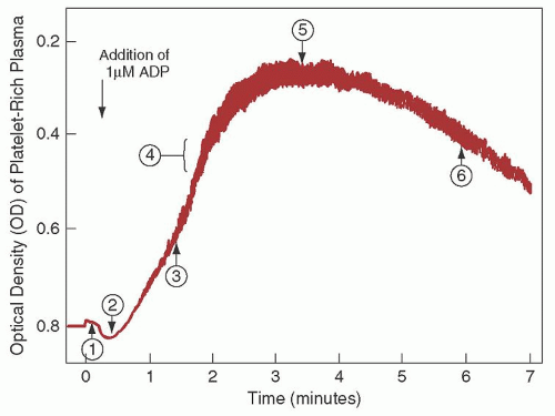
Since the 1960s, platelet aggregation using platelet-rich plasma has been the standard method to assess platelet function. This method uses aggregometers, which are modified nephelometers that permit measurement of changes in optical density of a platelet suspension under conditions of constant temperature and continuous agitation (Fig. 45.3). Most instruments measure
a combination of light scatter and absorption. Instruments have been developed that permit both nephelometric and photometric measurements and the simultaneous measurement of aggregation and nucleotide release.33


FIGURE 45.3. The interpretation of aggregometer tracings. Tracing of platelet aggregation produced by a low concentration of adenosine diphosphate (ADP), illustrating normal changes in optical density (OD)—that is, (1) a slight decrease caused by dilution with aggregating agent; (2) a transient increase caused by initial platelet swelling or shape change; (3) a rapid progressive decrease as platelet aggregates form, the size of which is roughly proportional to the amplitude of the oscillations in the tracing (4). The OD then reaches a nadir (5) from which maximal aggregation as a percentage of the initial OD may be calculated as follows: maximal aggregation (%) = OD at T0—minimum OD/OD at T0. After this (6), a slow increase in OD caused by disaggregation occurs under some conditions.

Platelet aggregation usually is studied in suspensions of citrated platelet-rich plasma, in which the size and dimensions of the stirring bar, variations in plasma citrate concentration attributable to variations in hematocrit, the pH, and the nature of the buffers are important variables. Platelet suspensions usually are prepared by differential centrifugation, but methods that use albumin density gradient centrifugation and gel filtration have also been described.34 Although harvesting platelets from the blood of thrombocytopenic patients is difficult, testing such platelets in the aggregometer is reproducible in suspensions containing as few as 50,000 platelets/µl. Methods for the study of platelet aggregation in whole blood35, 36 also have been described. Interfaced computer systems have been developed for calculating and expressing platelet function data.37

Adenosine diphosphate (ADP) in concentrations of 5 µmol/L or higher produces platelet aggregation directly that is independent of the release of platelet-contained ADP.38 Various other aggregating agents act mainly by inducing the release reaction, such as a suspension of connective tissue particles (collagen), epinephrine and norepinephrine, and thrombin. With epinephrine (5 µmol/L), a weak primary aggregating effect usually can be clearly distinguished from the subsequent release reaction, which produces a secondary wave of aggregation. Such primary and secondary waves of aggregation also may be seen with carefully titrated amounts of ADP (0.2 to 1.5 µmol/L).38

Ristocetin is an antibiotic that induces platelet agglutination (platelet metabolic activity not required) in the presence of von Willebrand factor (vWF). Patients deficient in vWF (vWD) or in the receptor for vWF (Bernard-Soulier syndrome) have an abnormal ristocetin response. Ristocetin is tested in concentrations of 0.6 to 1.2 mg/ml; the lower concentrations are helpful in identifying specific variants of vWD, type 2B and platelet-type vWD (see Chapters 52 and 53).

The release reaction is measured only indirectly by routine aggregometry—that is, the aggregation associated with the release of ADP from the platelets (release-induced aggregation or secondary aggregation). Methods for the quantitation of various substances released from platelets have been described. For example, the amounts of ADP or serotonin released/unit of time serve as indices of dense body release39; the amount of various hydrolytic enzymes or platelet factor 4 released is a measure of the extent of α-granule release.40 Suggested guidelines for standardization of platelet aggregation methods have been proposed.41, 42

Sensitive methods have been developed for the determination of platelet-derived substances in plasma that may serve as markers of intravascular platelet activation,43 including platelet factor 4, β-thromboglobulin, stable prostaglandins (6-keto prostaglandin F1α and thromboxane A2), and leukotrienes.43 These measurements may have diagnostic value in thromboembolic disorders and syndromes characterized by intravascular platelet aggregation.


New Assays of Platelet Function

An appreciation of the limitations of the bleeding time test has led to the development of newer assays to evaluate platelet function.44 Some of these are point-of-care tests. The clinical use and predictive value of these tests to identify patients with hemostatic disorders remain to be established. One assay, the platelet function analyzer (PFA-100), has been investigated for several years, and many published reports using this assay are available. In this method, citrated blood samples are exposed to high shear rates in a capillary flowing through an aperture within a membrane coated with collagen and either ADP or epinephrine.45 The closure time to hemostatic plug formation within the aperture is the endpoint of the test. A large study using the PFA-100 found that prolonged closure times could be attributed to specific quantitative or qualitative abnormalities in platelet function or vWF (or both) in 93% of patients tested.46 However, the International Society on Thrombosis and Haemostasis has taken the position that the PFA-100 is insufficiently sensitive and specific to be used as a screening device for platelet disorders.47 It has been suggested that optimal use of the PFA-100 in evaluation of hemostasis would use an algorithmic approach, evaluating not only PFA-100 closure times, but also a complete blood count, blood smear, and assays for vWD and platelet aggregation to further evaluate abnormal closure times. A recent addition to the PFA repertoire is the INNOVANCE PFA P2Y test designed to assess P2Y12-receptor blockade.

Several additional platelet function analyzers are available on the market, though they are not as well studied as the PFA-100.48 The ICHOR II-Plateletworks system (Helena Laboratories, Beaumont, TX) compares impedance-derived platelet counts in samples with and without added platelet agonists to assess platelet function. This system has historically been used to evaluate cardiopulmonary bypass patients, but more recently has been applied to patients undergoing coronary stent placement. The Impact-R (Matis Medical, Beersel, Belgium) is an automated cone-and-plate research analyzer that assesses platelet adhesion and aggregation on a polystyrene surface under laminar flow conditions. The VerifyNow system analyzes platelet agonist-induced aggregation of fibrinogen-coated microparticles to assess platelet function. Agonist cartridges are designed to evaluate the effects of aspirin, clopidogrel, and GPIIb/IIIa platelet receptor inhibitor administration on platelet function. Platelet mapping, a modification of thromboelastography, measures the platelet contribution to clot strength in the presence of specific agonists.49 To date, no platelet function analyzer assay has been sufficiently studied or validated to warrant routine clinical use.48, 50, 51


Tests of Coagulation Phase

In general, meticulous performance of coagulation tests is more important than the exact technique chosen. Blood samples obtained by traumatic venipunctures or from indwelling catheters often are inadequate for coagulation studies.52 A poorly collected blood sample is a far more common cause of inaccurate results than is technical error.

With the exception of one assay for fibrin degradation products (FDPs), all coagulation tests are performed on citrated plasma, most commonly obtained using blue-top vacuum blood collection tubes that pull in nine parts of blood to one part citrate. The International Society for Thrombosis and Haemostasis recommends the routine use of 3.2% sodium citrate. A pool of freshly frozen citrated plasma from several normal donors is a suitable control for screening procedures in most laboratories. Lyophilized control plasma and borderline abnormal control plasmas are available commercially to standardize coagulation assays and to provide reference standards.

The citrate ion does not enter the erythrocyte. Consequently, the plasma citrate concentration is abnormally high when blood with a high hematocrit (>55%) is collected in usual concentrations of this anticoagulant. This may produce artifactual prolongation of one-stage screening tests of coagulation, such as the PTT.53 To obtain interpretable data on such samples, tubes containing citrate concentrations appropriate for the hematocrit must be prepared by removing an aliquot of the citrate anticoagulant contained in standard blue-top tubes.


Activated Partial Thromboplastin Time

The activated partial thromboplastin time (PTT) is a simple test of the intrinsic and common pathways of coagulation. When a mixture of plasma and a phospholipid platelet substitute is recalcified,
fibrin forms at a normal rate only if the factors involved in the intrinsic pathway (prekallikrein, high-molecular-weight kininogen, and factors XII, XI, IX, and VIII) and in the common pathway (factors X and V, prothrombin, and fibrinogen) are present in normal amounts (Fig. 45.4). Platelet substitutes of various kinds may be used, such as chloroform extract of brain54 and other crude cephalin fractions as well as soybean phosphatides (inosithin). In the PTT, such platelet substitutes are provided in excess, and the test is unaffected by the number of platelets remaining in the plasma (unless the sample contains antiphospholipid antibodies). Platelet substitutes are only partial thromboplastins, however, and they are incapable of activating the extrinsic pathway, which requires complete tissue thromboplastin (tissue factor). Thus, the PTT bypasses the extrinsic pathway and is unaffected by a deficiency of factor VII. The PTT assay is used to detect factor deficiency, screen for the lupus anticoagulant, and monitor heparin anticoagulation.

Only gold members can continue reading. Log In or Register to continue

Stay updated, free articles. Join our Telegram channel

Oct 21, 2016 | Posted by in HEMATOLOGY | Comments Off on Diagnostic Approach to the Bleeding Disorders

Full access? Get Clinical Tree

Get Clinical Tree app for offline access