(1)
State University of New York at Buffalo Kaleida Health, Buffalo, NY, USA
Long-term central venous access is an essential technique in surgical oncology, required either for hyperalimentation of a malnourished patient before or after an oncologic procedure, or for periodic access to a central vein—usually the superior vena cava (SVC)—in order to infuse various chemotherapeutic agents. The latter indication is the more common one, as peripheral veins are often small and difficult to cannulate, or they become thrombosed and sclerotic after a few infusions of chemotherapeutic agents, presenting a risk of extravasation of these drugs, some of which are highly toxic to the infused tissues, with resultant necrosis and slough.
The most common method of central venous access is the percutaneous puncture of the subclavian vein and the placement of a catheter, the tip of which is advanced to the SVC just above its entry to the right atrium. Use of the left subclavian vein is slightly preferred because the smaller size of the left lung would make respiratory distress less likely in the event of pneumothorax. Factors such as a prior axillary dissection or use of the subclavian on one side may make use of the other side preferable, however.
Seldinger Technique
For cannulation of the subclavian vein, a skin puncture point is selected below the junction of the middle and lateral third of the clavicle, or over the estimated position of the cephalic vein in the deltopectoral groove. The point should be far enough below the clavicle for the needle, aimed toward the jugular notch, to be inserted in the space between the clavicle and first rib and thus to enter the subclavian vein.
Before cannulation of the subclavian vein is attempted, the patient should be in a good state of hydration or should be given extra intravenous fluids immediately prior to the operation. The patient should be placed in a Trendelenburg position on the operating table until successful cannulation occurs, so that the vein will be distended and easy to cannulate.
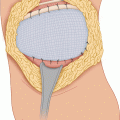
Local anesthesia is injected at the selected skin puncture point and in the subcutaneous tissue all the way to the clavicle in the direction the long cannulating needle is estimated to follow. At the proposed puncture point, a nick is made in the skin with a #11 blade and the long introducer needle connected to a 10-mL syringe (provided in the kit) is inserted in the subcutaneous fat for 1–2 cm, aiming toward the jugular notch. The syringe is then depressed so that the tip of the needle assumes a more anterior position as it is advanced, in order to enter the space between the clavicle and the first rib and go into the subclavian vein. One aspirates with the syringe frequently until a free flow of venous blood occurs, which indicates entry into the vein. The syringe is then disconnected and the guide wire is placed in the needle with the wire-straightener and is advanced to almost its full length if no resistance is encountered. The position of the wire tip is checked with fluoroscopy and, if necessary, is adjusted to be in the SVC. The patient’s position is made horizontal. The skin puncture point is made larger and the dilator-sheath is inserted over the guide wire to its full length. The guide wire and the dilator are removed and the permanent catheter, flushed with heparin solution, is inserted so that its tip lies in the SVC just above the right atrium. The peel-away catheter is then removed. The site for the creation of a subcutaneous pocket is chosen. A common site is the anterior end of the second to third ribs adjacent to the sternum. After injection of a local anesthetic, a transverse incision is made over that site, a little longer than the diameter of the port, and is deepened to the fascia. With the use of Metzenbaum scissors, a pocket sufficiently large to accept the entire port is made, the anterior surface of the port being about 0.5 cm from the skin. The external end of the indwelling catheter is attached to a tunneler, which is passed from the initial skin puncture point through the subcutaneous tissue to the incision for the port. The position of the tip of the indwelling catheter is checked again with fluoroscopy. A Huber needle is used to penetrate the plastic anterior surface of the chamber and is inserted until the tip of the needle hits the posterior metallic surface; the chamber of the port is flushed with heparinized saline. The catheter is trimmed and its external end is pushed over the tubular projection of the port, making for a tight connection, and the ring provided around the permanent catheter is pushed over the connection to make it more secure and leakproof. The port is kept in the same position by passing a suture between the hole in the flange on each side of the connecting point and the adjacent fascia. Alternatively, the opening of the pocket is closed around the port with two or three interrupted, absorbable sutures, approximating the subcutaneous fat in front of and behind the port at the opening of the pocket. The skin incision is closed with absorbable sutures for the subcutaneous layer and a subcuticular continuous suture followed by adhesive strips (e.g., Steri-Strips). The initial skin puncture point is approximated with a single absorbable suture, taking a bite through the dermis on each side and burying the knot. A Steri-Strip also may be used.
A pocket for the placement of the port may also be had by making a skin incision in continuity with the initial skin puncture and creating a pocket on top of the adjacent pectoralis major. The pocket should be made close enough to the skin (about 0.5 cm) to be easily palpable and accessed through the overlying skin. On the other hand, the tissue layer (skin and adipose tissue) over the port should not be too thin and liable to undergo pressure necrosis with exposure of the port in the weeks following the procedure.
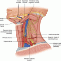
The internal jugular vein has also been used to access the SVC through the percutaneous (Seldinger) technique. This technique has been used primarily only for temporary access to the central venous system because the puncture point in the skin of the neck is in the visible part of the neck, and the curving catheter is visible beneath the skin of the lower neck on its way to the port.
Alternatives to the Seldinger Technique
There are other alternatives to the Seldinger technique for cannulation of the subclavian vein. Some surgeons prefer a cutdown approach. The reasons for this preference are aversion to performing a “blind” procedure and a higher rate of complications (especially pneumothorax) with the Seldinger technique. In a recent article, 3 % of patients developed a complication with a cutdown approach, compared with 9 % in the Seldinger technique group; the complications included pneumothorax, nerve palsy, and hematoma [1]. In various series, the rate of pneumothorax with the Seldinger technique has varied between 0 and 3.2 %. The success rate of the cutdown at the deltopectoral groove (cephalic vein) is about 83 % (range, 70–94 %) [1]. Proponents of the cutdown approach have suggested that the incorporation in the cephalic vein cutdown of technical elements of the percutaneous approach should increase the rate of successful cannulation of the vein [1]. Thus, with the use of a J guidewire to pass the junction of the cephalic vein to the subclavian vein, advancement of the catheter into the SVC was reported in six patients without complications [2]. The main impediment to successful cannulation of the cephalic vein is its often small diameter, which is unable to accept the port catheter.
Stay updated, free articles. Join our Telegram channel

Full access? Get Clinical Tree


