Bleeding Disorders Caused by Vascular Abnormalities


Bleeding Disorders Caused by Vascular Abnormalities


George M. Rodgers

Matthew M. Rees


This chapter discusses causes of bleeding that are not the result of thrombocytopenia, coagulation factor deficiency, or qualitative platelet defects. Bleeding disorders caused by these various “vascular” abnormalities represent a heterogenous group of diseases, and in considering the differential diagnosis, it is convenient to consider cutaneous causes, connective tissue abnormalities, vascular lesions, and intravascular causes.


MECHANICAL PURPURA

External pressure such as blunt trauma results in ecchymoses when the force is sufficient to disrupt vascular integrity and allow extravasation of red blood cells. The size of the resulting lesion is dependent on the durability of the tissue traumatized, the vascularity of the region, the density of the surrounding tissue, and the time elapsed.2, 3 The extent of bruising can increase over time, and tracking through tissue planes can occur, resulting in bruises in areas remote from the area of trauma.4 The color of the lesion is in part dependent on the location of the red cells—lesions near the surface have a more reddish color, and deeper lesions appear bluish. This finding is due to optical scattering in the dermis, and the fact that blue wavelengths scatter and reflect more than red.5

Bruises change color with time, and the color of bruise has been used to determine the age of lesions. However, evidence suggests that there is more variability in color changes than previously recognized.4, 6

Mechanical purpura can be seen on occasion with minor trauma such as blood pressure cuff monitoring in anticoagulated patients3 or even vigorous scratching. Young adults playing active sports such as basketball can develop calcaneal petechiae resulting from relatively minor, but repetitive, heel trauma.7 Periorbital, face, or neck purpura can occur after a sudden increase in intravascular pressure with a Valsalva maneuver8 and has been described after bungee jumping.9

Suction purpura occurs when negative pressure is applied to the skin in sufficient force to result in extravasation of erythrocytes. Young healthy people develop petechiae with 350 to 400 mm Hg negative pressure, but the required amount of pressure to induce purpura decreases with age to as low as 100 mm Hg.10 The application of rubber suction devices to the forehead can result in circumscribed purpura. This seems to occur mostly when new parents apply children’s suction toys to their foreheads and has been termed cyclops purpura.11 A similar, relatively common cause of suction purpura is seen in adolescents who place a drinking glass over their chin and suck out the air to form a vacuum, ultimately causing chin or perioral purpura.12 Adolescents are also prone to develop small purpuric lesions in the neck region from “hickeys” or “love bites,” another form of suction purpura.

Large areas of contusions develop with blunt force injuries. Certain patterns of contusion can help identify the type of device causing the injury. Skin bruises corresponding to the sites of seat belts are known as “seat belt mark” signs. It is important to recognize these, as the presence of this sign should alert physicians to the higher likelihood of internal injuries.13 A direct blow from a linear or cylindric object causes parallel lines (“tram-line” appearance) with central clearing, as the blood beneath the site of highest impact is displaced laterally. Similarly, a bruise in the shape of a circle with central clearing may occur as a result of a blow from the circular edge of a pipe or spherical objects.14 Caution, however, should be used in interpreting these types of bruises, as simple bruises can sometimes occur with “ring resolution.”15 Circular contusions 1 to 1.5 cm in diameter may be due to fingertip pressure and grab marks or bite marks, and they show semicircular arch contusions, but are highly variable.14 Certain bruising patterns can suggest child or elder abuse, but results should be interpreted carefully.16, 17

Larger ecchymotic areas also may demonstrate other patterns that can be a clue to underlying pathology. The well-known Cullen sign refers to bluish discoloration around the umbilicus, and Grey-Turner sign is flank ecchymosis. Both may indicate hemorrhagic pancreatitis or a rectus sheath hematoma.18 Scrotal ecchymosis may be a clue to intraperitoneal hemorrhage, and perianal ecchymosis has been described as a manifestation of aneurysmal rupture into the sigmoid mesocolon.19, 20 “Raccoon eyes” and mastoid ecchymosis (Battle sign) may indicate a basilar skull fracture after head trauma.21 Ecchymosis occurring in patients with swollen calves suggests a ruptured popliteal cyst with gravitational spread of the bruise down to the ankle and occasional linear appearance (“tide mark” appearance) with sparing of the foot and lower ankle due to the pressure of footwear.22


STRUCTURAL MALFORMATIONS OF VESSELS


Hereditary Hemorrhagic Telangiectasia

Hereditary hemorrhagic telangiectasia (HHT) was first described in 1864 by Sutton23 and later recognized and reported by Rendu,24 Osler,25 and Weber,26 and it is thus also known as Osler-Weber-Rendu syndrome. It is an autosomal dominant disorder characterized by multiple telangiectatic lesions involving the skin and mucous membranes associated with epistaxis and other bleeding complications. HHT has an estimated prevalence of 1 in 8,000,27 with complete penetrance by 40 years of age.28

Genetic studies have identified several mutations responsible for the vascular malformations. Mutations in the endoglin gene on chromosome 9 (HHT1) or in the activin receptor-like kinase
(ALK1) gene on chromosome 12 (HHT2) account for ˜85% of the cases. Currently more than 500 mutations in the ALK1 or endoglin genes have been identified, and each family studied appears to have a unique mutation.27 Endoglin is an integral membrane glycoprotein expressed on endothelial cells in arterioles, venules, and capillaries. This glycoprotein and ALK1 serve as a binding protein for transforming growth factor-β (TGF-β).

TGF-β regulates many transcriptional targets and plays a crucial role in vascular development and homeostasis. Recently identified mutations in the MADH4 gene abnormalities are associated with juvenile polyposis and HHT,29 and linkage studies suggest additional mutations exist on other chromosomes.30, 31 An HHT mutation database is available at http://www.hhtmutation.org.


TABLE 50.1 COMMON PHYSICAL FINDINGS AND ASSOCIATIONS OF VASCULAR DISORDERS


Pattern and Usual Location


Etiology


Associated Features


Subconjunctival and axillary petechiae


Fat emboli


Dyspnea


Periorbital, facial, and neck petechiae


Valsalva maneuvers


Periorbital ecchymosis (“black eye”)


Trauma; in penetrating globe injuries without periorbital trauma, suggests posterior rupture


“Raccoon eyes”


Basilar skull fracture


“Battle sign” (mastoid ecchymosis)


Basilar skull fracture


Palatal petechiae


Viral upper respiratory infection


Ecchymosis of limbs and face of infant


AIHE


Inflammatory edema


Eyelid and periorbital edema and purpura, often after dependency; purpura in other areas with minimal trauma


Amyloid


Glove and stocking petechiae with areas of confluence and sharp line of demarcation between normal and abnormal areas


PPGSS


Palm and sole petechiae


Rat bite fever and other infections


Purpuric lesions on the forearm in elderly patients


Solar or steroid purpura


Bluish discoloration of the umbilicus (Cullen sign)


Hemorrhagic pancreatitis or rectus sheath hematoma


Flank ecchymosis (Grey-Turner sign)


Hemorrhagic pancreatitis


Periumbilical purpura with “thumbprint” signing


Strongyloides infection


Scrotal/perineal ecchymosis


Intraperitoneal hemorrhage or iliac aneurysmal rupture (rupture into sigmoid mesocolon)


Telangiectatic lesions along the face, lips, nares, tongue, and nail beds


HHT


Lower extremity palpable, nonblanching purpura


Vasculitis


Perifollicular lower extremity, with areas of confluence


Scurvy


Gingival, intramuscular bleeds


Urticarial lesions followed by purpura


Urticarial vasculitis


Proximal thigh involvements often with a “kissing” or symmetric lesion


Calciphylaxis


Mucosal oozing and easy bruising


EDS


Skin hyperextensibility, joint immobility


Hemorrhagic bulla


Bacterial infections (Clostridium, Vibrio, Aeromonas); hypersensitivity to insect bites, snakebites; pemphigus


“Tram lines”


Blunt force with cylindrical or linear object


“Ring” bruises


Blunt force with circular end or sphere; may also be seen in normal bruise resolution


AIHE, acute infantile hemorrhagic edema; EDS, Ehlers-Danlos syndrome; HHT, hereditary hemorrhagic telangiectasia; PPGSS, papular-purpuric gloves and socks syndrome.


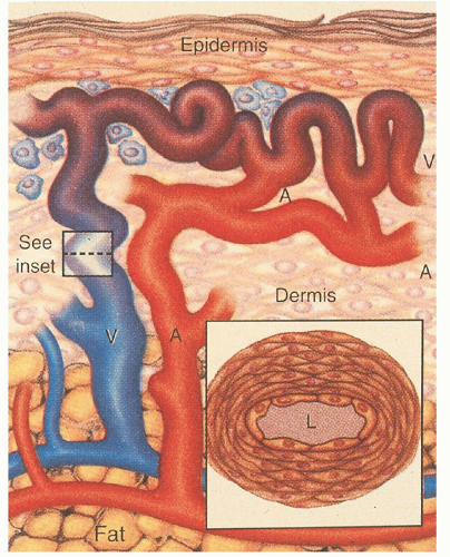
The mechanisms by which these genetic defects result in telangiectatic lesions have not been identified. Ultrastructural analysis of cutaneous HHT lesions suggests that postcapillary venule dilation is the earliest identifiable morphologic abnormality.32, 33 As the venules enlarge, they become convoluted and interconnect with arterioles through capillary segments. The capillary segments eventually disappear, and direct arteriolar-venular communications are established (Fig. 50.1). An infiltrate of mononuclear cells appears in the perivascular region of the HHT lesions.

The bleeding manifestations are thought to occur because of mechanical fragility of these vessels. Common abnormalities in the hemostatic system do not seem to represent a major factor in the underlying bleeding tendency. HHT patients manifest a variety of other complications including shunting, emboli, and thrombosis.


Clinical Manifestations

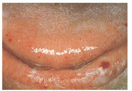
The cutaneous lesions usually appear in affected persons by 40 years of age, and they increase in number with age. The lesions measure 1 to 3 mm in diameter and are sharply demarcated in appearance (Fig. 50.2). They blanch with pressure, but the blanching may be incomplete as a result of “strangulation” of coiled loops of vessels.34, 35 The telangiectatic lesions are most commonly found on the face, lips, nares, tongue, nail beds, and hands. Some patients have only a few lesions, necessitating a thorough search in anyone suspected of having HHT. Bleeding from these cutaneous telangiectasias is uncommon and rarely of clinical importance. One report suggests that capillaroscopy of the dorsal hands can detect morphologic changes in the skin microcirculation, including microscopic telangiectatic lesions, but the clinical utility of this is undefined.36


FIGURE 50.1. Arteriovenous malformations in hereditary hemorrhagic telangiectasia. In a fully developed cutaneous telangiectasia, the venule (V) and its branches have become dilated and convoluted throughout the dermis. The connecting arterioles (A) have also become dilated and communicate directly with the venules without intervening capillaries. A perivascular infiltrate is present. The thickened wall of a dilated postcapillary venule is also shown. L, lumen of the vessel. (From Guttmacher AE, Marchuk DA, White RI. Hereditary hemorrhagic telangiectasia. N Engl J Med 1995;333:918-924, with permission.)

Epistaxis is the presenting complaint in up to 90% of patients with HHT. This symptom results from bleeding telangiectatic lesions over the inferior turbinates and nasal septum. Symptoms usually occur before 35 years of age and are highly variable. Approximately one-third of patients have mild symptoms requiring no treatment, and another third have moderate symptoms requiring only outpatient treatment. The remaining third have severe symptoms often requiring inpatient treatment, transfusions or chronic iron replacement therapy, and surgery.35 The nosebleeds may become more difficult to control as the patient ages.


FIGURE 50.2. Telangiectasias of the tongue and lower lip in a patient with hereditary hemorrhagic telangiectasia. (From Guttmacher AE, Marchuk DA, White RI. Hereditary hemorrhagic telangiectasia. N Engl J Med 1995;333:918-924, with permission.)

Pulmonary arteriovenous malformations (PAVMs) occur in 30% of patients with HHT, and 85% to 90% of people with PAVM are found to have HHT.32, 34, 35, 37 Genetic linkage studies have found that patients with endoglin mutations have significantly higher rates of PAVMs (40%) than HHT patients with other mutations (14%).38, 39 The PAVMs are primarily located in the lower lung lobes and are multiple. These PAVMs may result in a significant right-to-left shunt, and patients may develop dyspnea, cyanosis, clubbing, fatigue, decreased exercise tolerance, migraine headaches, and polycythemia. Paradoxic emboli can occur and result in brain abscesses, transient ischemic attacks, and strokes. The prevalence of cortical infarcts has been reported to be as high as 14% in patients with a single PAVM and increases to 27% in patients with multiple PAVMs.40 These lesions may also bleed and result in hemoptysis or hemothorax; pregnant women with PAVMs appear to be at increased risk.41

The detection of PAVMs may be difficult in patients with few or no symptoms. Physical examination may uncover an end-inspiratory bruit.34 A chest radiograph may detect a coin lesion but often misses smaller lesions. Gravitational shifts in blood flow to the lung bases result in increased right-to-left shunting in the sitting or standing position. Physiologic tests such as measuring O2 saturation in the supine and standing position (on room air and 100% O2) can detect the positional change in shunting and can be used to screen patients for PAVMs. However, the best screening test for PAVM appears to be contrast echocardiography.42 Patients with a positive screening test should undergo an unenhanced spiral computed tomography (CT) to confirm and further characterize the PAVM.43 Pulmonary angiography is a less sensitive study but is necessary in treatment planning.

Approximately 20% of patients with HHT develop significant upper and lower gastrointestinal (GI) tract hemorrhage. Bleeding is rare before the fifth decade of life. Approximately 40% of the bleeding episodes occur from upper GI tract lesions, whereas only 10% occur in the colon, and a full one-half are indeterminate after evaluation.35 Spontaneous regression of GI bleeding is rare, and steady progression or chronic intermittent bleeding is the norm.

Hepatic involvement occurs in 70% of patients, but symptoms and complications are rare.44 Patients may have hepatomegaly, a hepatic bruit or thrill, or elevated liver function studies.45 Types of intrahepatic shunting include hepatic artery to hepatic vein (arteriovenous), hepatic artery to portal vein (arterioportal), and portal vein to hepatic vein (portovenous). These shunts can lead to clinical complications of high-output heart failure, portal hypertension, encephalopathy, biliary ischemia, and nodular regenerative hyperplasia. The dominant type of shunt identified on multiphasic CT does not correlate strongly with the clinical syndrome.46 Nodular regenerative hyperplasia is found with a 100-fold increased prevalence compared to the general population, and along with portal hypertension can lead to a misdiagnosis of cirrhosis (pseudocirrhosis).44 It is important to consider this diagnosis in the setting of a liver mass, as biopsy should generally be avoided when suspected. Additionally, if patients with known focal nodular hyperplasia are being treated with estrogen or progesterone therapy, close monitoring is indicated with discontinuation of hormones in the case of symptomatic tumor enlargement.44 Hepatic AVMs can be detected by dynamic CT,45 color Doppler ultrasound, magnetic resonance imaging/magnetic resonance angiography (MRI/MRA), or celiac angiography. CT evidence for AVMs includes both the characteristic heterogenous enhancement of the entire liver as well as a dilated common hepatic artery.46

The neurologic manifestations of HHT result from PAVM in up to two-thirds of cases.32 The remainder of the neurologic symptoms are the result of cerebrovascular telangiectasias, AVMs, aneurysms, and cavernous hemangiomas. Ten to twenty percent of patients with HHT have cerebral AVM, but only 10% of people who have cerebral AVM are found to have HHT.47 The cerebral
AVMs are often multiple.47 The annual risk of bleeding from cerebral AVM is low, reported as 0.41% to 0.72%/year (compared to 2% to 4% risk for sporadic, non-HHT AVM).47, 48 MRI is recommended for detecting these lesions.41


TABLE 50.2 CURACAO CRITERIA FOR THE DIAGNOSIS OF HEREDITARY HEMORRHAGIC TELANGIECTASIA (HHT)


  1. Epistaxis—spontaneous and recurrent


  2. Telangiectasias—multiple, at characteristic sites (lips, oral cavity, fingers, nose)


  3. Visceral lesions—with or without bleeding (gastrointestinal, pulmonary, cerebral, hepatic)


  4. Positive family history—a first-degree relative with HHT


The presence of three of the above criteria indicate definite HHT; the presence of two criteria are suspicious for HHT.


From Shovlin CL, Guttmacher AE, Buscarini E, et al. Diagnostic criteria for hereditary hemorrhagic telangiectasia (Rendu-Osler-Weber Syndrome). Am J Med Genet 2000;91:66-67.


The clinical diagnostic criteria (Curacao criteria) are listed in Table 50.2.49 Genetic testing is now available, but requires sequencing the entire coding regions of ALK1 and endoglin, and is complex and expensive.27 Consultation with a medical geneticist is recommended.


Management

Asymptomatic patients with HHT should be screened with a thorough history, careful physical exam, complete blood count (CBC), and stool guaiac studies. A brain MRI to screen for cerebral AVMs is recommended, and contrast echocardiography to screen for PAVMs should be performed in all patients at least once after the age of 10. Children younger than 10 should be screened with oxygen saturations in the sitting and supine position every 1 to 2 years, with further testing for saturations <97%.41 Hepatic AVM screening is controversial, but if considered, an ultrasound is the preferred study.44

Recurrent epistaxis can be a perplexing problem, and few trials exist that compare various treatment modalities. Recent efforts to classify the nasal vasculature pattern50 and develop an epistaxis severity scoring system should pave the way for randomized trials of the various approaches. Prophylactic measures include humidification and saline nose drops. Nasal trauma from vigorous nose blowing, straining, and finger manipulation should be avoided. Antihistamines should also be avoided to prevent drying of the nasal mucosa. Mild bleeding can be treated with absorptive packing and direct pressure. Cautery is commonly used to stop persistent bleeding, but repeated cauterizations can result in necrosis and septal perforation, and should be avoided.51 Fibrin glue spray was of benefit in a small study.52 The neodymium:yttrium-aluminum-garnet (Nd:YAG) laser system has been shown to be effective treatment for epistaxis, and analysis of the vascular pattern appears to correlate with the success rates of laser therapies.50 Argon plasma coagulation also appears promising, and application of topical estrogens may be useful.53 Arterial embolization or ligation is effective in some patients. Septal dermoplasty is a technique of removing diseased nasal mucosa and the subepithelial telangiectasias and replacing abnormal tissue with an enduring barrier. In refractory cases, rhinotomy with forehead flap reconstruction may be required.54 The use of estrogen and e-aminocaproic acid is discussed in the following section.

PAVMs are treated with transcatheter embolotherapy55, 56 to diminish the risk of paradoxic emboli and other complications. PAVMs with feeder artery diameters >3 mm should be treated. This procedure is effective at decreasing the right-to-left shunt and improving oxygen saturation, and it has a low complication rate. In cases in which embolotherapy is technically difficult, surgical resection should be used. After embolotherapy or surgery, small AVMs may enlarge and become clinically significant. For this reason, patients should undergo screening helical CT scans every 5 years.32 Because brain abscesses and septic emboli occur in 1% to 20% of patients with HHT and PAVM, these patients should receive prophylactic antibiotic therapy before dental or surgical procedures.57

Bleeding GI vascular malformations can be treated with endoscopic thermal devices including bipolar electrocautery and laser techniques. The mucosa coagulates and sloughs, leaving a small ulcer in the place of the vascular lesion.58 The ulcer re-epithelializes over the next few days. These treatments are rarely effective for the long term, however, because new lesions continue to develop and small intestinal lesions are not accessible. Estrogen and progesterone have been effective in decreasing the bleeding episodes.

Cerebral AVMs have been treated with surgery, stereotactic radiosurgery, and embolotherapy. A follow-up angiogram should be repeated at 1 year, followed by periodic MRI. Hepatic malformations resulting in high-output heart failure, portal hypertension, or cholangitis should be treated with intensive medical management. Refractory cases have been treated with transcatheter embolization, but the complication rate is significant.59 The mortality rate of this procedure in HHT has been calculated to be as high as 25% to 40%.58 Consensus recommendations suggest that the procedure be used only “as a last resort in patients who are not candidates for liver transplant,” and should be absolutely avoided in patients with biliary signs or symptoms.44 Other treatments that have been successfully used include hepatic artery ligation for localized vascular malformations59 and liver transplant in patients with extensive lesions.60


Medical Therapy

Observations in the 1950s that epistaxis decreased during pregnancy and increased after menopause led to the use of estrogens as therapy for HHT. Estrogens in large doses result in metaplasia of the nasal mucosa, resulting in thick layers of squamous epithelium, and electron microscopy studies indicated that estrogen reestablished endothelial cell continuity.61 A small randomized trial of 3 months’ duration showed no benefit in reducing the number of bleeding episodes with estradiol valerate.62 However, another author reported 100% success in an uncontrolled series of 67 consecutively treated patients who continued with high-dose estrogen therapy.61 Several case reports and one small randomized controlled trial evaluated the use of low-dose estrogen-progesterone combination therapy in patients with severe GI bleeding. The bleeding episodes and transfusion requirements significantly diminished in treated patients.63 Patients on tamoxifen have also been noted in case reports to have decreased episodes of bleeding. Recent reports suggest that either oral or intranasal tranexamic acid may be useful.64, 65, 66

Several intriguing case reports describe regression of telangiectatic lesions and decreased bleeding in patients treated for unrelated reasons with interferon, sirolimus, and bevacizumab,67, 68, 69 suggesting a possible role for angiogenic inhibitors in managing HHT.

Virtually all patients with HHT have iron deficiency anemia. The treatment of iron deficiency anemia in this setting usually requires more than oral iron replacement. Patients with significant blood loss and anemia who do not respond or do not tolerate maximal doses of oral iron should be given intravenous iron therapy. Three products are recommended for parenteral iron therapy: low-molecular-weight iron dextran, iron gluconate, and iron sucrose.70 Patients who have toxicity with iron dextran should receive one of the other products. With the currently available options for iron replacement in anemic HHT patients, red cell transfusion should rarely be necessary.


Genetic counseling should be part of the treatment, and referral to a designated HHT center should be considered in most cases. Seventeen U.S. and Canadian HHT centers currently exist and are listed on the HHT Foundation International, Inc. Web site (www.hht.org). Additional centers exist in Europe, South America, Israel, and Asia. A recent review summarizes all aspects of HHT diagnosis and management.71


Vascular Malformations

Vascular malformations result from localized errors of angiogenic development. The underlying molecular genetics of angiogenesis are complex (see Chapter 19). Mutations in angiogenic pathways can result in a variety of malformations, including capillary or venous angiomas, cerebral cavernous malformations (CCMs), and AVMs, either as isolated or multifocal lesions. Bleeding problems occur from rupture or leakage from these vascular anomalies, and the clinical sequelae depend on their location.

Skin lesions are the most common vascular malformation and include a variety of birthmarks. Hemoptysis can result from pulmonary vascular anomalies including AVM and a rare disease known as pulmonary capillary hemangiomatosis. Hematuria may rarely be due to genitourinary AVMs or hemangiomas.

GI bleeding may be due to vascular malformations such as the blue rubber nevus syndrome, gastric antral vascular ectasia, telangiectasias, and AVM. GI angiodysplastic lesions deserve special attention, given their frequency. They appear to be due to degenerative dysplasia and are the most frequent cause of obscure GI bleeding. The cause is thought to be intermittent obstruction of the submucosal veins where they penetrate the muscular layers of the colon, ultimately leading to dilated, tortuous submucosal veins and venules.72 Small intestinal angiodysplastic lesions can be particularly hard to detect, but wireless capsule endoscopy can facilitate this diagnosis. Endoscopic-based therapy or surgery may be required for treatment. This disorder is frequently associated with aortic stenosis, and there are reports of cessation of bleeding after aortic valve repair. Continuous estrogen-progestin treatment is not useful in the prevention of rebleeding from GI angiodysplasia.73

Central nervous system vascular lesions include AVMs, berry aneurysms, and cavernomas. Bleeding from these lesions may account for 30% of spontaneous intraparenchymal lobar bleeds.74 Autopsy studies show the presence of intracranial aneurysms in 1% to 5% of the adult population, and 20% to 50% of these aneurysms rupture. They are sporadically acquired, but patients with autosomal dominant polycystic kidney disease may have up to a 40% incidence. Screening is recommended for families with two immediate relatives with intracranial aneurysms and all patients with autosomal dominant polycystic kidney disease. Histologically, there is a decrease in the tunica media causing structural defects, leading to aneurysmal dilatation at branch points at the base of the brain. Rupture leads to subarachnoid hemorrhage with symptoms ranging from headache and nuchal rigidity to drowsiness, stupor, and coma.75

CCMs are vascular lesions characterized by abnormally enlarged capillary cavities without intervening brain parenchyma. The most common presentation is with seizures and cerebral hemorrhage. Sporadic and familial forms exist, with the familial form showing autosomal dominant inheritance with incomplete penetrance. The familial forms usually have multiple CCM lesions. Three CCM loci have been identified, and the clinical and neuroradiologic features compared.76 CCM3 carriers have an earlier age of onset of cerebral hemorrhage. CCMs are not usually visible on angiography, but T1- and T2-weighted images on MRI show characteristic findings.

Patients with brain AVMs present between the ages of 10 and 40. The risk of bleeding in patients with untreated AVMs is 2.8% per year, but varies between 1% in low-risk patients to 30% in high-risk patients.77 Genetic abnormalities have been identified in CCMs; these defects result in abnormal blood vessel architecture, and interestingly, may be reversible by statin therapy (reviewed in Ref. 78).


Other Vasculopathies


Amyloidosis

Patients with multiple myeloma or systemic amyloidosis may have light chain deposits in the cutaneous blood vessels. These vessels are particularly fragile, and purpura can occur as a result of minor trauma (“pinch purpura”). The eyelids and periorbital regions are particularly prone to developing purpura (Fig. 50.3), and a classic sign is postproctoscopic periorbital purpura occurring after proctoscopies (for diagnostic rectal biopsies done in the past) or after Valsalva maneuvers. Purpura also commonly develops in other flexural skin areas such as the nasolabial folds, neck, axillae, and umbilicus.79 Biopsies of the cutaneous vascular lesions demonstrate amyloid deposits in the dermis and subcutaneous tissues, and inflammatory cells are scarce. The diagnosis and treatment of these disorders are discussed in detail in Chapter 99.


Moyamoya Disease

Moyamoya disease is a chronic cerebral vasculopathy initially described in Japan. The disease is characterized by occlusion of the terminal portion of the internal carotid arteries, or the proximal aspects of the middle or anterior cerebral arteries. An abnormal vascular network of collaterals develops in the regional area of occlusion. Cerebral infarcts are common in children, but adults have a higher propensity for intracranial hemorrhage. The risk of bleeding appears to be highest in adult Asians. Features of moyamoya phenomenon differ in U.S. patients, with women aged 30 to 50 most commonly affected, predominantly with ischemic symptoms.80 The etiology is unknown, and diagnosis rests on characteristic angiographic findings.


Cerebral Small Vessel Disease

Cerebral small vessel disease usually refers to arterioles <100 µm in size consisting of an internal elastic membrane and a tunica media one to two layers thick, or small arteries measuring 100 to 400 µm in size with tunica media composed of smooth muscle cells three to four layers thick. These vessels are usually end arteries. A constellation of syndromes is now recognized as causing cerebral small vessel disease, and these are mostly characterized
by recurrent ischemic strokes with progressive cognitive impairment. Bleeding, however, is a recognized complication of most of these disorders, and it is important to recognize that many disorders have a substantially increased risk of bleeding with anticoagulants.


FIGURE 50.3. Periorbital purpura in a 58-year-old woman with immunoglobulin A κ plasma cell dyscrasia associated with secondary amyloidosis. (This photograph was kindly provided by Drs. Theresa Scholz and Pamela Nemzer, Department of Dermatology, University of Utah Health Sciences Center.)

Degenerative cerebral microangiopathy is characterized by lipohyalinosis of small vessels associated with aging and increasing in severity with vascular risk factors such as hypertension, diabetes, and hyperhomocystinemia. The vessel walls thicken due to sclerosis, hyalinosis, and lipid deposition. Most of the bleeding appears to occur at or near the bifurcation of affected arteries where prominent degeneration of the media and smooth muscles is most appreciated. Occlusion leads to lacunar infarcts, and rupture can lead to cerebral microbleeds and lobar intracranial hemorrhages.

Cerebral amyloid angiopathy typically presents with lobar hemorrhages in patients older than 70 years, and is due to several types of mutations that lead to the accumulation of b-amyloid material in the media and adventitia of small cortical and leptomeningeal vessels. This leads to a “vessel in vessel” appearance on pathology studies. Because of the higher risk of bleeding, strict avoidance of anticoagulation and antiplatelet agents are recommended.

Cerebral autosomal dominant arteriopathy with stroke and ischemic leukoencephalopathy (CADASIL) is a cerebral vasculopathy occurring in patients usually between the ages of 40 and 60. Patients present with subcortical strokes and a slowly progressive dementia and may have mood disorders, migraine headaches, and in the later stages, pseudobulbar palsy. The diagnosis is suggested by a characteristic finding of widespread leukoencephalopathy on MRI. White matter lesions located in the temporal poles of the brain are considered pathognomonic for CADASIL syndrome.81 Recent studies suggest that cerebral microbleeds are found in 31% to 69% of patients with CADASIL syndrome and intracranial hemorrhaging occurs in 25% of patients.82 The vascular defect lies in the smooth muscle cells, with electron microscopy showing exocytosis of granular osmophilic material from the vascular smooth muscle cells and pericytes.

Type 4 collagen is a component of the vascular basement membrane in the brain, and recent mouse studies indicate that mutations can lead to microvascular fragility and result in intracranial hemorrhage. Some familial forms of intracerebral hemorrhage also appear to be due to mutations in the COL4A1 gene.83

Oct 21, 2016 | Posted by in HEMATOLOGY | Comments Off on Bleeding Disorders Caused by Vascular Abnormalities

Full access? Get Clinical Tree

Get Clinical Tree app for offline access