Anemias During Pregnancy and The Postpartum Period
Robert T. Means, Jr.
OVERVIEW/EPIDEMIOLOGY
By the second trimester of pregnancy, at least a mild degree of maternal anemia (defined as a hemoglobin concentration or hematocrit below the lower limit of normal for healthy adult females) is almost universal.1 Significant anemia (defined as a hemoglobin concentration <10 g/dl) occurs with a prevalence ranging between 2% and 26%, depending upon the population studied.2, 3, 4 In all populations, but particularly in the less-developed countries of the world, anemia is a major contributor to maternal and fetal morbidity and mortality.2, 3, 4, 5, 6
Anemia in pregnancy represents a combination of various potential etiologies. The common underlying mechanism, encountered routinely, is a consequence of the physiology of pregnancy itself (physiologic anemia of pregnancy). Superimposed on this can be deficiency of nutrients (typically in the context of a preexisting deficiency exacerbated by childbearing), hemolysis, or marrow failure syndromes. It should also be remembered that pregnant women remain susceptible to etiologies of anemia unrelated to their pregnancy.7 In some cases, such as sickle cell anemia, the underlying condition has major implications for the management of pregnancy.
MATERNAL ERYTHROPOIESIS
There is an increase in maternal erythropoiesis during the third trimester preceded by an increase in serum erythropoietin concentrations, presumably on the basis of reduced oxygen delivery to maternal renal tissues during the late second and early third trimesters.8 Similarly, serum erythropoietin concentrations are elevated in fetuses with anemia, and correlate with hypoxemia.9
The biology of erythropoietin and erythropoiesis in the fetus is discussed in Chapter 43. Erythropoietin is also present in human breast milk in concentrations of 10 to 20 mU/ml, essentially the same as in amniotic fluid.10 Erythropoietin concentrations in the mother’s milk do not correlate with erythropoietin concentrations in her blood. In fact, over the first weeks of lactation, maternal serum erythropoietin concentrations fall, whereas milk erythropoietin concentrations increase, reaching the highest concentrations in women breast-feeding for a year or more. The source of erythropoietin in breast milk appears to be mammary gland epithelium. Erythropoietin in human breast milk, like that in amniotic fluid and colostrum, is relatively protected from proteolytic digestion in the fetal and neonatal gastrointestinal tract.10
PHYSIOLOGIC ANEMIA OF PREGNANCY
Expansion of the plasma volume is the cause of the physiologic anemia of pregnancy (Table 42.1). Expanding the plasma volume reduces the volume-dependent indicators of anemia (the hematocrit, the blood hemoglobin concentration, and the circulating erythrocyte count) but does not reduce the absolute amounts of hemoglobin or of erythrocytes in the circulation as a whole. The mechanisms underlying these changes are uncertain. It has been speculated that the physiologic anemia of pregnancy serves the purpose of reducing maternal blood viscosity, thereby enhancing placental perfusion and facilitating oxygen and nutrient delivery to the fetus. Beginning in approximately the sixth week of pregnancy, the plasma volume increases disproportionately to the red cell mass. Plasma volume generally reaches a maximum value at approximately 24 weeks’ gestation11 but may continue increasing as late as the 37th week.12 At its peak, the plasma volume is about 40% higher among pregnant women than among nonpregnant women.13, 14 The reduction in the hematocrit, hemoglobin concentration, and circulating erythrocyte count generally occurs by the seventh to eighth week of pregnancy,11, 14 and continues until a new equilibrium is reached at the 16th to 22nd week.13, 15 As a result of this new physiologic equilibrium, it has been suggested that a hemoglobin concentration <11 g/dl in the late first trimester and <10 g/dl in the second and third trimesters are the lower limits below which a cause other than the physiologic anemia of pregnancy should be sought.16 In patients with β-thalassemia trait who have only modest anemia at baseline, hemoglobin concentration may drop significantly after the first trimester of pregnancy.17
During pregnancy, a 15% to 25% increase in the red cell mass generally occurs but is concealed by the dilutional effect of the increase in plasma volume.12, 13 A greater increase in red cell mass occurs if the mother takes iron supplements.1 Ferrokinetic studies demonstrate accelerated erythropoiesis during pregnancy, supporting the concept of an increase in red cell mass.18, 19 Choi and Pai investigated erythropoiesis during pregnancy among 342 women by examining reticulocyte subpopulations using flow cytometry and also by measuring serum transferrin receptor concentrations. They found no differences between prepregnancy and those during the first and second trimesters. However, during the third trimester of pregnancy, reticulocyte maturity index and serum transferrin receptor concentrations increased twofold. After delivery these values decreased, falling to nonpregnant values about 5 weeks after delivery. They concluded that maternal erythropoiesis increases late in gestation and returns to normal by about 1 month after delivery.20
Maternal plasma volume generally decreases during the final weeks of pregnancy, and consequently the hematocrit, hemoglobin, and circulating erythrocyte count increase.21, 22 However, studies of maternal blood volume in late pregnancy yield conflicting results depending on whether the patient is supine or in the decubitus position, and the effects of the gravid uterus
compressing blood vesssels are an additional source of variation. As a result, the results observed may represent only variations within the error of measurement or random occurrences.13, 21, 23 The maternal blood volume generally returns to prepregnancy levels within 1 to 3 weeks after delivery, in part reflecting blood loss at delivery.19, 24, 25
compressing blood vesssels are an additional source of variation. As a result, the results observed may represent only variations within the error of measurement or random occurrences.13, 21, 23 The maternal blood volume generally returns to prepregnancy levels within 1 to 3 weeks after delivery, in part reflecting blood loss at delivery.19, 24, 25
TABLE 42.1 FEATURES OF THE PHYSIOLOGIC ANEMIA OF PREGNANCY | |
|---|---|
|
IRON DEFICIENCY DURING PREGNANCY
The physiologic anemia of pregnancy is normochromic and normocytic. Therefore, if a pregnant woman has a microcytic hypochromic anemia, nonphysiologic causes must be considered. Iron deficiency is common in women of childbearing age and is certainly the most common cause of nonphysiologic anemia during pregnancy.1 It is particularly common in economically and socially disadvantaged populations.26, 27 The prevalence of iron deficiency ranges from 16% to 55% in pregnant women during the third trimester. This partially reflects utilization of iron by the fetus, and partially reflects pre-existing iron deficiency.1, 26, 28 In a Cochrane review, Milman et al. observed that among fertile women, 20% have presumed iron reserves of >500 mg (defined as a serum ferritin concentration >70 µg/L), which is stated as the required minimum during pregnancy; 40% have iron stores of 100 to 500 mg (defined by a serum ferritin concentration 30 to 70 µg/L), and 40% have undetectable iron stores (serum ferritin <30 µg/L).29 The fetus is a privileged recipient of the nutrients required for hemoglobin synthesis. Thus, the hemoglobin concentration of infants born to mothers who have severe iron deficiency anemia is normal, and the serum iron, transferrin saturation, and serum ferritin levels are unrelated to maternal iron status.30, 31, 32
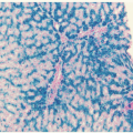
In a study by De Leeuw et al., 84% of women not supplemented with oral iron lacked stainable iron in bone marrow aspirates after delivery at term, whereas 81.5% of women treated with iron had stainable iron present at term.1 The demand for absorbed iron increases from 0.8 mg/day in early pregnancy to 7.5 mg/day in late pregnancy. An iron supplement of 65 mg/day beginning at or before 20 weeks’ gestation generally is adequate to prevent iron deficiency during pregnancy.29
There is evidence that the risks of premature delivery, low birth weight, and infant death are increased by severe iron deficiency. However, it is difficult to distinguish the effects of the iron deficiency itself from the conditions that produced the deficiency. A large case-control study from Venezuela indicated that maternal iron deficiency was associated with increased risk of premature delivery (risk ratio, 1.70; 95% confidence interval, 1.18 to 2.57).33 However, when Mahomed reviewed 20 trials evaluating the issue of iron supplementation in pregnancy for the Cochrane database, he concluded that iron supplementation during pregnancy resulted in a substantial reduction in women with anemia (a hemoglobin below 10 g/dl late in pregnancy) but had no detectable effect on maternal or fetal outcome.34 Thus, whether maternal iron deficiency adversely influences pregnancy is controversial. In fact, it is not entirely clear whether maternal iron deficiency reduces the fetal iron supply. Harthoorn-Lasthuizen et al. found no significant differences in iron status of neonates born to iron-deficient versus iron-replete women and concluded that the fetal iron supply is not negatively influenced by iron deficiency in the mother.35 In contrast, Turgeon O’Brien et al. reviewed medical records of 202 pregnant women to examine the association of low- and high-ferritin levels and anemia with pregnancy outcome.36 They observed an inverse relationship between firsttrimester anemia and birth weight, but they observed no relationship in the second and third trimesters. They suggested that maternal iron deficiency in early pregnancy was associated with poor fetal weight gain but that maternal iron deficiency later in pregnancy was not. During iron deficiency, the placenta becomes hypertrophied, a possible cause of maternal hypertension. A specific variant of pica, namely (ingestion of baking powder) can lead to symptoms suggesting preeclampsia.37
Recombinant erythropoietin administration, combined with iron replacement, has been reported to be an effective treatment for moderate and severe iron deficiency anemia during pregnancy,38, 39 although a meta-analysis of this topic suggests that the benefits from erythropoietin are relatively modest.40
The usual criteria for diagnosing iron deficiency are valid during pregnancy (see Chapter 23), including a reduced mean corpuscular volume (MCV), a reduced serum transferrin saturation to ≤15%, and a reduced serum ferritin concentration. Measurement of serum transferrin receptors may be useful in complicated situations in which inflammatory disease makes the serum ferritin value less reliable.41 Interpretation of serum iron values can be complicated during pregnancy: Even in apparently iron-replete women, serum iron levels progressively decline, and values for serum total iron-binding capacity and free erythrocyte protoporphyrin increase,1, 42 findings that usually indicate iron deficiency. Expression of the iron-regulatory peptide hepcidin appears to be suppressed during preganancy, consistent with an increased iron demand.43
Dietary supplementation with 78 mg of elemental ferrous iron daily during pregnancy increased the hematocrit, hemoglobin concentration, and red cell mass, with the red cell mass rising to nearly twice that found in similar, apparently healthy women not supplemented.1 At term, the mean hemoglobin concentration averaged 12.4 g/dl in those who received the supplement and 10.9 g/dl in those who did not. In Bantu women, whose diet is habitually high in iron, a significant change in serum iron does not occur, and iron deficiency anemia does not develop during pregnancy.44 Some have recommended that all pregnant women receive iron supplements beginning at 18 to 20 weeks’ gestation in doses ranging from 30 to 60 mg/day,26 whereas others recommend that iron supplementation be provided selectively. A typical regimen in this second circumstance is as follows: No iron supplementation if the serum ferritin concentration is >70 µg/L; lowlevel iron supplementation (40 mg elemental iron/day) if serum ferritin is between 30 and 70 µg/L; and 80 to 100 mg elemental iron/day in women with lower serum ferritin concentrations.45
Actual iron deficiency anemia should be treated as in individuals who are not pregnant (see Chapter 23). Constipation may be an especially problematic side effect in pregnant women; therefore, gradual increases in dose until fully therapeutic levels are achieved and an emphasis on taking medication with meals are particularly important precautions. Rarely, parenteral treatment may be necessary.
DEFICIENCY OF FOLATE AND OTHER NUTRIENTS DURING PREGNANCY
Macrocytic anemia of pregnancy is typically megaloblastic and in most cases results from deficiency of folic acid.46, 47, 48, 49, 50 In a study from India, macrocytic anemia in pregnancy carried a more negative prognosis for both mother and child than did iron deficiency.51 Megaloblastic anemia during pregnancy begins most often in either the third trimester or shortly after delivery.52 Folate requirements increase during pregnancy, and the diets of many pregnant patients are insufficient to meet the increased need.53, 54 Although folate deficiency occurs most often in economically deprived patients, this consequence of inadequate eating habits is not confined to the poor.52, 53, 54, 55 Particularly in pregnant adolescents, the diet may provide an inadequate source of folate, regardless of economic status.54 In uncomplicated pregnancies, the gastrointestinal absorption of dietary folate (polyglutamate) and of folic
acid (monoglutamate) is normal.56, 57 Folate deficiency during pregnancy is relatively common, although its frequency depends on the population studied. The prevalence varies from 1% to 50% of pregnant patients.48, 52, 55, 58 In North America, the prevalence is 1% to 4%.50 Not all patients in whom the serum concentration of folate is low develop megaloblastic anemia. In those who do, often the serum folate concentration was low earlier in pregnancy.52
acid (monoglutamate) is normal.56, 57 Folate deficiency during pregnancy is relatively common, although its frequency depends on the population studied. The prevalence varies from 1% to 50% of pregnant patients.48, 52, 55, 58 In North America, the prevalence is 1% to 4%.50 Not all patients in whom the serum concentration of folate is low develop megaloblastic anemia. In those who do, often the serum folate concentration was low earlier in pregnancy.52
Pregnant women often have no symptoms with folate deficiency anemia and may be found to have blood hemoglobin levels of 6 to 9 g/dl in the third trimester or postpartum. Folate deficiency is clearly associated with fetal neural tube defects and cleft palate, but these defects are established very early in fetal development, long before maternal megaloblastic anemia is detected.50 The association between low serum folate during the first trimester of pregnancy and fetal neural tube defects has been known for over 25 years.50 Lumley et al. reviewed this issue for the Cochrane database, examining four trials involving 6,425 women treated with folate or placebo.59 Preconceptional folate supplementation reduced the incidence of neural tube defects to a relative risk of 0.28 (95% confidence interval, 0.13 to 0.58). Folate did not increase miscarriage, ectopic pregnancy, or stillbirth. Multivitamins without folate did not prevent neural tube defects, and adding multivitamins to folate supplementation did not produce additional preventive effects. Thompson et al. reported an unexpected but significant reduction in childhood acute lymphoblastic leukemia among offspring of women supplemented with folate during pregnancy.60
An association between bacteriuria and folate deficiency was noted in pregnant patients. In experimental systems, infection may induce folate-deficient megaloblastic anemia. Malabsorption (see Chapter 36) may be associated with megaloblastic anemia of pregnancy, even in nontropical areas.50
In rare cases, folate-deficient pregnant patients develop erythroblastopenia and bone marrow changes morphologically simulating acute leukemia.61 Application of current molecular, immunologic, and cytochemical tools for diagnosing leukemia will avoid a mistaken diagnosis.
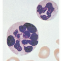
Megaloblastic anemia during pregnancy, as in the nonpregnant patient, is suggested by an increased MCV with oval macrocytes and hypersegmented granulocytes on blood smear, but a bone marrow examination can be done if doubt exists. Folate deficiency must be distinguished from vitamin B12 deficiency. The latter is rare in pregnancy, and the distinction can often be made on clinical grounds, especially on the basis of a careful nutritional history.
Related posts:
Stay updated, free articles. Join our Telegram channel

Full access? Get Clinical Tree


