Acquired Nonimmune Hemolytic Disorders


Acquired Nonimmune Hemolytic Disorders


Robert T. Means, Jr.

Bertil Glader


Hemolysis occurs when red blood cells (RBCs) are exposed to a variety of infectious agents, chemicals, or physical stresses. In some cases these effects are antibody mediated (Chapters 29 and 30). In other patients, such as those with glucose-6-phosphate dehydrogenase (G6PD) deficiency (Chapter 28) or those with unstable hemoglobins (Chapter 35), there is an underlying propensity of the RBCs to be more susceptible to injury. In still other cases, such as paroxysmal nocturnal hemoglobinuria (PNH), hemolysis occurs as a consequence of an acquired clonal abnormality in the RBC membrane (Chapter 31). Hemolytic anemia also occurs when otherwise normal red cells are injured directly by infectious agents, chemicals, thermal injury, mechanical stresses, or altered metabolites. These etiologies of hemolysis are the focus of this chapter.


HEMOLYSIS DUE TO INFECTION

A variety of infectious processes can lead to hemolytic destruction of normal RBCs. In some cases, such as in Mycoplasma pneumoniae infection or with infections related to paroxysmal cold hemoglobinuria, hemolysis is related to antibody-mediated cell destruction (Chapter 29). With the infections described in this section, hemolysis is largely the result of direct nonimmune effects on erythrocytes. Some of the infections discussed here are not major problems in North America or Europe; however, to the extent that there is significant international travel to and from endemic areas, recognition of these infections is important for medical personnel worldwide.


Malaria

Malaria is an acute, chronic, or recurrent febrile disease caused in humans by four species of Plasmodia: Plasmodia vivax, Plasmodia falciparum, Plasmodia malariae, and Plasmodia ovale. Infections with P. falciparum are the major form of malaria in Africa and Southeast Asia, whereas P. vivax is most common in Central America and India. These protozoan microorganisms are capable of parasitizing erythrocytes and other body tissues. Malaria is spread by female mosquitoes of the genus Anopheles (Fig. 32.1). The sexual phase of the Plasmodium life cycle takes place within the mosquito. The semitropical and tropical endemic distribution of malaria corresponds to the distribution of the vector.

On a worldwide basis, malaria is the most prevalent of all serious diseases; it has been estimated that approximately 2.5 billion people are at risk for malaria, and that approximately 500 million people are infected with P. falciparum.1, 2 Ninety percent of the deaths is in African children.3 Malaria has not been endemic in the United States since the 1940s, but approximately 1,000 cases have been reported each year since 1985, and there has been a steady increase in the number of cases reported annually.4 Malaria can also be transmitted by blood transfusions5 or by sharing needles among intravenous drug abusers.6, 7


Clinical Manifestations

After the initial exposure to malaria, some patients are completely asymptomatic, whereas others have nonspecific flulike symptoms that mimic a viral illness.8 Classically, the most prominent clinical manifestations are recurrent paroxysms of chills and fever with temperatures as high as 105° to 106°F (40.5° to 41°C) associated with malaise, headache, vomiting, and other systemic symptoms. The paroxysms tend to recur regularly every 36 to 72 hours. They are most prominent with P. malariae infections and much less with P. falciparum. Splenomegaly is noted in about one half of patients during early stages of disease8 and becomes more common later. Jaundice and hepatomegaly may develop in later stages of the illness.

Of the various malaria species, P. falciparum infection causes the most morbidity and mortality. In the acute stage, it can be associated with increasing parasitemia, hypotension, malignant hyperthermia, and death. In addition, P. falciparum malaria is associated with cerebral, pulmonary, and renal complications.
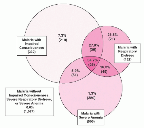
9 The overall mortality from a study of over 1,800 children with malaria in Kenya was 3.5%; in 84% of cases death occurred within 24 hours of admission.10 The most important prognostic factors for death were impaired consciousness and respiratory distress (Fig. 32.2). Severe anemia alone did not affect prognosis.3, 10

Anemia is common in malaria.11, 12, 13, 14 It is particularly characteristic of P. falciparum malaria because of the greater extent of red cell parasitization with this species. With uncomplicated P. falciparum malaria, moderately severe anemia is seen in approximately 20% of previously healthy patients during or after the first infection.15 Complete eradication of malaria parasites from the blood may take months to years, particularly in areas of high transmission; immunity to malaria is slowly acquired. In tropical areas, anemia tends to be most prevalent and most severe in children from 1 to 5 years of age,16 whereas only moderate anemia is usually noted in adolescents and adults.

In children, the circulating parasite count is inversely proportional to the hematocrit.17 Leukocyte numbers may be normal, but patients often have leukopenia. Thrombocytopenia has been observed in about two thirds of patients with P. falciparum malaria,9,16 often associated with splenomegaly.18

The most serious hematologic complication of malaria is acute intravascular hemolytic anemia (blackwater fever), which occurs as a rare event in the course of infection by P. falciparum. The clinical manifestations are fulminating, the intravascular hemolysis being associated with prostration, vomiting, chills, and fever. Hemoglobinemia, hemoglobinuria, and hyperbilirubinemia are consistent features, and in the most severe episodes, acute oliguric renal failure supervenes.


Pathogenesis

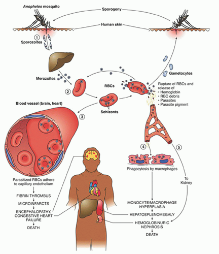
After a bite from the female Anopheles mosquito, sporozoites introduced into the circulation go to the liver parenchyma where they proliferate into thousands of merozoites (Fig. 32.1). The duration of this liver development stage varies between species. The infected hepatocytes next release merozoites into the bloodstream where they invade erythrocytes.

The ability of various Plasmodia to infect red cells is related to their attachment to specific membrane receptors. Of the species that infect humans, P. vivax and P. ovale invade only reticulocytes. P. malariae invades mature red cells, and P. falciparum invades erythrocytes of all ages. As a result, the proportion of cells parasitized in P. vivax malaria rarely exceeds 1%, whereas as many as 50% of cells may be affected in P. falciparum malaria. It is of interest that P. vivax invades only Duffy blood-group-positive red cells19; in West Africa where the Duffy antigen is missing on red cells, P. vivax malaria is nonexistent. P. falciparum apparently has two receptors: one that binds to sialic acid groups on the erythrocyte membrane protein glycophorin and another that
binds to a trypsin-sensitive, nonsialated ligand.20, 21, 22 Other proposed parasite receptors include surface heparinlike molecules23 and complement receptor type-1.
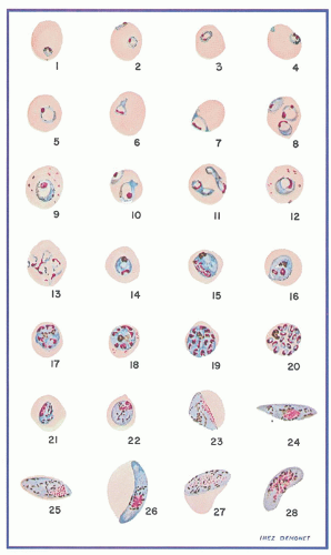
24


FIGURE 32.1. Life cycle of malaria. An Anopheles mosquito bites an infected person, taking blood that contains micro- and macrogametocytes (sexual forms). In the mosquito, sexual multiplication (“sporogony”) produces infective sporozoites in the salivary glands. (1) During the mosquito bite, sporozoites are inoculated into the bloodstream of the vertebrate host. Some sporozoites leave the blood and enter the hepatocytes, where they multiply asexually (exoerythrocytic schizogony), and form thousands of uninucleated merozoites. (2) Rupture of hepatocytes releases merozoites, which penetrate erythrocytes and become trophozoites, which then divide to form numerous schizonts (intraerythrocytic schizogony). Schizonts divide to form more merozoites, which are released on the rupture of erythrocytes and re-enter other erythrocytes to begin a new cycle. After several cycles, subpopulations of merozoites develop into micro- and macrogametocytes, which are taken up by another mosquito to complete the cycle. (3) Parasitized erythrocytes obstruct capillaries of the brain, heart, kidney, and other deep organs. Adherence of parasitized erythrocytes to capillary endothelial cells causes fibrin thrombi, which produce microinfarcts. These result in encephalopathy, congestive heart failure, pulmonary edema, and frequently death. Ruptured erythrocytes release hemoglobin, erythrocyte debris, and malarial pigment. (4) Phagocytosis leads to monocyte/macrophage hyperplasia and hepatosplenomegaly. (5) Released hemoglobin produces hemoglobinuric nephrosis, which may be fatal. (From Rubin E, Farber JL. Pathology, 3rd ed. Philadelphia, PA: Lippincott Williams & Wilkins, 1999.)

Further development of the parasite within red cells is along one of two pathways, either asexual or sexual differentiation (Fig. 32.1). Sexual forms, or gametocytes, continue their development within mosquitoes. The asexual differentiation of parasites in red cells proceeds from young ring forms through trophozoites to produce schizonts containing 6 to 32 merozoites (Fig. 32.3). In the process, parasites use 25% to 75% of the hemoglobin of the cell.25 The intraerythrocytic phase lasts 24 to 72 hours, depending on the species. The schizonts then lyse, the cell ruptures, and the merozoites are released to invade other cells, thereby continuing the erythrocyte cycle. The simultaneous rupture of billions of schizonts from red cells is associated with the classic paroxysms of malarial fever.

Erythrocytes parasitized by certain strains of P. falciparum develop electron-dense knobs that mediate the attachment of the infected red cells to venules.26, 27, 28 Such sequestration of parasiteinfected RBC creates an obstruction to tissue perfusion. In addition, the sequestration in venules prevents parasitized cells from entering the splenic circulation, thereby evading destruction and enhancing merozoite development; this phenomenon may be a factor in the rapid development of anemia in severe infections.29

The anemia in malaria is due to a combination of factors that include parasite-mediated RBC destruction, splenic removal of
infected RBCs, and decreased red cell production (Table 32.1). Hemoglobin digestion and cell disruption by the parasite are clearly the major causes of hemolysis.11, 12, 30 The malarial pigment hemozocin, a product of hemoglobin degradation,25 also inhibits erythropoiesis.31, 32


FIGURE 32.2. Prevalence, overlap, and mortality for major clinical subgroups of severe malaria in 1,809 African children. In 1,027 of these children, malaria was present without signs of impaired consciousness, respiratory distress, or severe anemia. In a subset of all children presenting with malaria (782 patients; approximately 40% of total) there was evidence of severe anemia (hemoglobin <5 g/dl), severe respiratory distress, impaired consciousness, and/or some combination of these clinical findings. Almost all deaths occurred in the children who presented with combinations of these clinical abnormalities. Total numbers of patients are given in parentheses. Mortality is given as a percentage. (Modified from Marsh K, Forster D, Waruiru C, et al. Indicators of life-threatening malaria in African children. N Engl J Med 1995;332(21):1399-1404.)

The role of the spleen in red cell destruction is related to the decreased deformability of RBCs infected with Plasmodia,33, 34, 35 erythrocyte retention in the red pulp, prolonged exposure to splenic macrophages, and removal of parasitized cells or “pitting” of the parasite, with consequent damage to the cell.36, 37, 38 A similar process of macrophage-mediated destruction of parasitized RBCs occurs in the marrow sinusoids.39 Moreover, normal nonparasitized RBCs have a shorter survival in malaria, presumably a consequence of hypersplenism and hyperactive macrophages.40

Even after complete clearance of the parasites, hemolysis may persist for 4 to 5 weeks.41 A complement-mediated process may be responsible in part, and the direct antiglobulin test is often positive.42

Dyserythropoiesis with characteristic morphologic findings also occurs in malaria. It is thought that this contributes to the slow recovery seen after a single malarial attack and also to the persistence of anemia in individuals with chronic parasitemia.13, 43, 44

Anemia often persists for weeks following treatment of malaria, and this results in part from relative marrow failure, as occurs in association with other forms of infection (see Chapter 41).12, 41 Abnormalities in several of the cytokine mediators of the anemia of chronic disorders (tumor necrosis factor, interferon gamma) have been reported in patients with severe P. falciparum malaria.45, 46, 47, 48, 49

Serum erythropoietin levels often are inadequate for the degree of anemia.50, 51, 52 The bone marrow response to erythropoietin also appears to be impaired.53 Circulating concentration of the iron regulatory peptide hepcidin, a key mediator of the anemia of chronic disease, is elevated early in the course of malaria.54, 55 This results in impaired iron mobilization.56, 57


FIGURE 32.3. Plasmodium falciparum. 1: Very young ring form trophozoite. 2: Double infection of single cell with young trophozoites, one a marginal form, the other a signet ring form. 3,4: Young trophozoites showing double chromatin dots. 5-7: Developing trophozoites. 8: Three medium trophozoites in one cell. 9: Trophozoite showing pigment in a cell containing Maurer spots. 10,11: Two trophozoites in each of two cells, showing variation of forms that parasites may assume. 12: Almost mature trophozoite showing haze of pigment throughout cytoplasm. 13: Aestiveo-autumnal slender forms. 14: Mature trophozoite, showing clumped pigment. 15: Parasite in the process of initial chromatin division. 16-19: Various phases of the development of the schizont. 20: Mature schizont. 21-24: Successive forms in the development of the gametocyte, usually not found in the circulation. 25: Immature macrogametocyte. 26: Mature macrogametocyte. 27: Immature microgametocyte. 28: Mature microgametocyte. (Reproduced from Wilcox A. Manual for the microscopical diagnosis of malaria in man. National Institutes of Health Bulletin No. 180.)

The percentage of reticulocytes tends to be low during active infection and increases transiently after effective treatment. In P. vivax malaria, however, the low reticulocyte count may be explained in part by the increased affinity of the organism for immature erythroid precursors and reticulocytes.58 Infection by P. vivax appears to be retricted to reticulocytes.59

The pathogenesis of acute intravascular hemolysis (blackwater fever) remains uncertain, and currently this complication is less commonly seen, although it still occurs.60, 61 Blackwater fever does not reflect an unusual degree of parasitemia. In many historical cases, the acute intravascular hemolysis appears to have been precipitated by quinine ingestion62, 63; quinine, for many years the primary treatment agent for malaria, may act as
a hapten, becoming antigenic after interacting with the red cell. However, cases involving untreated individuals also have been reported.9 Some episodes thought to represent blackwater fever may have resulted from the use of primaquinelike drugs in G6PD-deficient people.


TABLE 32.1 FACTORS CONTRIBUTING TO ANEMIA IN MALARIA


Accelerated red blood cell (RBC) destruction


Direct parasite destruction of red cells


Decreased deformability of parasitized RBCs and destruction by splenic macrophages


Macrophage-mediated destruction of parasitized RBCs in marrow and liver sinusoids


Destruction of nonparasitized cells by immune mechanisms


Destruction of nonparasitized cells by hypersplenism and hyperactive macrophages


Hapten (quinine)-induced intravascular hemolysis (blackwater fever)


Decreased RBC production


Bone marrow suppression due to inflammatory cytokines


Inadequate erythropoietin production


Dyserythropoiesis


Modified from Menendez C, Fleming AF, et al. Malaria-related anaemia. Parasitol Today 2000;16(11):469-476.


Certain inherited red cell disorders appear to confer resistance to malaria, either by inhibiting parasitic invasion or by slowing intracellular growth. It is thought that these phenomena may contribute to increased prevalence of such inherited diseases because of their effects on survival (balanced polymorphism). These disorders include sickle cell trait,64, 65, 66 G6PD deficiency,67, 68, 69, 70 thalassemia,71, 72 hemoglobin E variants,70 hemoglobin C variants,73 ovalocytosis of the Melanesian (Malayan) type,74 and lack of the Duffy blood group antigen.19


Management

Therapeutic considerations for malaria include supportive medical care for anemia and other complications. Administration of folate to patients with malaria is controversial; its use is associated with higher hematocrits, but may also prolong parasitemia.79 Chemoprophylaxis should be recommended to all people traveling to an endemic area.80, 81 Because of the spread of drug-resistant strains of P. falciparum, however, no single regimen is completely effective. Knowledge of the characteristics of the malaria strains in the sites to be visited is essential. Because of changes in drug resistance and the development of new agents, before recommending a regimen to a prospective traveler, physicians should become familiar with current guidelines from the U.S. Centers for Disease Control and Prevention (CDC), Atlanta, Georgia. See the CDC website (http://www.cdc.gov/travel/malinfo.htm).


Babesiosis

Infection by tick-borne protozoans of the genus Babesia is rare in humans.82, 83, 84 The infection has been reported in Nantucket and other islands off the northeastern U.S. shore, as well as in neighboring coastal areas of New England. It also is found in north central states, Washington, and California.85 Most cases of babesiosis described in Europe have occurred in asplenic individuals.84, 86 In the United States, Babesia microti is the causative agent, whereas Babesia divergens is the species identified in Europe, and the latter cases are usually more severe.82, 87 Babesiosis also can be transmitted by blood transfusion.88

Babesiosis is characterized by an acute febrile illness and hemolytic anemia, very similar to malaria. In most cases it is a mild self-limited disorder that goes undiagnosed, and thus is not reported. It is likely the true incidence of babesiosis in healthy hosts is underrecognized. However, in asplenic individuals it can produce serious, often fatal illness with hemolytic anemia, renal failure, or pulmonary edema.82, 84, 89

Laboratory features include hemoglobinuria, hyperbilirubinemia, normocytic anemia, thrombocytopenia, and sometimes leukopenia.82 Both B. microti and B. divergens can be seen in RBCs on the peripheral blood smear and can be confused with malaria.82 Serologic antibody tests and polymerase chain reaction-based assays are available to aid in diagnosis.90

In mild cases of babesiosis no treatment may be necessary. In more severe cases, the combinations of atovaquone and azithramycin or of clindamycin and quinine are useful. The atovaquone/azithramycin is reportedly better tolerated.90, 91 Red cell exchange transfusions have been used also.90, 91, 92


Trypanosomiasis

Moderate to severe hemolytic anemia is a regular feature of African trypanosomiasis (sleeping sickness).93, 94, 95 This often fatal illness is caused by Trypanosoma brucei gambiense or T. brucei rhodesiense. The diseases induced by the two subspecies are similar except that T. brucei gambiense infection follows a more chronic course. The organisms are transmitted to humans and domestic animals by the bite of the tsetse fly.

Normocytic anemia with reticulocytosis is prominent. Red cell survival is shortened and autoagglutination of erythrocytes with accelerated erythrocyte sedimentation characteristically is observed. The results of the direct antiglobulin (Coombs) test may be positive. Erythrophagocytosis by macrophages is seen throughout the reticuloendothelial system.96

The toxic effects of the parasite and immunologic mechanisms both are implicated in the destruction of red cells. The intensity of the hemolytic anemia may fluctuate with the degree of parasitemia. Transient hepatosplenomegaly and decreased serum complement levels accompany the episodes. Marrow failure often supervenes during the terminal phases of the illness. Diagnosis
depends on serologic tests or demonstration of the parasite in the blood.97


Visceral Leishmaniasis (Kala-Azar)

Leishmaniasis is an infection caused by intracellular protozoan parasites transmitted by sandflies. There are three main forms of Leishmania infections in humans: cutaneous, mucocutaneous, and visceral. The major hematologic problems occur with the visceral infection (kala-azar) and involve the lymph nodes, liver, spleen, and bone marrow. The disorder is caused by Leishmania donovani, and is found throughout Asia and Africa, affecting individuals of all ages. A variant parasite, L. donovani infantum is the form that causes kala-azar in southern Europe and North Africa, and it primarily affects young children and infants. Visceral leishmaniasis mainly occurs in local endemic areas; however, it may be contracted on short-term visits. It has been recognized in Europe in travelers returning from Mediterranean holidays.98 Both cutaneous and visceral leishmaniasis have been reported in military personnel returning from deployment in the Persian Gulf.99, 100

Following an incubation period of 1 to 3 months, there is the insidious onset of fever, sweating, malaise, and anorexia, but these acute symptoms gradually abate. Next, hepatosplenomegaly gradually evolves, and this stage of illness is associated with anemia, neutropenia, and thrombocytopenia. In young children with acute visceral leishmaniasis, particularly in Mediterranean populations, the clinical and hematologic features may be more aggressive with a rapid onset of severe hemolytic anemia.101

The bone marrow is hyperplastic with dyserythropoietic changes, and the diagnosis can usually be made by finding macrophages containing intracellular parasites (Leishman-Donovan bodies). The overall hematologic picture is typical of hypersplenism. Red cell survival studies indicate that hemolysis is the major cause of anemia in leishmaniasis.101, 102

In most cases there is no evidence of immune hemolysis, although both immunoglobulin G (IgG) and complement occasionally are found on the red cells. Similar to what is seen in malaria, nonsensitized red cells are destroyed by macrophages recruited to the spleen and liver as part of the inflammatory response to the parasite.


Bartonellosis (Carrion’s Disease)

A severe, acute hemolytic anemia is produced in humans by Bartonella bacilliforms, a flagellated bacillus.103 The infection is limited to South America, particularly in the Andean valleys of Peru, Ecuador, and Columbia, at elevations of 500 to 3,000 m.104, 105, 106, 107 The bacillus is transmitted by the sand fly (Phlebotomus) and probably by other arthropods. After a 2- to 3-week incubation period, the acute phase of the illness, known as Oroya fever, begins. It is marked by malaise, headache, muscle pains, remittent fever, chills, and rapid onset of severe anemia. The disease has existed in Peru since pre-Incan times.103, 108 The highest rates of infection are in children.104, 107

The findings in the blood are characteristic of acute extravascular blood destruction.103, 109 As viewed in Wright- or Giemsastained blood smears, numerous Bartonella organisms are apparent in the erythrocyte.103 The organisms are rod shaped (1 to 2 m in length and 0.2 to 0.5 m in width) or round (0.3 to 1.0 m in diameter).

In patients who recover from the acute phase, a quiescent period ensues during which the organisms disappear from the blood. A chronic eruptive stage follows, verruca peruviana, a benign condition characterized by hemangiomalike lesions of the skin but without hematologic manifestations.109

Bartonella infection can be treated by antibiotic combinations containing chloramphenicol and other antibiotics, often a beta lactam. Verruca peruviana is typically treated with streptomycin-containing regimens. In the era prior to antibiotics, the only available treatment was blood transfusion.104, 110


Clostridial Sepsis

Clostridium perfringens septicemia occurs after septic abortion or in association with a diseased biliary tree, traumatic wound infections, cancer, leukemia, endocarditis, gastrointestinal arteriovenous malformations, or necrotizing enterocolitis of newborns.111, 112, 113, 114, 115, 116, 117 Sometimes no underlying disease is identified.118, 119, 120 Profound, often fatal hemolytic anemia is a regular feature of clostridial sepsis.118, 121 Signs of intravascular red cell destruction are prominent, and many microspherocytes are found in the blood. The hemolysis can be rapid and massive, with hematocrit values falling to very low levels in a matter of hours.119, 120 Hemolysis is thought to result from the elaboration of a clostridial toxin, a phospholipase that attacks erythrocyte membrane lipids to form highly lytic lysolecithins.122, 123 The diagnosis should be suspected when fever, jaundice, and intravascular hemolysis occur together in a patient with a history of previous gastrointestinal or genitourinary surgery, a recent wound, cancer, or other disease. Clostridial infections respond to antibiotic therapy, but in order to affect outcome, treatment must be started quickly, usually before culture results are available.124


Other Bacterial Infections

Acute hemolytic anemia with bacterial infection is common, especially in childhood, and has been reported with streptococcal, staphylococcal, or pneumococcal septicemia or endocarditis.125, 126, 127, 128 Intravascular hemolysis with hemoglobinuria has been reported in patients with cholera129 and with typhoid fever.130 Escherichia coli 0157 gastroenteritis can cause the hemolytic-uremic syndrome, but can also bring about hemolytic anemia with no renal involvement or red cell fragmentation.131 Severe hemolytic anemia not attributable to autoimmune mechanisms is observed occasionally in patients with miliary tuberculosis.126, 132 Certain spirochetal infections are also associated with hemolytic anemia, including relapsing fever caused by Borrelia recurrentis133 and leptospirosis.134

The pathogenesis of hemolysis in most cases cited above is uncertain. In some it is thought that anemia is due to direct action of the infectious agent or its products on erythrocytes. Adsorption of microbial antigens to red cells has been detected by immunofluorescent techniques,135 and this phenomenon may lead to phagocytosis or complement-mediated erythrocyte destruction. The capsular polysaccharide of Haemophilus influenzae type b, polyribosyl ribitol phosphate (PRP), is released from growing organisms during human infection and can be found in body fluids including red cells. It has been proposed that the hemolytic anemia that occurs during H. influenzae type b infection may be due to absorption of PRP to red cells and immune destruction of sensitized erythrocytes.136

In other cases, serious bacterial infections are associated with disseminated intravascular coagulation and a microangiopathic hemolytic anemia. Also, as discussed above, C. perfringens may release phospholipases that can lead to red cell membrane injury and cell destruction. Of interest, some bacteria release neuraminidase, an enzyme that cleaves red cell sialic acid residues, thereby exposing a cryptic “T antigen.”137 These “T-activated” red cells can react with anti-T IgM antibodies present in most human adult plasma, thereby resulting in RBC agglutination and possible hemolysis in vivo.137 The main bacteria that release neuraminidase are C. perfringens and Streptococcus pneumoniae; however, Bacteroides, E. coli, Actinomyces, and Vibrio cholerae also have been implicated.138


HEMOLYSIS DUE TO DRUGS AND CHEMICALS

Many drugs and chemicals injure normal red cells to cause hemolytic anemia. Some of the more common occurrences are summarized below. (Drug-induced immune hemolysis is discussed in Chapter 29.)


Oxidant Drugs and Chemicals

Certain chemical agents can bring about the oxidative denaturation of hemoglobin, leading to the sequential formation of methemoglobin, sulfhemoglobin, and Heinz bodies. In some cases, the chemical itself acts as an oxidizing agent; more often, however, it interacts with oxygen to form free radicals or peroxides. These free radicals or peroxides, if produced in quantities too great to be detoxified by the glutathione-dependent reduction system, denature hemoglobin and damage other cellular structures, such as the cell membrane.139 Individuals deficient in G6PD or other components of glutathione-dependent detoxification processes (Chapter 28) are particularly sensitive to the hemolytic effects of oxidant compounds (Table 32.2). These agents may also unmask otherwise insignificant defects in the metabolic pathways that defend the erythrocyte against oxidative stress.140 However, some of these agents are powerful enough to overcome the defense mechanisms of otherwise normal erythrocytes, and can cause hemolysis if given to healthy subjects in higher than usual doses or if renal failure leads to unusually high blood levels.

Hemolytic anemia caused by oxidant drugs varies considerably in severity. Usually the anemia is noted within 1 to 2 weeks after drug therapy is initiated with laboratory findings of low hemoglobin, reticulocytosis, hyperbilirubinemia, and low serum haptoglobin. Also, in some cases, hemoglobinemia and hemoglobinuria may be apparent. Cyanosis with methemoglobinemia or sulfhemoglobinemia is sometimes noted. The hemolytic process usually disappears within 1 to 3 weeks after use of the offending drug has been discontinued.


TABLE 32.2 DRUGS AND CHEMICALS THAT CAUSE HEMOLYTIC ANEMIA IN PATIENTS WITH NORMAL ERYTHROCYTES


Sulfonamides375


Sulfones376


Phenazopyridine (Pyridium)142


Nitrofurantoin (Furadantin)377


Phenacetin378


Acetylsalicylic acid379


Phenol380


Cresol (Lysol, penetrating oil)381


Naphthalene (mothballs)382


Nitrobenzene383


Aniline384


Phenylsemicarbazide385


Phenylhydrazine143


Chlorates386


Nitrates387


Oxygen388


Hydroxylamine389


Methylene blue (in infants)390


Hematin391


Pentachlorophenol392


Cisplatin, Carboplatin393


Morphologic findings characteristic of hemolytic anemia caused by oxidant drugs and chemicals include the following: Heinz bodies (seen with brilliant cresyl blue supravital stains of blood during hemolytic episodes) (Fig. 32.4A); “bite cells” (seen in routine Wright-stained blood smear) as erythrocytes that look as if a semicircular bite has been taken from one edge (Fig. 32.4B)141, 142; and hemighosts143 or eccentrocytes,144 erythrocytes that look as if the hemoglobin has shifted to one side of the cell, leaving the other side clear (Fig. 32.4C). These hemighosts also are referred to as “blister cells” and may appear to contain a large vacuole. These RBCs contain a coagulum of hemoglobin that has separated from the membrane, often leaving an unstained non-hemoglobin-containing cell membrane.145 Hemighosts appear only when hemolysis is brisk,141, 143 and probably indicate a particularly severe degree of oxidant damage. All of these morphologic alterations are consequences of oxidative assault on hemoglobin.

Although the treatment of drug-induced nonimmune hemolysis is largely supportive, erythropoietin has been used in cases associated with a blunted erythropoietic response, particularly in the hemolytic anemia that is observed in hepatitis C patients treated with ribavirin.146


Arsine Poisoning

Arsine (AsH3) is the most acutely toxic form of arsenic. It is a colorless, nonirritating, highly toxic gas that is produced by the action of water on a metallic arsenide. Arsine poisoning is associated most often with the use of acids in refining, extracting, or otherwise processing crude metals that contain arsenic as an impurity.147, 148 Industrial processes such as galvanizing, soldering, etching, and lead plating can expose workers to this noxious gas.149 Arsine is also used in the transistor industry to stabilize silicon, and leakage from cylinders in which the gas is transported can lead to accidental poisoning.150

Manifestations of poisoning appear 2 to 24 hours after exposure and include abdominal pain, nausea, and vomiting; the passage of dark-red urine; jaundice; anemia; reticulocytosis; leukocytosis; and other signs of acute hemolytic anemia. Hemoglobinemia and hemoglobinuria are found, and acute, oliguric renal failure may ensue. The antiglobulin test result is negative. The mortality rate can approach 20%.150, 151 The mechanism of red cell injury is not known for certain. Interactions between arsine and oxyhemoglobin may be involved.152, 153 Also, arsine-induced membrane injury with altered ion transport has been proposed.152, 154

Only gold members can continue reading. Log In or Register to continue

Stay updated, free articles. Join our Telegram channel

Oct 21, 2016 | Posted by in HEMATOLOGY | Comments Off on Acquired Nonimmune Hemolytic Disorders

Full access? Get Clinical Tree

Get Clinical Tree app for offline access