Introduction
Endogenous hypercortisolism is one of the most challenging diagnostic problems in clinical endocrinology. Chronic hypercortisolism, responsible for Cushing’s syndrome (CS), is usually of neoplastic origin and secondary to autonomous endocrine secretion from a tumor that originates from adrenocortical cells, pituitary corticotrophs, or various ACTH-secreting endocrine tumor cells located outside the pituitary [ , ]. However, chronic ACTH-dependant hypercortisolism can also result from “functional” sustained activation of the hypothalamic-pituitary-adrenal axis (HPA) due to various somatic and psychological causes [ ]. This condition has been previously defined as “pseudo-Cushing’s syndrome” (pseudo-CS) when functional hypercortisolism is associated with clinical features that suggest CS. The term “nonneoplastic hypercortisolism-NNH” which more precisely reflects the pathophysiology of this situation is now recommended [ ]. This condition is of interest since distinguishing between neoplastic (NH) and NNH can represent a significant diagnostic challenge [ ]. Furthermore, chronic exposure of tissues to cortisol excess, as is observed in NNH, can result in long-term morbidity. In this chapter, we will review the main clinical conditions responsible for NNH, the available tools to differentiate CS from NNH and the potential long-term issues related to NNH.
Pathophysiology of NNH
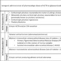
Several conditions may be responsible for NNH ( Table 6.1 ) and can be classified into two different groups [ ]. The first group includes pathologies whose clinical expression mimics NH and leads to diagnostic challenges. These include neuropsychiatric disorders, alcoholism, poorly controlled type 2 diabetes (T2D), obesity associated with insulin resistance and/or polycystic ovary syndrome, and end-stage kidney disease. In contrast, the second group of conditions responsible for NNH includes eating disorders and chronic intense exercise. These conditions do not pose diagnostic problems as the clinical presentation of the patients is not suggestive of CS and therefore will not be discussed further in this paper.
Table 6.1
Main causes of endogenous ACTH-dependant hypercortisolism.
| Endogenous ACTH-dependant hypercortisolism | |
|---|---|
| Neoplastic hypercortisolism | Nonneoplastic hypercortisolism |
| ACTH-secreting neoplasm | Possible Cushing phenotype |
| Pituitary | Alcohol-induced |
| Ectopic | Chronic kidney disease (stages 4–5) |
| Neuropsychiatric disorders | |
| Poorly controlled diabetes mellitus | |
| Pregnancy | |
| Obstructive sleep apnea | |
| Non-Cushing phenotype | |
| Eating disorders | |
| Negative energy balance | |
The main pathophysiological hypothesis for the occurrence of NNH is through activation of various neuronal pathways targeting the paraventricular nuclei of the hypothalamus, leading to hypersecretion of CRH or vasopressin, which then stimulate secretion of ACTH by pituitary corticotroph cells [ ]. This pathophysiological model appears particularly appropriate for NNH linked to neuropsychiatric disorders and alcoholism. In patients with metabolic syndrome, an additional mechanism, involving peripheral cortisol production, can be associated.
Alcohol-induced hypercortisolism
Alcohol-induced hypercortisolism is probably the most difficult differential diagnosis of CS as chronic alcohol consumption may cause clinical and biochemical features that are indistinguishable from NH. Chronic alcoholism has a catabolic effect and can induce proximal amyotrophy and android obesity. Furthermore, it is often associated with malnutrition, resulting in muscle wasting and depression. Arterial hypertension is also frequently found in these patients.
ACTH-dependent hypercortisolism has been documented in many studies and can be detected in hair, plasma, or urine of patients with alcohol abuse [ ].
The mechanism of alcoholism-induced hypercortisolism is thought to be mainly of central origin, due to an increase in hypothalamic corticotropin-releasing hormone (CRH) [ ] and vasopressin. In animal studies, alcohol administration induces an increase in CRH mRNA expression in the hypothalamic paraventricular nucleus of the rat [ ]. Furthermore, the alcohol-induced increase in circulating corticosterone concentration is abolished when CRH receptor antagonists are used and is not seen in hypophysectomized rats [ , ]. Alcohol-induced increases in hypothalamic vasopressin secretion may also contribute to hypercortisolism by increasing the sensitivity of ACTH to CRH [ ]. Alterations in peripheral cortisol metabolism by the liver may be an additional mechanism contributing to NNH [ ]. Increased cortisol half-life or increased hepatic cortisol production (via induction of 11-beta-hydroxysteroid dehydrogenase type 1 gene expression and activity) in patients with alcoholic liver disease has also been implicated, but in these cases, if glucocorticoid negative feedback remains intact, normal cortisol levels might be expected unless the stimulatory input to the hypothalamus persists [ , ]. Psychiatric pathologies, particularly depression, are very common in these patients, and may also contribute to the pathogenesis of functional hypercortisolism.
The diagnosis of alcohol-induced hypercortisolism should so be considered in all patients who drink more than 1 or 2 glasses of alcohol a day (>13 g/day), or when they have elevated liver function tests (elevated AST > AST), or MCV (mean corpuscular volume) that can be a clue to hidden alcohol consumption. Blood phosphatidylethanol (PEth) levels, as a means of quantifying chronic alcohol consumption, can help to confirm the diagnosis in this case [ ]. It should also be emphasized that increased cortisol secretion occurs during acute alcoholism (consumption of over 30 mg/dL) but can also occur due to the stress of acute alcohol withdrawal [ ]. Acute alcohol withdrawal can also induce ACTH-dependent hypercortisolism during in-patient biochemical investigations.
Neuropsychiatric disorders
There is evidence of aberrant HPA activity in many psychiatric disorders including depression, anxiety/panic disorders, obsessive/compulsive disorder, schizophrenia, and autism spectrum disorder [ ]. In rare “acute” cases, the increase in urinary free cortisol (UFC) may reach up to 4 times the upper limit of normal (ULN) [ ]. Obesity and metabolic syndrome are also common in these patients either spontaneously or due to antipsychotic drugs, so that the differential diagnosis from CS can be a real challenge from a clinical perspective.
ACTH-dependent hypercortisolism has been documented in many studies and the mechanism underlying functional hypercortisolism is thought to be mainly of a central origin [ ]. Evidence from both animal and human studies strongly supports the view that CRH may be over-secreted from both hypothalamic and extrahypothalamic neurons in depression. This increase in CRH mediates some of the behavioral symptoms of depression as shown by central nervous system administration of CRH to laboratory animals which produces physiological and behavioral changes almost identical to those observed in response to stress (sleep and appetite disturbances, reduced libido, psychomotor changes etc.) [ ]. Similarly, central administration of CRH or overexpression of CRH in transgenic mice produces several signs of increased anxiety [ ], while conversely, central administration of a CRH receptor antagonist produces anxiolytic effects in rats [ , , ]. A similar anxiolytic action has been reported in transgenic mice lacking CRF1 receptors [ , ]. Interestingly, in humans, HPA axis hyperactivity normalizes following successful antidepressant treatment [ ].
The increased activity of the HPA axis is also thought to be related, in part, to reduce feedback inhibition by endogenous glucocorticoids. Indeed, numerous studies have shown that HPA axis function is not suppressed by pharmacological stimulation of glucocorticoid receptors with an oral dose of the synthetic glucocorticoid dexamethasone [ , ]. Reduced glucocorticoid receptor function in peripheral tissues, such as peripheral blood mononuclear cells and skin cells, has also been described in patients with depression [ ]. Interestingly, successful antidepressant treatment is associated with resolution of the impaired negative feedback of the HPA axis by glucocorticoids [ ].
Finally, an alteration in glucocorticoid metabolism due to decreased 5-alpha-reductase and type 2 11-beta hydroxysteroid dehydrogenase activity has also been reported [ ].
Poorly controlled type II diabetes (T2D), obesity, insulin resistance
Cortisol plays a key role in the regulation of glucose metabolism and glucose intolerance is observed in 40%–80% of patients with endogenous CS [ ]. Conversely, opinions are divided as to whether patients with poorly controlled T2D have chronic activation of the HPA axis [ ]. Proponents of the view that patients with T2D or obesity have altered HPA axis activity suggest that there is increased cortisol production in the liver and visceral adipose tissue, due to increased expression of the 11β-HSD1 enzyme which locally converts cortisone to cortisol [ ]. The glucocorticoid receptor is present in subcutaneous adipose tissue and is over-expressed in visceral adipose tissue, leading to an autocrine action of locally produced cortisol. Administration of a selective inhibitor of 11β-HSD1 to obese subjects with T2D was able to improve hepatic insulin sensitivity and lower blood glucose, triglycerides, and blood pressure [ ]. Nonalcoholic fatty liver disease and sleep apnea syndrome (SAS), which very often accompany central obesity, may both also contribute to functional hypercortisolism. SAS is accompanied in particular by hypersensitivity of pituitary ACTH secretion to the action of hypothalamic CRH, and also bursts of HPA activation following episodes of apnea, which could explain the observed hypercortisolism [ ]. Finally, stress and depression may be important mediators and/or covariates in the development of NNH [ ].
Chronic kidney disease (CKD)
CKD leads to variable cortisol excess, depending on the severity of the disease and coexisting morbidities [ ]. The pathophysiology of CKD-related hypercortisolism is multifactorial. One of the main causes is a reduction in cortisol clearance and an increase in its half-life [ , ]. Reduced renal filtration of cortisol metabolites and their systemic accumulation appear to interfere with the enzymatic clearance of cortisol in the liver [ ]. In addition, loss of 11-beta-hydroxysteroid dehydrogenase type 2 expression in the kidney reduces the reversible inactivation of cortisol [ ]. Dialysis treatment is unable to completely reverse these changes [ ]. Alongside changes in cortisol clearance, there are likely changes in the regulation of the HPA axis. Cortisolemia is increased in renal failure, in contrast to normal or even increased pituitary ACTH concentrations [ ], suggesting an impairment of negative glucocorticoid feedback, a major player in the physiological control of the HPA axis. The impaired negative feedback in CKD is illustrated by dexamethasone suppression tests. Around 10% of patients with CKD fail to suppress cortisol secretion after an overnight 1 mg dexamethasone suppression test (ONDST), but do respond to higher doses of dexamethasone (low-dose dexamethasone suppression test—LDDST) [ ]. In addition, inflammation and metabolic acidosis both upregulate the HPA axis and could explain the variable activation of the HPA axis in people with advanced CKD [ , ].
Diagnosis of NNH
Identifying the clinical and biochemical differences between NNH and NH can be a significant challenge [ ]. Although the typical presentation of NNH is as a clinically mild CS associated with moderate hypercortisolism and the maintenance of diurnal cyclicity, the differential diagnosis between NNH and NH requires a comprehensive history and physical examination associated with biochemical testing ( Table 6.2 ). Guidance in this differential diagnosis is hampered by the heterogeneity of published data due to variable criteria for defining NNH, the different diagnostic thresholds used to analyze the results of biochemical tests, and the small number of well-characterized patients included in published studies.
Table 6.2
Clinical and laboratory differences between neoplastic and nonneoplastic hypercortisolism.
Adapted from
| Neoplastic | Nonneoplastic | |
|---|---|---|
| History | ||
| Alcohol ≥2/d | — | +++ |
| Hypertension | +++ | +++ |
| Diabetes mellitus type II | ++ | +/− |
| Low bone density with fractures | +++ | — |
| Physical examination | ||
| Cushingoid facies | +++ | ++ |
| Dorsocervical fat | ++ | + |
| Cutaneous wasting | +++ | + |
| Myopathy | +++ | + |
| Laboratory results | ||
| CKD 1–3 | + | + |
| CKD 4–5 | + | +++ |
| Liver ALT > AST | +++ | + |
| Liver AST > ALT | — | +++ |
| LNSC > 5ULN | +++ | + |
| Abnormal 1 mg DST | ++ | ++ |
| 24 hours UFC > 4ULN | +++ | – |
| Positive dDAVP stimulation | +++ | — |
Clinical symptoms of NNH
Although metabolic signs such as obesity, hypertension, and glucose intolerance are present in both NNH and NH, catabolic and muscular signs of CS such as large purple striae, proximal amyotrophy, and skin fragility with ecchymosis are more specific and consistent with the diagnosis of NH [ ]. Nevertheless, many confounding factors are possible: age or alcohol consumption can induce skin fragility or amyotrophy and CKD may be responsible for undernutrition, sarcopenia, osteoporosis, and fractures [ ]. Conversely, specific symptoms may be lacking in patients with CS of mild intensity.
In a study by Gatta et al., which examined two populations of NNH and mild NH patients matched for the (moderate) increase in UFC, central weight gain, hypertension, and hirsutism were equally prevalent in the NH and NNH groups [ ]. Ecchymoses, purple striae, and proximal myopathy were observed only in patients with Cushing’s disease (CD). However, despite this difference, catabolic symptoms were observed in only 50% of cases of CD of mild intensity [ ].
In the study of Arnaldi et al., patients with NNH and CD were equally affected by hypertension, hirsutism, purple striae, impaired fasting glucose/diabetes, dyslipidemia, oligomenorrhea, acne, overweight/obesity, and muscle weakness [ ]. Ecchymosis and osteoporosis were significantly more frequent in CD patients [ ]. Assessment of osteoporosis by bone densitometry could therefore be used as a diagnostic tool. Psychiatric problems were also more frequent in patients with NNH.
The clinical context, taking into account the history of comorbidities and associated conditions is, of course, essential in making a diagnosis of NNH [ ].
Baseline biochemical investigations
As discussed above, NHH results essentially from a central activation of the HPA axis and results in a state of ACTH-dependant hypercortisolism. ACTH and cortisol concentrations are clearly lower than those observed in ectopic ACTH syndrome and differential diagnosis between NNH and CD needs to be made. Although some studies report lower plasma ACTH concentrations in NNH compared with NH, ACTH concentrations are not useful for discriminating between these two entities [ , ].
In NNH, UFC is generally normal or moderately increased [ ], and only a single early study reported levels up to 4 times the upper limit of normal range (ULN) [ ]. Conversely, UFC may be normal, at least in some urine samples, in approximately 10% of CD cases. Consequently, UFC >1.0 X ULN permits a diagnosis of CD with a sensitivity ranging from 78% to 92% but with suboptimal specificity (40%–89%) [ ] ( Table 6.3 ). As mentioned above, 24-hour UFC values > 4.0 ULN exclude the diagnosis of NNH.
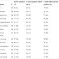
Table 6.3
UFC, ONDST, LDDST, and midnight plasma cortisol diagnostic performance for distinguishing NH from NNH. The data show the sensitivity and specificity of each test to diagnose CD depending on the criteria used.
| Study | n | CD | NHH | UFC | ONDST | LDDST | Midnight cortisol | ||||||||
|---|---|---|---|---|---|---|---|---|---|---|---|---|---|---|---|
| Threshold (nmol/24 hours) | Sens (%) | Spe (%) | Threshold (nmol/L) | Sens (%) | Spe (%) | Threshold (nmol/L) | Sens (%) | Spe (%) | Threshold (nmol/L) | Sens (%) | Spe (%) | ||||
| Moro et al. [ ] | 173 | 76 | 30 | >221 | 92 | 66 | >138 | 97 | 97 | >207 | 97 | 91 | |||
| Gatta et al. [ ] | 31 | 17 | 14 | >50 | 94 | 79 | >256 | 100 | 100 | ||||||
| >55 | 94 | 86 | |||||||||||||
| Pecori et al. [ ] | 55 | 32 | 23 | >220 | 97 | 44 | >50 | 98 | 58 | >50 | 94 | 74 | >50 | 98 | 18 |
| Reimondo et al. [ ] | 31 | 16 | 15 | >657 | 75 | 100 | >110 | 87.5 | 93 | >52 | 81 | 93 | >229 | 56.2 | 100 |
| Valassi et al. [ ] | 101 | 60 | 41 | >38 | 86 | 96 | |||||||||
| >50 | 86 | 96 | |||||||||||||
| Tibarassi et al. [ ] | 111 | 52 | 28 | >413 | 79 | 53 | >138 | 79 | 95 | >207 | 94 | 83 | |||
| >50 | 90 | 66 | |||||||||||||
| Tibarassi et al. [ ] | 60 | 30 | 18 | >413 | 83 | 40 | >138 | 73 | 90 | >207 | 90 | 73 | |||
| >50 | 97 | 50 | |||||||||||||
| Alwani et al. [ ] | 54 | 35 | 19 | >50 | 94 | 84 | |||||||||
| Rollin et al. [ ] | 124 | 68 | 56 | >248 | 90 | 79 | >138 | 92 | 77 | >138 | 77 | 86 | >207 | 98 | 84 |
| >50 | 100 | 51 | >50 | 95 | 61 | ||||||||||
CD , Cushing’s disease; LDDST , low-dose dexamethasone suppression test; NNH , nonneoplastic hypercortisolism; ONDST , over-night dexamethasone suppression test; Sens , sensitivity; Spe , specificity; UFC , urinary free cortisol.
Analysis of serum or salivary cortisol at the expected time of its physiological nadir is an important biochemical investigation for the differential diagnosis between NH and NNH. Indeed, with the exception of cyclic cases, mild CD with normal UFC is almost always associated with increased late-night values [ ]. In contrast, lower levels of cortisol are found in NHH patients due to the persistence of a degree of nychthemeral cycle in CRH secretion. In the study of Gatta et al. , which involved groups of patients with a similar mild increase in 24-hour UFC, all patients with CD had a midnight cortisol concentration greater than 256 nmol/L whereas all NNH patients had a concentration lower than this threshold [ ]. However, most published studies have used a midnight serum cortisol threshold of 207 nmol/L which permits a diagnosis of CD with a sensitivity and specificity that varies from 82.0% to 97.4% and 65% to 90.6%, respectively [ , ]. In one study, a midnight serum cortisol threshold of 353 nmol/L gave a diagnosis of CD with a sensitivity and specificity of 89% and 100% respectively [ ]. These thresholds need to be carefully considered owing to the lower cortisol concentrations currently measured with newer assays. However, in one prospective study, a midnight plasma cortisol threshold of 243 nmol/L, measured by liquid chromatography coupled to tandem mass spectrometry (LC-MS/MS), permitted the diagnosis of CD with a sensitivity of 98% and a specificity of 95% in 73 patients [ ] ( Table 6.3 ).
The use of midnight cortisol/8:00 a.m. cortisol ratio, or repeated measurements of serum cortisol during the day (diurnal cycle), has also been proposed to discriminate between NNH and NH but does not appear to offer superior performance to midnight serum cortisol concentration alone [ , ].
Late night salivary cortisol (LNSC) is an easier test to perform in outpatients but has been much less studied by far in NNH patients. Moreover, wide variability in salivary cortisol thresholds between assays has been reported for the diagnosis of CD [ ], making it difficult to establish clear recommendations regarding the threshold to choose. Alwani et al. reported in a 2014 study that only 33/70 patients with CD and 11/20 patients with NNH had measurements of salivary cortisol. In these patients, a threshold of 9.3 nmol/L permitted the diagnosis of CD with 100% sensitivity and 83% specificity [ ]. Finally, as the test assumes that cortisol levels reach a nadir in the late evening, it may be appropriate to adapt the time of sampling to bedtime for shift workers or for patients whose bedtimes vary [ ] ( Table 6.3 ).
Dynamic testing
Several dynamic tests may help to differentiate NNH from CD in patients with mild ACTH-dependent hypercortisolism [ ]. These tests include: ONDST and LDDST, the Dex-CRH, and the desmopressin stimulation tests (dDAVP). To understand their diagnostic performance, it is important to keep in mind that, in CD, ACTH secretion originates from ACTH-secreting adenomatous cells which have reduced sensitivity to negative feedback of cortisol, meaning that they can maintain ACTH secretion despite hypercortisolism. Conversely, in NNH, ACTH secretion likely originates from normal corticotroph cells that are overstimulated by CRH and/or hypothalamic vasopressin, but then retain normal sensitivity to glucocorticoid negative feedback. Conceptually, NNH patients with normal corticotrophs would be expected to have both greater sensitivity to dexamethasone inhibition and reduced sensitivity to acute stimulation by exogenous CRH compared with CD patients with adenomatous corticotrophs [ ]. Furthermore, as 81%–100% of adenomatous cells aberrantly express the vasopressin V2 receptor and overexpress the V3 receptor, it is expected that only patients with CD would show an exaggerated ACTH and cortisol response to dDAVP administration [ ].
ONDST and LDDST
As mentioned above the rationale for these tests is to obtain inhibition of cortisol production by exogenous glucocorticoid administration (dexamethasone) which, at least in theory, should be stronger in NNH cases than in CD cases. During the ONDST, a 1 mg oral dose of dexamethasone is administered between 11:00 p.m. and midnight, and cortisol levels then measured at 8–9:00 a.m. the following morning [ ]. In the LDDST, a total of 2 mg oral dose of dexamethasone is given at six-hourly intervals for 2 days and cortisol is measured 6 hours after the final dose was given [ ].
Pecori-Giraldi et al. [ ] studied the diagnostic performance of ONDST and LDDST in a cohort of 52 patients, including 23 with NNH. Using a cortisol concentration threshold of 50 nmol/L, the sensitivity and specificity of ONST for the diagnosis of CD were 98% and 58%, while the corresponding values for LDDST were 94% and 74% ( Table 6.3 ). These results emphasize the low specificity of the ONDST which may be improved by the use of higher doses of dexamethasone.
Rollin et al. [ ] reported the diagnostic performance of ONDST and LDDST in a cohort of 124 patients, including 56 with CD. Using a cortisol concentration threshold of 138 nmol/L, the sensitivity and specificity of ONST for the diagnosis of CD were 92% and 77% respectively, while the corresponding values for LDDST were 77% and 86% respectively. The use of a more restrictive threshold of 50 nmol/L, increased the sensitivity of the two tests to 100% and 95%, respectively, but as expected the specificity was decreased to unacceptable levels (51% and 61%, respectively) ( Table 6.3 ).
In our opinion and practice, LDDST performs better than ONDST for identifying CD patients, by inducing stronger negative feedback of the pituitary axis; however in the absence of extensive comparative studies, this opinion remains to be confirmed.
Dex-CRH test
The combined DEX-CRH test was introduced to better show the difference between adenomatous and normal pituitary corticotrophs in response to acute inhibition and stimulation of ACTH secretion by dexamethasone and CRH, respectively. This cumbersome test consists of IV injection of 1 mg/kg human CRH at 8:00 a.m. following the LDDST, the latter being performed using a slightly different schedule (patients receive 0.5 mg dexamethasone orally, every 6 hours for 2 days, starting at 12.00 hours).
Even though the first published study reported an excellent diagnostic performance in NNH (100% sensitivity and specificity with a cortisol threshold <38 nmol/L), subsequent studies have not confirmed these results and the improved performance, compared with the LDDST test alone, has not in fact been demonstrated. The sensitivity and specificity of the Dex-CRH test for the diagnosis of CD vary from 86% to 100% and 50%–100% respectively, depending on the criteria used [ , , , ] ( Table 6.4 ). Gatta et al. reported that no patient in the 19 CD patients in their study had a plasma cortisol concentration that was less than 110 nmol/L, 15 minutes after CRH infusion, whereas only two patients out of 14 with pseudo-CS had cortisol concentrations above this threshold [ ]. Thus, a threshold of 110 nmol/L gave a sensitivity of 100% and specificity of 86% for the diagnosis of CD in this study. Valassi et al. [ ], reported sensitivity and specificity of 93% and 92% respectively, using a post-CRH cortisol threshold of 38 nmol/L, in a population of 60 CD and 41 NNH patients. Despite its high cost and cumbersome protocol, this test has been used by many centers as a second-line test for the diagnosis of NNH, but is no longer used in practice due to the shortage of CRH.
Table 6.4
Dex-CRH diagnostic performance for distinguishing NH from NNH. The data show the sensitivity and specificity of dex-CRH to diagnose CD depending on the criteria used.
| Study | n | CD | NHH | Cortisol T15 | ACTH T15 | ||||
|---|---|---|---|---|---|---|---|---|---|
| Threshold (nmol/L) | Sens (%) | Spe (%) | Threshold (pmol/L) | Sens (%) | Spe (%) | ||||
| Gatta et al. [ ] | 31 | 17 | 14 | >38 | 100 | 50 | >3,5 | 100 | 85 |
| >110 | 100 | 86 | |||||||
| Pecori et al. [ ] | 55 | 32 | 23 | >38 | 100 | 63 | |||
| >100 | 100 | 82 | |||||||
| Reimondo et al. [ ] | 31 | 16 | 15 | >44 | 94 | 80 | |||
| Valassi et al. [ ] | 101 | 60 | 41 | >38 | 93 | 92 | >3,5 | 85 | 96 |
| >50 | 90 | 96 | >5,9 | 73 | 96 | ||||
| >70 | 86 | 96 | |||||||
| Alwani et al. [ ] | 54 | 35 | 19 | >87 | 94 | 100 | >19 | 94 | 100 |
Related posts:
 Endogenous Cushing’s syndrome: Causes and genetics
 Identification of Cushing’s syndrome (CS) and its history from Harvey Cushing to today
 Cushing syndrome in pediatrics: From molecular pathogenesis to therapeutic management
 Cushing syndrome in pregnancy
 Adrenal tumors causing Cushing syndrome: Genetics, molecular advances, diagnosis, and treatment
 Surgical issues for a patient with Cushing’s syndrome: Techniques, complications, and recovery
Stay updated, free articles. Join our Telegram channel

Full access? Get Clinical Tree


