Viral Hepatitis


Viral Hepatitis


Kenrad E. Nelson

David L. Thomas


INTRODUCTION

Hepatitis is inflammation of the liver, which may be caused by viral or other infections, toxins, and a number of other conditions. This chapter considers the epidemiology of the five diverse viruses that cause hepatitis as their primary clinical syndrome and, therefore, share the name hepatitis virus. Two of these viruses—hepatitis A virus (HAV) and hepatitis E virus (HEV)—are transmitted by fecal-oral exposure from an infected to a susceptible individual, or, in the case of HEV genotype 3 or 4, from a foodborne infection that is acquired from a zoonotic reservoir. The other hepatitis viruses—hepatitis B virus (HBV), hepatitis C virus (HCV), and hepatitis delta virus (HDV)—are parenterally transmitted by exposure to infected blood, or through sexual or perinatal contact.

Other viruses, such as cytomegalovirus (CMV), Epstein-Barr virus (EBV), yellow fever virus, Lassa fever virus, Ebola virus, and other agents, may also infect the liver and cause hepatitis (Table 23-1). However, the clinical pathology of these viruses includes infection of other tissues, so they are not considered to be hepatitis viruses.

As early as the 1600s, epidemics of jaundice and other manifestations of liver disease were associated with military campaigns and were especially problematic during World War II.1 Hepatitis following use of glycerinated human lymph to prevent smallpox was described in 1885.2 Outbreaks of jaundice related to the administration of pooled human serum to prevent mumps3 or to prepare a yellow fever vaccine4 suggested that a transmissible agent was present in human blood. Blood transmission of hepatitis was also suggested by the frequent recognition of hepatitis in syphilis patients treated in clinics where injection equipment was not sterilized between patients.5 Conversely, hepatitis was uncommon in patients attending clinics with good infection control practices.5

Along with the appreciation that human blood could transmit hepatitis came the recognition that many cases of hepatitis, including epidemics of the disease, did not follow blood exposure.
The suspicion that there were at least two different epidemiologic types of hepatitis virus was confirmed by studies conducted by Krugman et al. at the Willowbrook State School in New York.6 These studies showed that viruses the researchers labeled MS-2 strains were exclusively transmitted parenterally, whereas other MS-1 strains could be transmitted orally. Furthermore, heat-inactivated convalescent sera obtained after MS-2 infection could prevent MS-2 infection. These studies laid the foundation for the classification of hepatitis into serum hepatitis (MS-2 viruses) and infectious hepatitis (MS-1 viruses).


Table 23-1 Conditions that Cause Hepatitis in Humans


Hepatitis viruses


Hepatitis A virus


Hepatitis B virus


Hepatitis C virus


Hepatitis D virus


Hepatitis E virus


Other viruses


Epstein-Barr virus


Human immunodeficiency virus


Lassa fever virus


Yellow fever virus


Adenovirus


Herpes simplex virus


Human herpes-6 virus


Ebola virus


Nonviral infectious agents


Pneumococcal pneumonia


Leptospirosis


Syphilis


Coxiella burnetti


Toxoplasmosis


Noninfections


Alcohol


Medications


Dilantin


Isoniazid


Ritonavir


Chlorpromazine


Rifampin, etc.


Anesthesia (halothane)


In the 1950s and 1960s, numerous attempts were made to isolate the viruses or infect experimental animals with the agents responsible for hepatitis. In 1965, the “Australia antigen” was isolated from the blood of Australian aboriginals by Blumberg and associates.7 This antigen was subsequently found to be the hepatitis B surface antigen (HBsAg).8

Within a decade of this discovery, the HBV virus and its major antigens had been fully characterized, and serologic tools became available for epidemiologic studies. After the introduction of the routine screening of blood donors for HBsAg in 1973, the incidence of post-transfusion hepatitis decreased by approximately 50%—yet post-transfusion hepatitis that was not due to hepatitis B continued to occur, indicating that another parenterally transmitted hepatitis virus existed.9 HCV was discovered in 1989 and was shown to be the cause of most parenterally transmitted non-A, non-B (PT-NANB) hepatitis worldwide.10, 11 and 12

The hepatitis delta virus (HDV) was discovered by Rizzetto in 1977 and was initially described as a new antigen detectable in patients with HBV-associated chronic liver disease.13 Studies in chimpanzees subsequently established that HDV was a unique RNA virus that was transmissible but dependent on the presence of active HBV infection to cause infection in humans.14

Viral particles of HAV were identified in stool samples of patients with infectious hepatitis in 1973.15 Over the next several years, the immunologic and virologic aspects of HAV and its natural history were more clearly defined. In 1979, HAV was first grown in tissue culture16; subsequently, vaccines were developed from cell culture-derived virus and shown to be effective.17, 18

However, large epidemics of waterborne hepatitis—most notably a very large epidemic that occurred in New Delhi, India, in 1955—were found not to be due to HAV.18 Researchers found that convalescent sera from persons who were infected during this large epidemic did not have HAV antibodies.19 The agent of these outbreaks was called enterically transmitted non-A, non-B hepatitis (ET-NANB).20 Virus-like particles were identified in the feces of patients by immune electron microscopy in the early 1980s and named hepatitis E virus (HEV). Subsequently, an animal model of hepatitis E was developed in cynomolgus monkeys.20, 21


BIOLOGIC BASIS FOR TRANSMISSION

The five hepatitis viruses differ markedly in their genetic composition and biology. The viral characteristics can explain differences in transmission routes. HBV, HCV, and HDV are not transmitted through fecal-oral exposure, as their lipid envelopes are unstable in the biliary excretory tract, rendering them noninfectious in stool. HAV and HEV do not have envelopes, are infectious in stool, and are relatively stable under environmental conditions. In contrast, the capability of hepatitis viruses to be spread by blood relates to the total time and viral levels in serum. All hepatitis viruses can cause infection if they are percutaneously inoculated. However, HAV and HEV exist in blood for very brief intervals and, therefore, only rarely contaminate percutaneous transmission vehicles, such as blood products or needles. In contrast, HBV, HCV, and HDV may be detected in the blood of asymptomatic carriers for decades.


CLINICAL SYNDROME

All hepatitis viruses may cause the same general syndrome (Table 23-2). After exposure, there is an incubation period lasting from 2 to 10 weeks for HAV and HEV, from 4 to 10 weeks for HCV, and from 6 to 20 weeks for HBV. This may be followed by a flulike prodromal illness with fever, chills, anorexia, vomiting, and fatigue. A few patients with HBV infection develop an urticarial rash, arthralgia, arthritis, or glomerulonephritis during the acute illness that are caused by immune complexes. As the systemic symptoms improve, jaundice may occur, followed by a period of convalescence. The hallmark of hepatitis is an elevation of liver enzymes in the blood—specifically, alanine aminotransferase (ALT) and aspartate aminotransferase (AST). The blood bilirubin may also rise, with levels greater than 3.0 mg/dL causing jaundice. Rarely, patients will develop acute fulminant hepatic necrosis and liver failure. This outcome is less frequent with HCV than with the other hepatitis viruses.


Table 23-2 Characteristics of Hepatitis Viruses


Virus


Nucleic Acid


Routes of Transmission


Mortality


Risk of Chronic Illness


HAV


Non-enveloped single-stranded RNA


Fecal-oral


Low


None


HBV


Enveloped double-stranded DNA


Parenteral (sex, perinatal)


Moderate-High


High


HCV


Enveloped single-stranded RNA


Parenteral (sex, perinatal)


Moderate-High


High


HDV


Enveloped single-stranded RNA


Parenteral (sex)


High


High


HEV


Non-enveloped single-stranded RNA


Fecal-oral


Low-moderate


Low


HEPATITIS A VIRUS


Virology

The hepatitis A virus (HAV) is a small (27 nm) non-enveloped RNA virus belonging to the family Picornaviridae. The virus, which has icosohedral symmetry and contains approximately 7478 nucleotides, is composed of at least four major structural polypeptides, designated as VP1 to VP4. The genomic organization and replication of HAV are similar to that of poliovirus and other picornaviruses. However, HAV has little nucleotide or amino acid homology with other enteroviruses, and there is less evidence that HAV replicates in intestinal tissues. HAV was originally classified in the genus Enterovirus, but it is now classified in a separate genus, Hepatovirus.

HAV is quite stable in the environment after it is shed in the feces, retaining infectivity for at least 2 to 4 weeks at room temperatures. The virus is resistant to non-ionic detergents, chloroform, or ether, and it retains infectivity at pH 1.0 at 38°C for 90 minutes. It is only partially inactivated at 60°C for 1 hour. Temperatures of 85°C to 95°C for 1 minute are required to inactivate HAV in shellfish. The virus is also relatively resistant to free chlorine, especially when it is associated with organic matter. These features explain the occurrence of HAV outbreaks from consumption of shellfish and other foods or beverages and outbreaks associated with swimming pools.

HAV exists as a single serotype, and HAV infection—whether symptomatic or not—confers lifelong immunity in people infected with strains from any location worldwide. Only humans and several nonhuman primates (e.g., marmosets, tamarins, owl monkeys, and chimpanzees) are known to be naturally infected with HAV.

In 1979, HAV was cultured in fetal rhesus monkey kidney cells after the virus had been passaged multiple times in marmosets.16 Since then, HAV has been cultivated directly from clinical or environmental samples, but adaptation periods of 4 to 10 weeks have been required for detection of significant amounts of HAV antigen in infected cells. Generally, HAV isolates do not produce cytopathology in tissue cultures, although cytopathic variants have been isolated that produce plaques in cell culture. The isolation of the virus has allowed comparative virologic studies and diagnostic reagents to be developed to confirm current or past infection and immunity.


Transmission Routes

The principal means of HAV transmission is by ingestion of infectious feces. Infection can occur by direct person-to-person transfer of virus on hands or fomites, or by consumption of contaminated food or water. As noted earlier, bloodborne transmission is uncommon, as HAV is present in the blood only from the middle of the incubation period until early in the clinical illness. Infectivity titers in the stool are very high, as much as 108 infectious units per gram of feces in the late incubation period and first week of the illness.26 Virus can also be present in saliva at titers of 102 and 3 or less per milliliter.27 Household or sexual contact with a person with hepatitis is the most common exposure reported to the Centers for Disease Control and Prevention (CDC), accounting for approximately 22% of cases 28 To be counted as a secondary case, the most recent exposure to a case of hepatitis should have been 2 to 6 weeks before onset of illness. Transmission among homosexual men has been well documented; whether this occurs through sexual contact or simply by nonsexual intimate contact is not clear.29, 30

Transmission of HAV by blood occurs infrequently.31, 32 However, a large outbreak of parenterally transmitted HAV was reported in 1994 among European hemophiliacs infected by contaminated clotting factor concentrates.33 The clotting factor concentrate was purified from a large pool of plasma donors who had not yet developed neutralizing antibodies, so the HAV in the preparation was infectious.34 Injection-drug users are believed to be at increased risk of infection, but the route of transmission could be parenteral or fecal-oral transmission from poor hygienic practices.29, 35, 36

Common-source outbreaks have occurred from contamination of food and water supplies.37, 38 Foodborne outbreaks usually result from contamination of food by an infected food handler. Uncooked foods, such as salads, fruit, lettuce, sandwiches, glazed or iced pastries, and some dairy products, are particularly susceptible to such contamination.39, 40 Some foodborne outbreaks have involved consumption of shellfish harvested from sewage-contaminated waters that have been eaten with little or no cooking.41, 42 Bivalve mollusks, such as clams, oysters, and mussels, are particularly risky because they filter large volumes of water and, therefore, concentrate infectious HAV and other viruses in their digestive system.43 In addition, outbreaks of HAV have been reported from consumption of foods contaminated at the time of harvest or processing that were subsequently served raw, such as lettuce, onions, strawberries, and raspberries.39, 40 Nevertheless, hepatitis traced to contaminated food or water accounts for only some 8% of HAV cases reported to the CDC.44, 45

Daycare settings, especially those that enroll children in diapers, may have hepatitis A outbreaks. Adult contacts of 1- to 2-year-old children are at highest risk of infection, and adults are more likely to be symptomatic overall. In daycare centers not enrolling children in diapers, outbreaks are much less common.46 In addition to the absence of symptoms in infected children, prolonged viral secretion in infants has been linked to transmission in daycare centers and hospitals. More than 15% of all HAV infections reported to the CDC have been related to daycare center transmission.

International travel to developing countries with potentially contaminated food or poor water sanitation infrastructure may also result in HAV infection. The risk of HAV infection during international travel is highest among long-term residents of developing countries, such as missionaries, Peace Corps volunteers, and military and peacekeeping force personnel.49, 50 and 51 Although short-term tourists are at increased risk of HAV, the risk is not great, as most of these travelers can avoid potentially contaminated water and foods during short travel periods.


Worldwide Epidemiology

The epidemiology of HAV infections varies greatly in different populations throughout the world (Figure 23-1). Seroprevalence studies in various countries have been used to define the prevalence of prior infection to classify areas into those with high, intermediate, or low endemicity. Areas with high endemicity for HAV include countries in Africa, Asia, Central and South America, and the Middle East. In these areas, the prevalence of HAV antibody reaches 90% among adults, and most children become infected by age 10. However, persons in the upper socioeconomic classes may not become infected until
they reach adolescence or adulthood. Paradoxically, prevention of disease among young children increases the risk of morbidity.51


Figure 23-1 Global distribution of Hepatitis A. Data from the World Health Organization. Global Alert and Response, Hepatitis A: Surveillance and Control. http://www.who.int/csr/disease/hepatitis/whocdscsredc2007/en/index4.html#endemicity. Accessed October 25, 2012.

In more developed countries in Europe and Asia, the endemicity of HAV is intermediate, and the prevalence of HAV antibody varies widely. In countries such as Italy, Greece, Thailand, Taiwan, and Korea, the prevalence of HAV antibody in adults reaches 80% or higher, but in children younger than age 10, antibody prevalence is only 20% to 30%, and the major increase in antibody occurs in persons between the ages of 10 and 20 years (Figure 23-2).52, 53, 54 and 55 These data indicate a cohort effect in which older adults belong to cohorts that were infected in childhood.

In Europe and the United States, HAV antibody prevalence in adults varies from 30% to 50% but is less than 10% in children under age 10. However, low socioeconomic status is associated with high rates of infection.56

In some northern European countries and in Japan, HAV infection has become rare. The antibody prevalence is less than 10% in children and adolescents in such areas. By comparison, adults older than age 40 have antibody prevalence of 30% to 60%, indicating a cohort effect where older persons were infected as children when HAV infections were more common.

Despite the fact that the United States is considered to be a country where the endemicity of HAV is low, there is considerable geographic variation among HAV incidence rates within the country. In the United States, low socioeconomic status is associated with high rates of infection.57 Counties with significant minority populations have high incidence rates. Studies have demonstrated that counties with more than 10% American Indian residents or 15% or more Hispanic residents had 3.5 and 2.1 times the rate of HAV found in other counties, respectively.56, 57

In countries with high endemicity of HAV infection, most acute hepatitis in children younger than age 15 is due to HAV; however, HAV is rarely the cause of hepatitis in adults. In areas of intermediate endemicity, studies have shown that a relatively high proportion (50% to 60%) of adult cases of hepatitis are due to HAV. In low-endemicity areas in Western Europe and the United Sates, the majority of acute hepatitis cases in children are caused by HAV, but in adults, the proportion varies from 10% to 50%.56

In the United States and many other countries with low or intermediate endemicity, HAV incidence is cyclical, with 7- to 10-year peaks in the number of reported cases (Figure 23-3). Recent peak years include 1971 (59,000 cases; 29/100,000), 1989 (36,000 cases; 14/100,000), and 1995 (31,582 cases; 12/100,000).44, 58, 59 and 60 The cyclical pattern of HAV is even more apparent in surveillance data from Shanghai, China (Figure 23-4). These data show a substantial epidemic of HAV in 1989—more than 300,000 cases—due to consumption of contaminated raw clams.42 This large foodborne epidemic illustrates the potential for explosive epidemics of HAV in a country where the endemicity has been reduced in recent years by improvements in the hygiene. Low transmission results in the accumulation of a large susceptible population in which a large outbreak can occur.


Figure 23-2 Age-specific hepatitis A (HAV) antibody prevalence (with 95% confidence intervals) among school children in Bangkok: A, rates measured in 1987 (group A) and 1988 (group B); B, combined antibody prevalence rates for groups A and B contrasted to those in the group school in 1977. Reproduced from BL Innis, R Snitbhan, and CH Hoke et al., The Declining Transmission of Hepatitis A in Thailand, Journal of Infectious Diseases, Vol. 163, pp. 989-995, © 1991, by permission of Oxford University Press.


Figure 23-3 U.S. hepatitis A incidence by year, 1966-2003. Reproduced from Wasley et al. Incidence of Hepatitis A in the United States in the Era of Vaccination. JAMA. Vol. 294 No. 2. 194-201. Copyright © 2005 American Medical Association. All rights reserved.


Figure 23-4 Distribution of the estimated numbers of persons who ate clams from 9 December 1987 to 3 January 1988 and the number of cases of hepatitis A between 14 January and 18 March 1988 in 12 districts of Shanghai. Reproduced from Halliday et al. An Epidemic of Hepatitis A Attributable to the Ingestion of Raw Clams in Shanghai, China. J Infect Dis. 164 (5): 852-859. Figure 1. © 1991 by permission of Oxford University Press.


HEPATITIS B VIRUS


Virology

Hepatitis B virus is a partially double-stranded DNA virus that is a member of the family Hepadnaviridae. This family includes several other animal viruses, including woodchuck hepatitis virus, duck hepatitis
virus, and ground squirrel hepatitis virus74. Humans are the only natural host for HBV infection. The chimpanzee is the primary experimental model for infection, but the disease has also been studied in gibbons, marmosets, and other primates.74 HBV can retain infectivity for at least 1 month at room temperature and much longer when frozen. Heating to 90°C for 1 hour renders HBV noninfectious.

The HBV genome has approximately 3200 nucleotides and replicates through an RNA intermediate that is transcribed by a gene product with reverse transcriptase activity. The proteins encoded by the HBV genome are the envelope, the nucleocapsid, the X protein, and a DNA polymerase. The envelope proteins encoded by the HBV genome includes the surface proteins, pre-S, pre-S2, and the HBsAg. HBsAg is a glycosylated lipoprotein that contains the major site for binding of neutralizing antibody. It can circulate as a part of the complete virion (called the Dane particle) or independently of viral particles. Four subtypes of HBsAg have been identified, which are designated adw, ayw, adr, and ayr. The “a” epitope, which is common to all HBsAg subtypes, is the binding epitope for neutralizing antibodies. Therefore, antibodies to HBsAg are protective for all subtypes.75 Nonlethal mutations can also occur in the S gene, sufficiently altering the expression of this protein to permit viral escape from neutralizing antibodies. The occurrence of the other subtype determinants varies geographically, such that HBsAg subtyping has been used in epidemiology studies to establish patterns of transmission.76 The subtypes do not appear to differ in infectivity or virulence.

The HBV nucleocapsid proteins—HBeAg and HBV core antigen (HBcoreAg)—are also used to delineate between important disease states. HBeAg is a marker for current active viral replication. However, mutations can occur that truncate expression of “e” antigen without substantially altering virion production. The resultant viral phenotype results in clinical infection with HBV DNA and HBsAg but no detectable HBeAg in the blood. Some individuals infected with these HBeAg-negative mutant viruses have developed fulminant hepatitis.77, 78, 79 and 80 The HBcoreAg is the major nucleocapsid protein and is not detected in the serum but is present in the liver. The cellular immune response to HBcoreAg in the liver is believed by some investigators to be responsible for hepatic necrosis associated with chronic liver disease in HBV carriers.80, 81, 82, 83 and 84 Persons infected with HBV form antibodies to HBcoreAg that are persistent, making them useful in the diagnosis of current or previous infection.


Transmission Routes

HBV can be transmitted by percutaneous blood exposure, sexual intercourse, and from a mother to her infant (Figure 23-9). The risk of transfusiontransmitted HBV infections in the United States declined substantially starting with HBsAg screening of blood donors in 1973, followed by exclusion of high-risk populations for HBV, HCV, and HIV
infection as blood donors and in 2010 routine utilization of nucleic acid testing by PCR to detect HBV DNA among blood donors. Also, blood centers in the United States and Europe routinely test donated blood for anti-HBc. These testing procedures have virtually eliminated transfusion-transmitted HBV in the countries adopting this policy. However, screening donated blood for anti-HBc is not feasible in many countries in Asia or Africa because the rate of anti-HBc is so high that 40% to 70% of possible donors would be excluded. Therefore, transfusiontransmitted HBV is more frequent in these areas.105 Also, parenteral transmission still occurs in some situations. Persons who inject illicit drugs commonly share injection equipment and have a very high prevalence and incidence of HBV infection.108, 109 Persons receiving pooled blood products may likewise be at risk high rates for HBV infection because very large pools may include a rare donor who was in the seronegative window period or had a false-negative test for HBsAg or very low levels of HBV DNA at the time of donation.


Figure 23-9 Percent of Hepatitis B Cases by Transmission Risk Category. Data from the Centers for Disease Control and Prevention (2009). MMWR Surveillance for Acute Viral Hepatitis- United States, 2007. Vol. 58, No SS-3.


Table 23-3 Interpretation of Hepatitis B Serologic Test Results


HBsAg


negative


Susceptible


anti-HBc


negative


anti-HBs


negative


HBsAg


negative


Immune due to natural infection


anti-HBc


positive


anti-HBs


positive


HBsAg


negative


Immune due to hepatitis B vaccination


anti-HBc


negative


anti-HBs


positive


HBsAg


positive


Acutely infected


anti-HBc


positive


IgM anti-HBc


positive


anti-HBs


negative


HBsAg


positive


Chronically infected


anti-HBc


positive


IgM anti-HBc


negative


anti-HBs


negative


HBsAg


anti-HBc


anti-HBs


negative


positive


negative


Interpretation unclear; four possibilities:


  1. Resolved infection (most common)


  2. False-positive anti-HBc, thus susceptible


  3. “Low level” chronic infection


  4. Resolving acute infection


Reproduced from the Centers for Disease Control and Prevention (2012). Interpretation of Hepatitis B Serologic Test Results. http://www.cdc.gov/hepatitis/HBV/PDFs/SerologicChartv8.pdf. Updated August 1, 2011. Accessed November 19, 2012.


In many developing countries, where the HBsAg carrier rate is very high (i.e., 10% or higher) in the general population and disposable injection equipment is not available, HBV transmission by medical injections continues to be common. Also, parenteral exposures, such as acupuncture, tattoos, and body piercing, are risks for HBV transmission. Because of the very high concentration of virus in some persons with acute or chronic HBV infection (as much as 1010 virions/mL), exposures to minute amounts of blood that occur from activities such as sharing of toothbrushes, razors, wash cloths, or towels and the presence of eczematous skin lesions that exude serum can result in transmission in the household setting.110, 111 and 112 HBV transmission has also been demonstrated to occur between children and teachers in a classroom.113 Other populations who are at high risk of HBV infection include clients of institutions for the developmentally disabled and prisoners.

HBV is also transmitted commonly by sexual intercourse. Numerous studies in various populations have shown that the prevalence of HBV increases with the number of sex partners.114, 115 and 116 Populations with large numbers of sex partners, such as commercial sex workers and both homosexuals and heterosexuals with multiple partners, can have a high prevalence of HBV infection.114, 115 and 116 In households, the sexual partners of an HBsAg carrier are at greater risk of HBV infection than are others in the household.114, 115 and 116 Virologic studies have shown that HBV is present in semen and other secretions, although at levels 100- to 1000-fold lower than in the blood.

In many parts of the world, HBV infection is acquired during the perinatal period or in early childhood. Infants born to a mother who is an HBsAg carrier and is HBeAg positive have a 90% risk of acquiring an HBV infection if they are not given hepatitis B immunoglobulin (HBIG) and HBV vaccine soon after birth.86, 87 However, if HBIG is given within a few hours of birth along with an initial dose of HBV vaccine, the transmission rate is reduced by as much as 90%. HBV vaccination alone reduces the risk of transmission by 70%. Limited data also suggest that antiviral therapy of the pregnant woman with high HBV DNA levels (more than 108 IU/mL) may further reduce transmission to infants. Infants of women who are HBsAg positive but HBeAg negative have a lower risk.


Worldwide Epidemiology

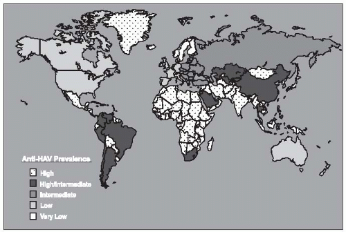
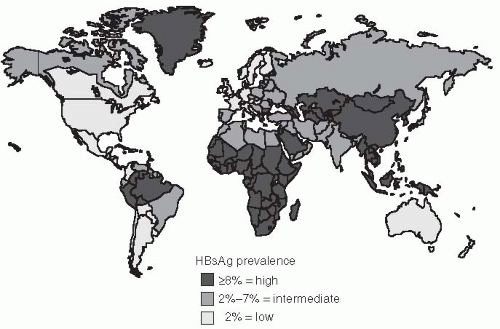
The prevalence of HBV infection varies greatly worldwide. In some areas of the world, HBV infections are highly endemic, and 8% or more of the total population are chronic carriers of HBsAg. These areas encompass 45% of the global population and include China and Southeast Asian countries, sub-Saharan Africa, and several areas in the Arctic, including Alaska, northern Canada, and Greenland (Figure 23-6). The First Nation people of Canada and the United States have high rates of HBV. In most of these areas, primary liver cancer is also very common and is often the most frequent cancer in adult males. Nearly all HBV infections occur during the perinatal period or in early childhood in these areas. Because acute HBV infections in infancy and early childhood result in a high rate of chronic carriage, the high endemicity is perpetuated from one generation to the next. The perinatal transmission of HBV among Alaska natives has been reduced dramatically in the last several years by the implementation of an effective program to vaccinate newborns immediately after they were born.117

In developed countries of North America, Western Europe, Australia, and some areas of South America, the rates of HBV infection are much lower. Less than 2% of the population are chronic carriers, and the overall infection rate, measured by the prevalence of anti-HBc, is 5% to 20%. In these areas, which constitute 12% of the global population, the frequency of perinatal or early childhood infection is low; however, they may account for a disproportionately high number of chronic HBV infections. Some subgroups also have higher rates, such as those living in the Amazon
basin, and immigrants from areas with higher population prevalence. These immigrant populations may contribute substantially to the subset of HBV infections that are transmitted during the perinatal and early childhood period.118, 119 and 120 In these areas, most infections occur among high-risk adult populations, including injection-drug users; homosexual men; persons with multiple sex partners; patients with multiple exposures to pooled blood products, such as hemophiliacs; and healthcare workers.115, 119, 121

The remaining parts of the world, which constitutes 43% of the global population, have an intermediate rate of HBV infection. The prevalence of HBsAg positivity ranges from 2% to 8%, in these regions and serologic evidence of past infection is found in 20% to 60% of the population. In these areas, there are mixed patterns of infant, early childhood, and adult transmission. In many countries in sub-Saharan Africa, for example, transmission of HBV commonly occurs during childhood rather than the perinatal period. Another risk for populations in developing countries is that posed by contaminated blood products or medical equipment. Nosocomial transmission in developing countries where HBV is highly endemic remains common.


Table 23-4 Number and Percentage of Patients with Acute Hepatitis B who Reported Selected Epidemiologic Characteristics, by Age Group, United States, 2007


Age group (yrs)


<45


≥45


Total


Characteristic


No.


(%)


No.


(%)


No.


(%)


Cases reported with risk factor data


Injection-drug use


229/1,200


(19.1)


55/688


(8.0)


284/1,888


(15.0)


Sexual contact with hepatitis B patient


62/851


(7.3)


22/505


(4.4)


84/1,356


(6.2)


Household contact of hepatitis B patient


19/851


(2.2)


12/505


(2.4)


31/1,356


(2.3)


Homosexual activity (male)§


46/400


(11.5)


16/189


(8.5)


62/589


(10.5)


Medical employee with blood contact


5/1,236


(0.4)


6/719


(0.8)


11/1,955


(0.6)


Hemodialysis


1/1,032


(0.1)


2/589


(0.3)


3/1,621


(0.2)


Had >1 sex partner


322/744


(43.3)


118/405


(29.1)


440/1,149


(38.3)


Heterosexual


293/708


(41.4)


110/390


(28.2)


403/1,098


(36.7)


Homosexual or bisexual (male)


29/36


(80.6)


8/15


(53.3)


37/51


(72.5)


Blood transfusion


1/1,221


(0.1)


8/709


(1.1)


9/1,930


(0.5)


Surgery


102/1,165


(8.8)


112/671


(16.7)


214/1,836


(11.7)


Percutaneous injury (e.g., needlestick)


52/1,080


(4.8)


21/631


(3.3)


73/1,711


(4.3)


Unknown


757/1,363


(55.5)


483/775


(62.3)


1,240/2,138


(58.0)


Cases reported with no risk factor data available


1,468


893


2,361


Total cases reported


2,831


1,668


4,499


* The percentage of cases for which a specific risk factor was reported was calculated on the basis of the total number of cases for which any information for that exposure was reported. Percentages might not total 100% because multiple risk factors might have been reported for a single case.


Exposures that occurred during the 6 weeks-6 months before onset of illness.

§ Among males, 18% reported homosexual behavior.


Reproduced from the Centers for Disease Control and Prevention (2009). MMWR Surveillance for Acute Viral Hepatitis- United States, 2007. Vol. 58, No SS-3.


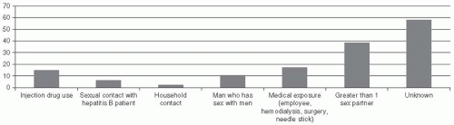
In the United States, the CDC has conducted a detailed assessment of behaviors in the Sentinel Counties Study of viral hepatitis to estimated the risk factors for acute HBV infection. The CDC estimate of the proportion of persons with acute HBV infections associated with known risk factors in 1992-1993 was as follows: heterosexual contact, 41%; injectiondrug use, 15%; homosexual contact, 9%; household contact, 2%; healthcare employment, 1%; other, 1%; and unknown, 31% (Table 23-4, Figure 23-9). It is likely that the relative contribution of injection-drug
use to HBV infections may be higher in populations with a high burden of drug use.

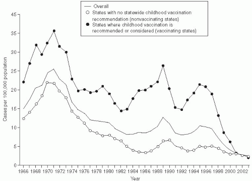
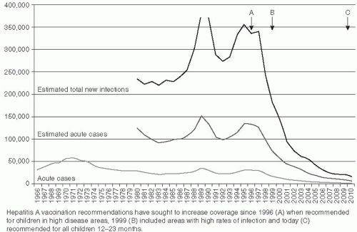
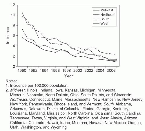
The incidence of hepatitis B infection has declined in the United States since the mid-1980s. In 2007, it was estimated to be 1.5 cases per 100,000 population, which was the lowest rate since active surveillance began in 1996 and represents an estimated decline of more than 80% from the rate in 1990, when a national strategy was implemented to reduce the incidence of HBV infections.118 (Figure 23-10) CDC has estimated that 38,000 new HBV infections occurred in the U.S. population in 2008, which represents a sharp decline from an estimated 73,000 cases in 2003. The greatest decline in incidence has occurred among the cohort of children in whom the recommendations for routine infant and adolescent vaccination have been implemented. Vaccination coverage among young adults (19-35 years of age) is estimated at 93%. The incidence among children younger than age 15 years declined 98% from 1.2 cases per 100,000 population in 1990 to 0.02 cases per 100,000 in 2007.118

The incidence of HBV among adults has also declined, albeit to a lesser extent than among infants and children. Few cases are now reported in certain populations that previously were considered to be at high risk, e.g. dialysis patients and health care workers. These declines are a result of improvements in infection control and routine use of hepatitis B vaccination in these populations. A high proportion of the new HBV infections occur among injection drug users, MSM, and persons with multiple sex partners117, 118, 119, 120, 121, 122 and 123


Figure 23-10 Incidence of Acute Hepatitis B, by Region and Year—United States, 1990-2007. Reproduced from the Centers for Disease Control and Prevention (2009). MMWR Surveillance for Acute Viral Hepatitis- United States, 2007. Vol. 58, No SS-3.

In recent years, however, a new high-risk group has been identified. During 1999-2007, 16 outbreaks of hepatitis B were detected and investigated in the United States, involving 157 persons. In nearly all of these outbreaks, the cases involved diabetics in nursing or assisted care facilities sharing glucosemonitoring equipment.124 and 125 CDC has estimated that 38,000 new HBV infections occurred in the U.S. population in 2008, which declined from an estimated 73,000 cases in 2003. Overall, 4.35% to 5.6% of the U.S. population is estimated to have been infected with HBV, which gives an estimated 800,000 to 1.4 million persons with HBV in the United States118

The risk of transfusion-transmitted HBV infections in the Untied States has declined substantially in the last few decades. Screening of blood donors for HBsAg was instituted in 1972. More effective screening of blood donors for drug use or sexual risk behavior and exclusion of men who have sex with men was implemented to reduce the risk of HIV transmission in the early 1980s. Most blood collection centers in the United States implemented routine nucleic acid testing to detect HBV DNA among blood donors in 2010. Also, blood
centers in the United States and Europe routinely test donated blood for antibodies to anti-HBc. Anti-HBc is a very sensitive test for HBV as any persons ever infected would be removed from the blood supply, but a less specific test as not all persons with anti-HBc are viremic for HBV. These testing procedures virtually eliminated transmission-transmitted HBV. However, screening donated blood for anti-HBc is not feasible in many countries in Asia or Africa because the rate of anti-HBc is so high that 40% to 70% of possible donors would be excluded. Therefore, transfusion-transmitted HBV is more frequent in these areas.114, 121, 122

Although the incidence of HBV has declined in the United States, the prevalence of chronic HBV infection has not changed; this steady state is largely related to immigration of individuals from countries with endemic HBV.117 Chronic HBV infections are recognized especially among several populations in the United States—namely, First Nations people from Canada and the United States, injection-drug users, persons with multiple sex partners, immigrants from Asia118 and Africa , and immunocompromised patients with AIDS and other causes of immunosuppression whose HBV infection has reactivated.114, 118, 121

A recent study of the incidence of hepatocellular carcinoma in the United States estimated that the number of cases increased by 46% between 1976-1980 and 1991-1995.126 This increased incidence of liver cancer probably reflects the effects of an increased rate of chronic HBV and HCV infections, combined with the effects of alcohol use among carriers of these viruses. The incidence of hepatocellular carcinoma tripled in the United States between 1975 and 2005, increasing from 1.6 to 4.9 cases per 100,000 population.129 Worldwide, primary cancer of the liver is the third leading cause of cancer mortality among men and the sixth leading cause among women.126


Screening Hepatitis B Virus Carriers for Cirrhosis and Hepatocellular Carcinoma

It is particularly important to periodically screen persons who are chronic HBV carriers for hepatocellular carcinoma (HCC). In 2007, an expert committee of the American Association for the Study of Liver Disease (AASLD) published recommendations for screeing of HBV carriers, i.e., persons who are HBsAg positive.127 The committee recommended screening the following HBsAg positive patients at 6 month intervals with ulstrasound to detect early liver cancer: Asian over 40 years of age, African patients over 20 years of age, all patients with cirrhosis, and patients with a family history of HCC. These recommendations were based, in part, on a randomized control trail in Shanghai which found a reduction of mortality from liver cancer by 37% from 131.5 to 83.2 per 100,000 population in a population of 18,816 HBsAg carriers whose HCC was detected at an earlier stage by screening.128 Most of these patients, in addition to screening would be candidates for treatment of their chronic HBV infection with anti-viral drugs to suppress their viral replication. More recent studies have found that pre-core mutations in the X gene of HBV may precede the occurrence of HCC by several years.129, 130 These data suggest that it might be possible to prevent or delay the onset of HCC by screening chronic carriers of HBV to detect high-risk mutations and treating such patients to reduce their viral load.

Although these screening recommendations may appear straightforward, it is sometimes difficult to determine whether a patient with chronic HBV infection might have cirrhosis. Many would argue that liver biopsy is the gold standard for diagnosis, and although histologic examination of a liver biopsy may be the best method to detect cirrhosis, a biopsy is invasive and occasionally causes complications. Moreover, it represents only a small sample of the liver, so it may not always detect early cirrhosis. Recently, other noninvasive diagnostic methods have become available, including fibroscan, which allows for more frequent assessment. Whereas fibroscan abnormalities reliably detect advanced fibrosis or cirrhosis they are less sensitive in detecting early stages of fibrosis. In addition, biochemical methods have been used to diagnose cirrhosis; these include measurements of elevated liver enzymes, AST/platelet ratio, haptoglobin, apolipoprotein-A1 and other methods that evaluate the functional status of the liver.129


Risk Factors and Biomarkers for Hepatocellular Carcinoma

Several viral, host, and environmental factors have been found to be associated with an increased risk of hepatocellular carcinoma among persons with chronic HBV infection. Host factors include older age, male sex, the presence of cirrhosis, a family history of liver cancer, and coinfection with HIV, HDV, or HCV. Virus factors include high levels of HBV replication, infection with HBV genotype C versus genotype B or A, a common double mutation in the core-promoter region of the virus (nucleotides 1762 A to T and 1764 G to A mutation), and several other mutations in the x gene, or pre-core genome of HBV. Environmental factors include frequent high alcohol
consumption, aflatoxin exposure, cigarette smoking, diabetes, and obesity.130

Among these risk factors, the association between the acquired double mutation in the HBV genome 1762T/1764A may be especially useful as a biomarker for the prediction of increased risk of subsequent liver cancer, as prospective studies have detected such mutations 5 or more years prior to the appearance of a cancer.131

Although the presence of cirrhosis is an important risk factor for the subsequent development of liver cancer, it is important to recognize that HCC may sometimes develop among HBV chronic carriers in the absence of cirrhosis. Only 40% to 50% of liver cancer among chronically HBV-infected patients in Africa have cirrhosis, whereas cirrhosis precedes liver cancer in 90% of such patients in Asia, the United States, and Europe.132

An increased rate of HCC among males has been found in every population. In a study of more than 2 million pregnancies between 1983 and 2000 in Taiwan, 17% of the women were HBsAg positive at the time of delivery. By 2005, 294 of these women had died from HCC.133 The relative risk (RR) of HCC among HBsAg carriers was 42% lower (RR = 0.58; 95% confidence interval [CI], 0.42-0.74) in women having two pregnancies and 52% lower (RR = 0.48; 95% CI, 0.34-0.72) in women having three or more pregnancies.136 In addition to these data among women, a nested case-control study among men with chronic HBV infection who subsequently developed HCC found higher levels of androgenic hormones among the men several years prior to the diagnosis of HCC.134 These data lend support to the hypothesis that the levels of female and male hormones may be cofactors in the pathogenesis of HCC among persons with HBV infections and may partially explain the sex difference in HCC.


Staging of Liver Disease

The natural history of chronic HBV infection is now recognized to have four phases, although not all patients go through all phases. These phases of HBV infection are most prominent when infection is acquired in infancy or childhood.

The first phase is the “immune tolerant phase,” which is characterized by the presence of HBeAg, high levels of serum DNA, normal serum transaminases, and minimal or no inflammation on liver biopsy. During this phase, which may last several decades in patients with infection acquired perinatally, spontaneous or treatment-induced HBeAg clearance is unusual and patients rarely progress to cirrhosis or liver cancer.130 In patients who acquire HBV infection during late childhood or as adults, this phase may be short-lived.

The second phase, immune clearance, is characterized by the presence of HBeAg, elevated HBV DNA levels, persistent or intermittent elevation in serum liver enzymes, and active inflammation in the liver. The elevated liver enzymes reflect immunecytolysis of infected hepatocytes. Some of the flares in liver enzymes may lead to hepatic decompensation. The immune response in this phase may lead to clearance of HBeAg from the serum.

The third phase, known as the “immune control” phase, is characterized by the absence of HBeAg, presence of anti-HBe, normal aminotransferase levels, and low levels of serum HBV DNA (i.e., less than 105 IU/mL). Liver biopsy at this stage may show mild hepatitis and minimal fibrosis. However, some patients may have cirrhosis if the liver inflammation during the immune clearance phase was severe or persistent. The use of sensitive assays to detect HBV DNA has revealed that replication is ongoing,138 but when this immune control stage persists, the patient is at minimal risk of progression to severe liver disease or cirrhosis.

Some inactive carriers have “reactivation” of their HBV infection, a fourth stage. This may occur spontaneously or as a result of immunosuppression.

A study of 283 patients in Taiwan who were followed for approximately 9 years after they developed antibodies to HBeAg (the third phase) found that 67% had a sustained remission, 4% had HBeAg revision to positivity, and 24% had HBeAgnegative chronic hepatitis.135 Cirrhosis developed in 8% and HCC in 25% of these patients; these conditions occurred more frequently in those patients with active hepatitis after their loss of HBeAg. In fact, the core-promoter double mutation131 and a stop codon mutation in the pre-core region of the HBV genome, NT/896,131 will turn off production of HBeAg. The double mutation commonly precedes the onset of hepatocellular carcinoma in patients with HBeAg-negative chronic HBV infection.131

The data from follow-up of patients after HBV infection indicate that the virus is not generally directly cytopathic, but that the sequelae of cirrhosis and liver cancer are primarily immune mediated. Furthermore, HBV infections are not curable. Thus the goal of antiviral therapy of chronic HBV infection is the prevention of liver fibrosis, cirrhosis, and liver cancer. The main stages of the infection when inflammation, fibrosis, and cirrhosis develop are during the immune clearance and reactivation stages. Antiviral therapy should be used in patients during the immune clearance or reactivation stages of this infection when
inflammation and fibrosis of the liver may occur. The goal of treatment during these stages is to suppress viral replication and liver inflammation and fibrosis.

Jul 8, 2016 | Posted by in INFECTIOUS DISEASE | Comments Off on Viral Hepatitis

Full access? Get Clinical Tree

Get Clinical Tree app for offline access