, Joong Mo Ahn2 and Yusuhn Kang2
(1)
Department of Radiology, Seoul National University College of Medicine, Seoul National University Bundang Hospital, Seongnam, South Korea
(2)
Department of Radiology, Seoul National University, Bundang Hospital, Seongnam, South Korea
7.1 Hemangioma
Overview
Hemangioma is a common benign bone tumor, characterized by newly formed thin-walled capillary or cavernous type blood vessels.
Epidemiology
Hemangiomas may occur in any age, but most cases are diagnosed between the fourth and seventh decades of life. A slight female predilection has been reported (male: female = 2:3).
Common Locations
The vertebral bodies and craniofacial bones are the two most commonly affected sites. Multifocal involvement is frequent in the spine. When they occur in a long bone (femur, tibia, humerus), the metaphysis is commonly affected, and can occur with a medullary, intracortical, or periosteal location.
Imaging Features
Radiograph
Vertebral hemangiomas present as radiolucent lesions with coarse trabeculations or striations (corduroy pattern, polka-dot pattern). The mixed osteolytic and trabeculated appearance of hemangiomas may be found in flat bones and tubular bones, sometimes exhibiting a “honeycomb” pattern (Fig. 7.1). In flat bones, hemangiomas may cause significant expansion of the bone and may accompany radially oriented striations.
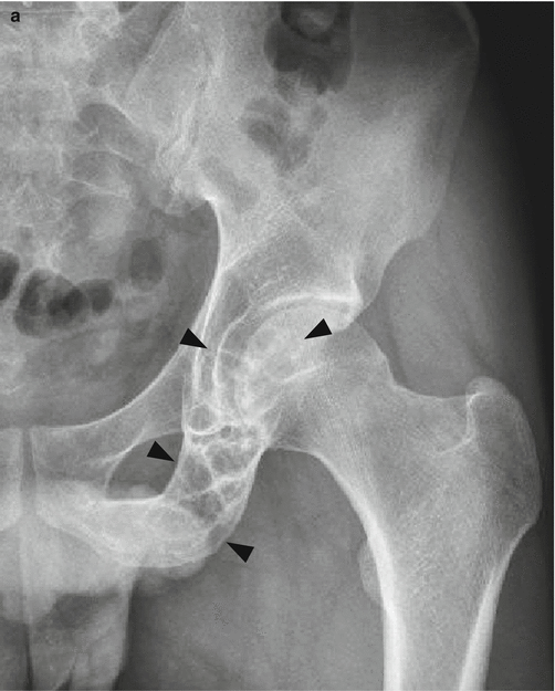
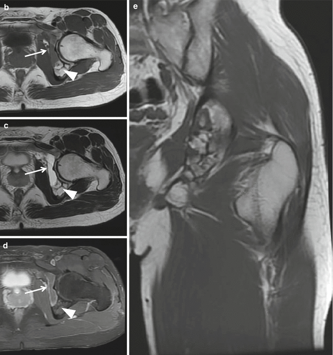


Fig. 7.1
Hemangioma in the left acetabulum and inferior pubic ramus. (a) Anteroposterior radiograph of the left hip shows an expansile osteolytic lesion in the left acetabulum and inferior pubic ramus (black arrowheads). Multiple thickened trabeculations are noted within the mass. (b) Axial T1-weighted image, (c) axial T2-weighted image, (d) axial contrast-enhanced T1-weighted fat-suppressed image, and (e) coronal T1-weighted image show a multilobulated septated intramedullary lesion in the left acetabulum and inferior pubic ramus. Some portions exhibit fluid-equivalent signal intensity and show thin peripheral rim enhancement (arrows), whereas some portions have fat-equivalent signal intensity (white arrowheads)
Magnetic resonance imaging
The classic appearance of hemangioma of the bone is hyperintense on T1-weighted images due to the presence of it within the lesion, but the signal may be variable depending on the amount of fat. Hemangiomas are hyperintense on T2-weighted images and fluid-sensitive sequences, due to the vascular channels within the mass (Fig. 7.2). The thickened trabeculation exhibits dark signal intensity on both T1-weighted and T2-weighted images. Most lesions show significant contrast enhancement after intravenous gadolinium injection.
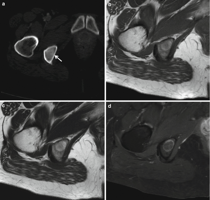

Fig. 7.2
Hemangioma in the right ischium. (a) Axial contrast-enhanced CT image shows a hyperattenuated lesion in the right ischium (arrow). This lesion is hyperintense on both (b) axial T1-weighted image and (c) axial T2-weighted images, and shows peripheral enhancement on (d) contrast-enhanced T1-weighted fat-suppressed image. Hyperintensity on both T1-weighted and T2-weighted images probably corresponds to the fat deposition within the hemangioma
Differential Diagnoses (for Lesions in Long Bone)
Aneurysmal bone cyst
Hemangiomas that exhibit expansile osteolytic appearance with striations may appear similar to aneurysmal bone cysts on radiograph.
7.2 Angiosarcoma
Overview
Angiosarcoma is a vascular tumor of high-grade malignancy, which is histologically characterized by cells showing endothelial differentiation.
Epidemiology
Angiosarcoma accounts for <1 % of primary malignant bone tumors. It has been reported to occur in patients between the third and ninth decades with a peak incidence in the third to fifth decades. It has a male predominance, with a male-to-female ratio of 2:1.
Common Locations
Angiosarcomas more commonly involve the skin and soft tissue, and rarely occur in the bone. It has a wide skeletal distribution, with a majority involving the long tubular bones (tibia, femur), pelvic bones, and spine.

