, Joong Mo Ahn2 and Yusuhn Kang2
(1)
Department of Radiology, Seoul National University College of Medicine, Seoul National University Bundang Hospital, Seongnam, South Korea
(2)
Department of Radiology, Seoul National University, Bundang Hospital, Seongnam, South Korea
4.1 Desmoplastic Fibroma of Bone
Overview
Desmoplastic fibroma of bone is a rare, locally aggressive benign tumor, characterized by the presence of spindle cells and abundant collagen produced by the tumor cells. It is the intraosseous counterpart of the soft tissue desmoid tumor. It does not metastasize and is considered benign, but is locally aggressive, and recurrences are common after curettage.
Epidemiology
Desmoplastic fibroma of bone accounts for <0.1 % of primary bone tumors, and 0.3 % of benign bone tumors. It can be seen at any age, but it is common in adolescents and young adults, with most cases occurring in the second and third decades of life. It distributes equally in both genders.
Common Locations
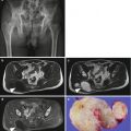
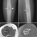
Desmoplastic fibromas are most common in the mandible, followed by femur, pelvic bone, radius, and tibia. In the long bones, desmoplastic fibromas are usually located at the metadiaphysis (Fig. 4.1). They may extend to the epiphysis in patients with closed physis (Fig. 4.2).
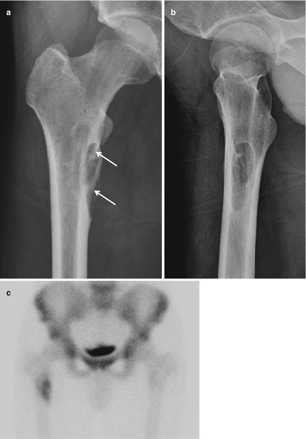
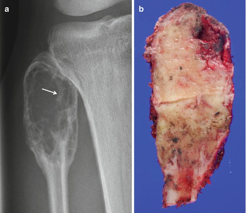
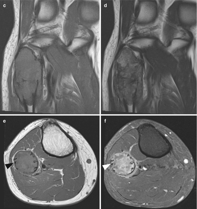

Fig. 4.1
Desmoplastic fibroma of bone. (a) Anteroposterior and (b) lateral radiographs show a well-defined osteolytic lesion at the proximal metadiaphysis of the femur. Intralesional trabeculations are seen within the lesion (white arrow in a). (c) The lesion shows increased uptake on bone scan


Fig. 4.2
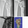
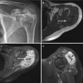
Desmoplastic fibroma of bone. (a) A well-defined expansile osteolytic lesion is seen at the proximal epimetaphysis of the fibula on radiograph. Internal trabeculations or ridges (white arrow) are seen within the lesion, giving the lesion a soap-bubble appearance. (b) Section through the specimen of resected proximal fibula shows expansile lobulated solid fibrous tumor. The lesion appears isointense to adjacent muscle on T1-weighted images (c, e) and heterogeneous on T2-weighted images (d), and exhibits heterogeneous enhancement (e). Areas of hypointensity are noted on T2-weighted images due to the rich collagen content. Internal ridges are noted at the periphery of the lesion on axial images (arrowheads in e, f)
Imaging Features
Radiograph
Desmoplastic fibromas are well-defined osteolytic lesions noted at the metadiaphysis of a long bone. They may have a sclerotic border and usually expand the bone. The internal trabeculations or ridges within the lesion, along with bone expansion, give a soap-bubble appearance on radiograph (Fig. 4.2). Periosteal reaction is rarely seen with desmoplastic fibromas.
Magnetic resonance imaging
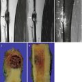
Desmoplastic fibromas usually appear hypointense on T1-weighted images, and heterogeneous isointense to hypointense on T2-weighted images, with heterogeneous enhancement. The signal intensity of desmoplastic fibroma on MR imaging is attributable to its collagen content, as for soft tissue desmoid tumors. Therefore, the signal intensity of the desmoplastic fibromas may vary according to the proportion of collagen content and cellular component. Acellular lesions with rich collagenous stroma exhibit hypointensity on T2-weighted images, whereas more cellular lesions with relatively small amount of collagenous stroma exhibit isointensity to hyperintensity on T2-weighted images (Fig. 4.3).
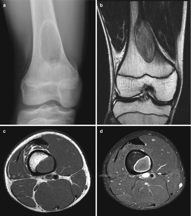

Fig. 4.3
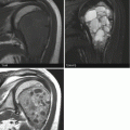
Desmoplastic fibroma of bone. (a) A geographical osteolytic lesion with a thin sclerotic rim is noted at distal femoral metadiaphysis on anteroposterior radiograph. (b) The lesion appears heterogeneous on T2-weighted image with areas of hypointensity corresponding to the collagenous stroma. The lesion shows intermediate signal intensity on T1-weighted image (c) and shows mild enhancement on contrast-enhanced T1-weighted fat-suppressed image (d)
Differential Diagnoses
- 1.
Aneurysmal bone cyst
The radiographic appearance of desmoplastic fibromas and aneurysmal bone cysts may be similar; expansile osteolytic lesions with well-defined borders occur in the metadiaphysis. However, the two lesions are readily differentiated on MRI; aneurysmal bone cysts appear as multiloculated cystic lesions with multiple fluid–fluid levels.
- 2.
Chondromyxoid fibroma
Chondromyxoid fibromas exhibit soap-bubble appearance on radiograph, similar to desmoplastic fibromas. However, on MR imaging, chondromyxoid fibromas are distinguished from desmoplastic fibromas by their predominantly high signal intensity on T2-weighted imaging.
- 3.
Other differentials may include fibrous dysplasia and giant cell tumor
4.2 Fibrosarcoma of Bone
Overview
Fibrosarcoma of bone is a rare malignant spindle cell neoplasm of the bone. Fibrosarcomas were originally reported to show a characteristic histological appearance, “herringbone” pattern. However, more recent studies have revealed that the “herringbone” pattern is not a finding specific to fibrosarcoma and that it can be seen in a variety of other primary bone tumors. Moreover, the advances in immunohistochemistry and molecular genetic analysis have resulted in the reclassification of tumors previously diagnosed as fibrosarcomas, into other primary bone tumors. Nowadays, fibrosarcoma is less commonly used as a specific diagnostic category and is considered a diagnosis of exclusion.
Epidemiology
Fibrosarcoma of bone has been reported to account for <5 % of all primary malignant bone tumors. It usually occurs in the third to sixth decades of life and has an equal distribution between genders.
Stay updated, free articles. Join our Telegram channel

Full access? Get Clinical Tree


