, Joong Mo Ahn2 and Yusuhn Kang2
(1)
Department of Radiology, Seoul National University College of Medicine, Seoul National University Bundang Hospital, Seongnam, South Korea
(2)
Department of Radiology, Seoul National University, Bundang Hospital, Seongnam, South Korea
Case 1
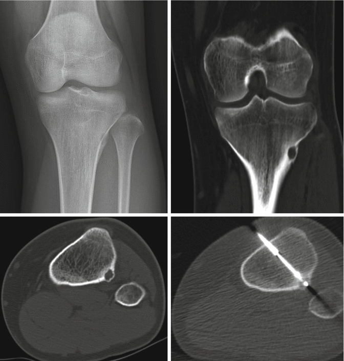
Fig. 12.1
- 1.
List the differential diagnoses of this condition.
- 2.
What is the most likely diagnosis?
- 3.
How would you treat this patient?
- 4.
What procedure does this patient have?
- 5.
What are the advantages of the treatment methods you mentioned?
- 6.
List the relative contraindications of this procedure.
- 7.
What are the complications of this procedure?
Case 2
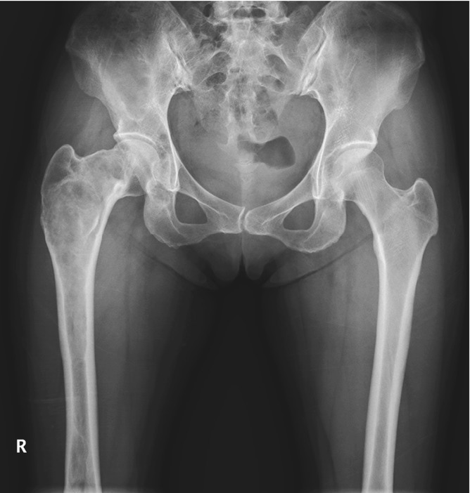
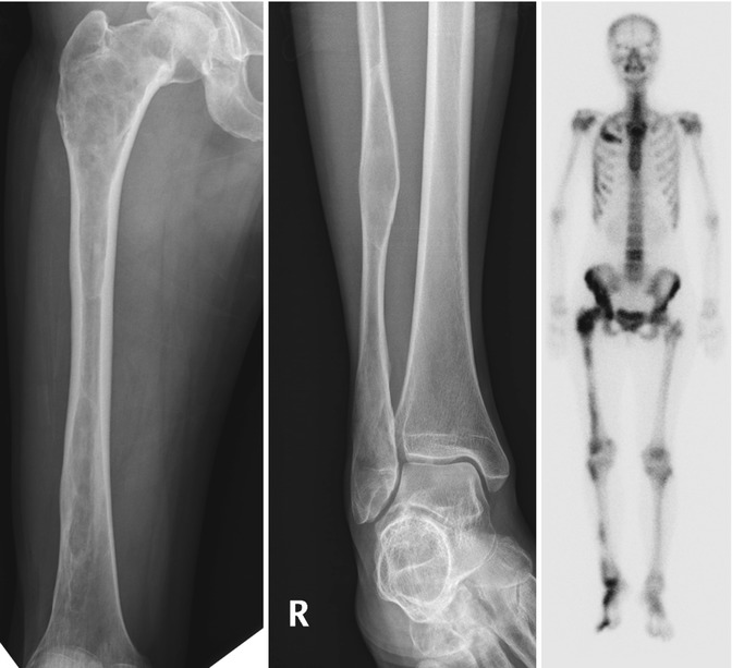
Fig. 12.2
- 1.
What is the likely diagnosis?
- 2.
What is the McCune-Albright syndrome?
- 3.
What is meant by a ground-glass appearance?
- 4.
What is the diagnosis if this patient also has multiple soft tissue myxomas?
- 5.
What is the most common cause of an expansile focal rib lesion?
Case 3
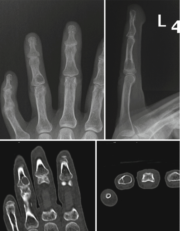
Fig. 12.3
- 1.
What observation did you make?
- 2.
What is the most common tumor of the hand?
- 3.
Can enchondroma be purely lytic in appearance on radiographs?
- 4.
What is the most likely diagnosis?
Case 4
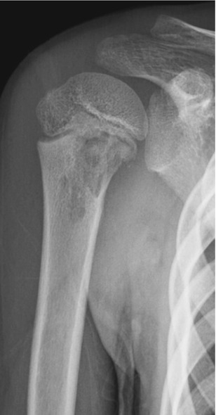

Fig. 12.4
- 1.
List your differential diagnoses.
- 2.
What is the most likely diagnosis?
- 3.
Where are the common locations of this lesion?
- 4.
At what age does this lesion occur?
- 5.
List benign musculoskeletal lesions which may show misleading aggressive appearance.
Case 5
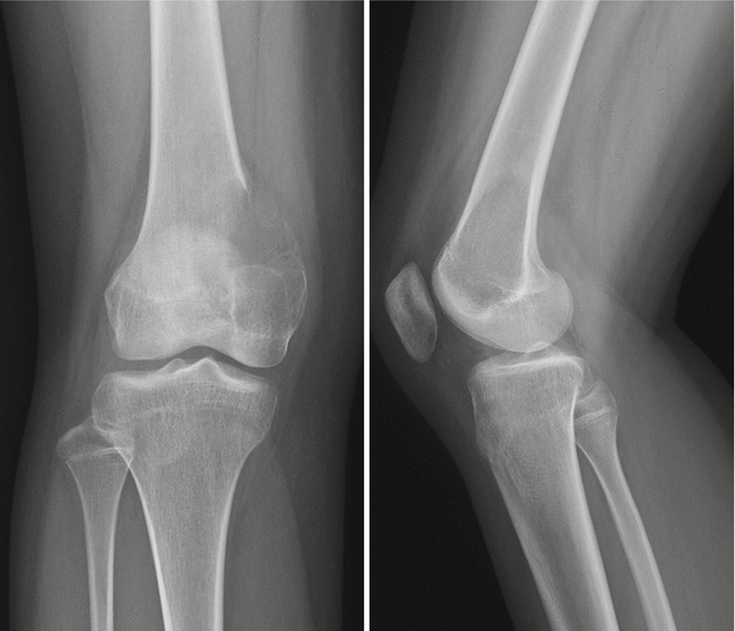
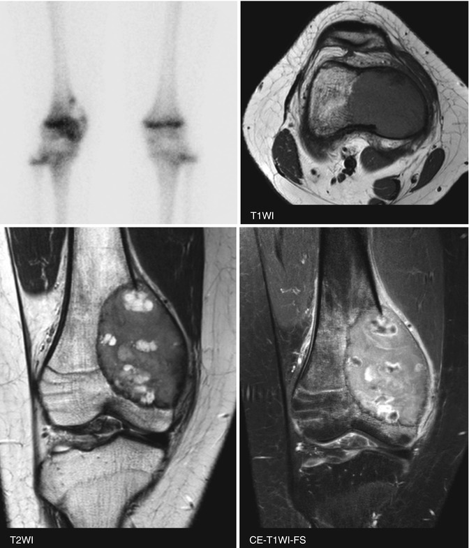
Fig. 12.5
- 1.
What is the likely diagnosis?
- 2.
Most bone tumors show equal sex predilection or slight male predominance. List the tumors with female predilection.
- 3.
What is the characteristic MR appearance on fluid-sensitive sequences?
- 4.
List the osseous lesions with T2-hypointense tumor matrix.
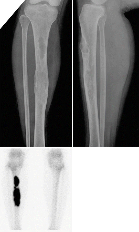
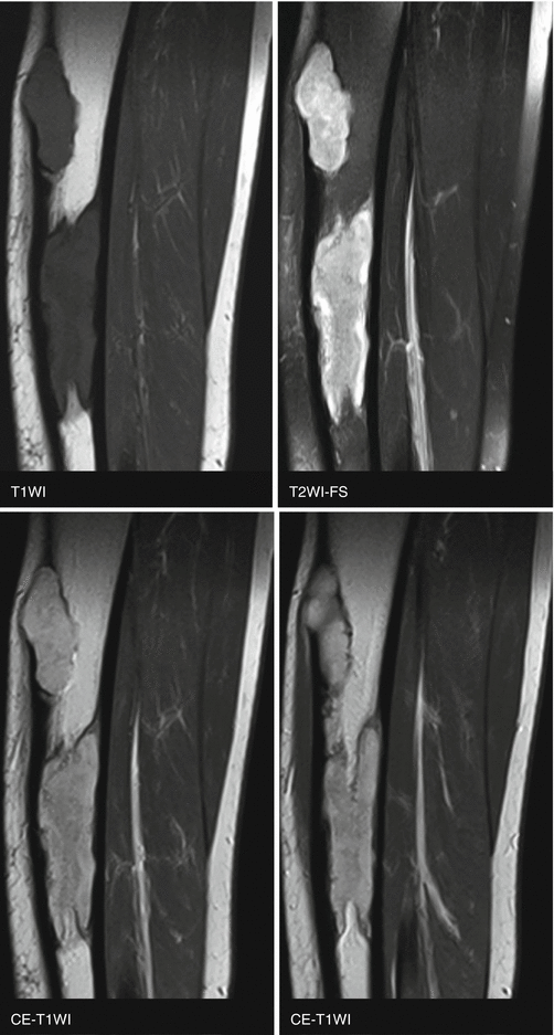
Case 6
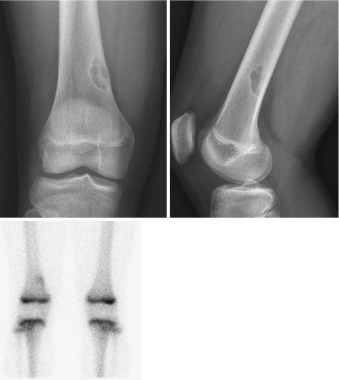
Fig. 12.6
- 1.
List the bubbly lesions of bone.
- 2.
What is the most likely diagnosis?
- 3.
What is a finding that would represent a benign process?
- 4.
Does the differential diagnosis include a nonossifying fibroma, if the patient is a 50-year-old man?
- 5.
In which syndrome could the multiple nonossifying fibromas be seen?
Case 7
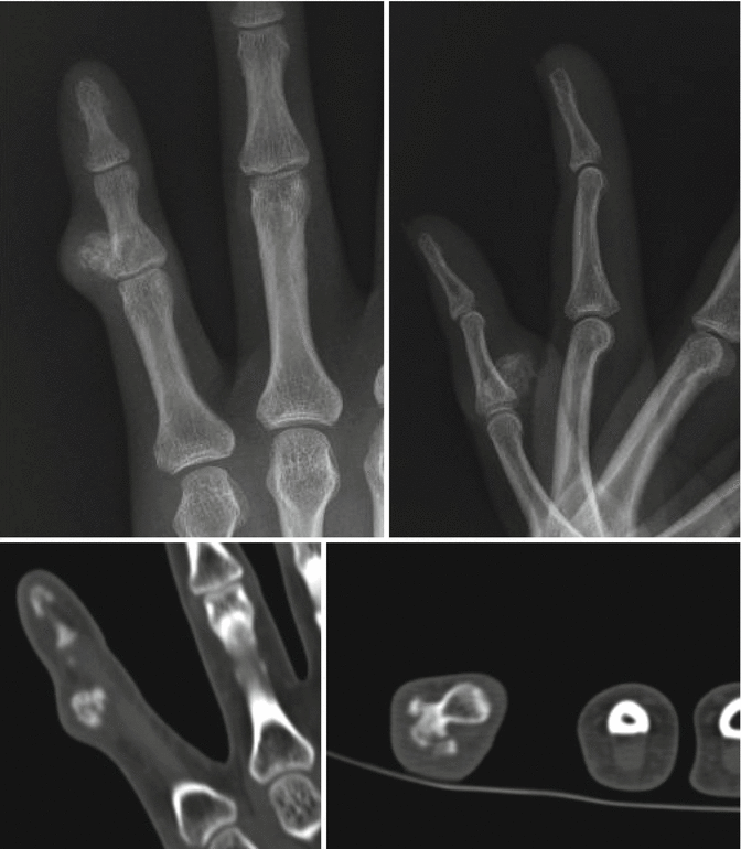
Fig. 12.7
- 1.
What observation did you make on radiographs?
- 2.
List the differential diagnoses.
- 3.
What is the characteristic CT finding to help to distinguish this condition from an osteochondroma?
- 4.
What is the most likely diagnosis?
Case 8
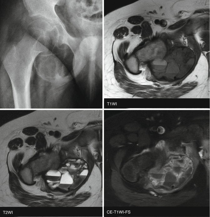
Fig. 12.8
- 1.
What observation did you make on radiographs?
- 2.
What are the characteristic observations on MR images?
- 3.
What is the most likely diagnosis?
- 4.
What is the definition of the secondary aneurysmal bone cyst?
- 5.
What are the typical MR imaging findings of the primary aneurysmal bone cyst?
Case 9
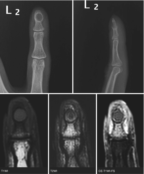
Fig. 12.9
- 1.
What observation did you make on radiograph?
- 2.
List your differential diagnoses.
- 3.
What is the most likely diagnosis?
- 4.
Which is the most common location for an epidermal inclusion cyst?
- 5.
What MR imaging findings can distinguish this condition from a glomus tumor?
Case 10
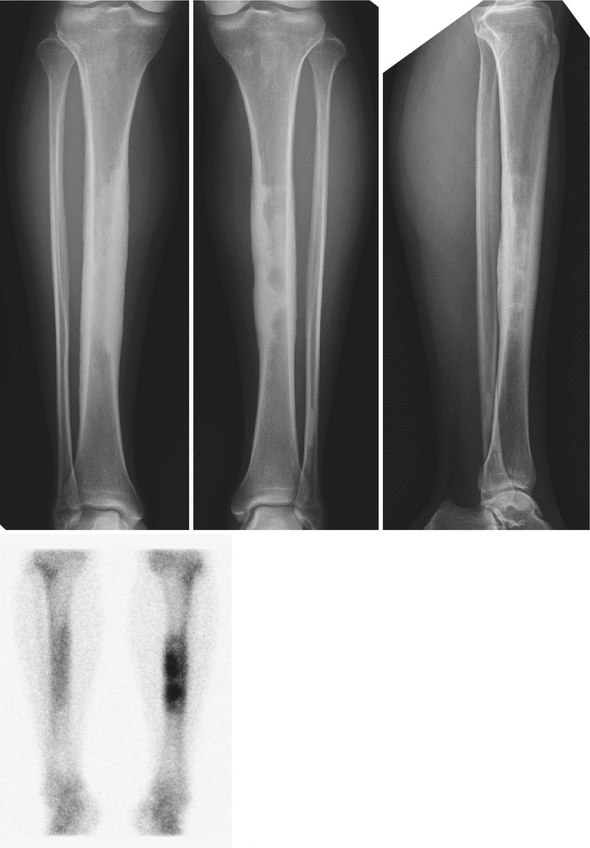
Fig. 12.10
- 1.
What are the findings?
- 2.
What is the likely diagnosis?
- 3.
Dose this disorder involve epiphysis?
- 4.
Can this condition be unilateral?
- 5.
List the differential diagnoses if this lesion involved a single bone.
Case 11
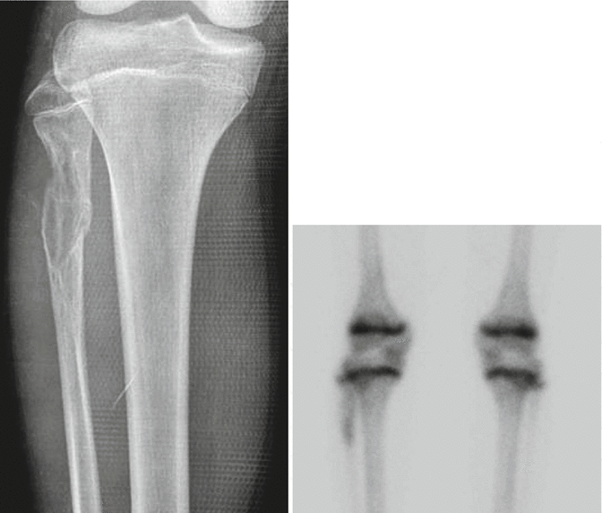
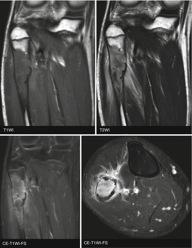
Fig. 12.11
- 1.
List the differential diagnoses on radiograph.
- 2.
What are the radiographic findings that suggest aggressive tumor in this patient?
- 3.
What is the likely diagnosis?
- 4.
What radiographic finding can help to distinguish low-grade central osteosarcoma from conventional osteosarcoma?
Case 12
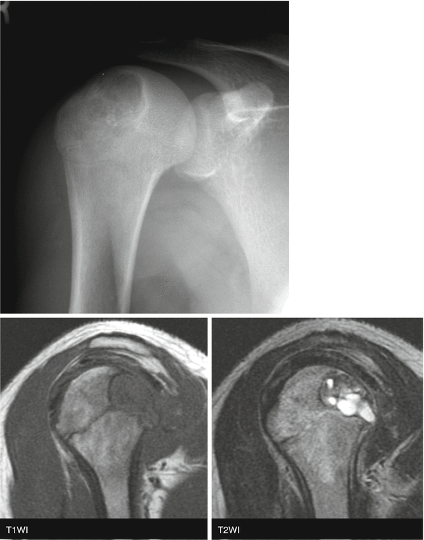
Fig. 12.12
- 1.
List the differential diagnoses of this lesion.
- 2.
What is the most likely diagnosis?
- 3.
What is a characteristic observation on MR imaging?
- 4.
Where are the preferred sites of this lesion in the feet?
Case 13
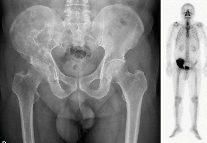
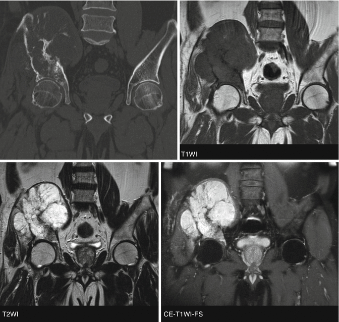
Fig. 12.13
- 1.
What observation did you make on CT?
- 2.
What observation did you make on MR images?
- 3.
What is the most likely diagnosis?
- 4.
What radiographic findings in this condition would suggest high-grade lesion in histology?
Case 14
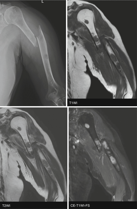
Fig. 12.14
- 1.
What is the likely diagnosis?
- 2.
List the skeletal locations where multiple myeloma preferentially involves.
- 3.
Explain the reason above.
- 4.
Can multiple myeloma and plasmacytoma be sclerotic?
- 5.
What does POEMS stand for?
Case 15
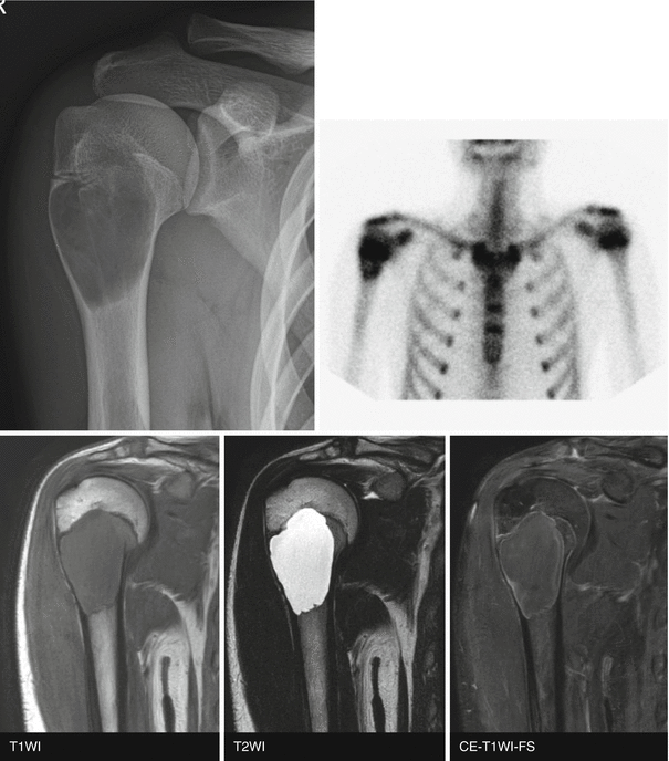
Fig. 12.15
- 1.
What is the likely diagnosis?
- 2.
Where is it most common?
- 3.
What is a “fallen fragment sign”?
- 4.
Could the fallen fragment sign be seen in fibrous dysplasia?
- 5.
List a short differential diagnosis for a well-defined lucent lesion in the anterior aspect of the body of the calcaneus.
Case 16
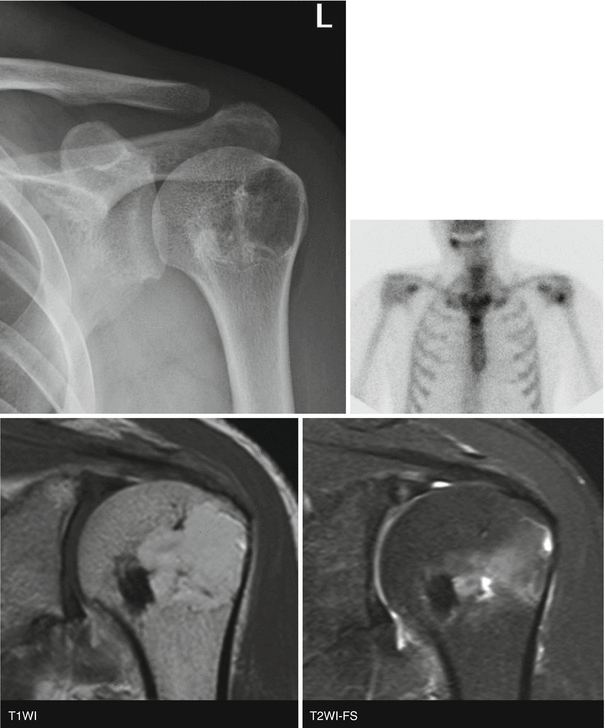
Fig. 12.16
- 1.
What observation did you make on radiograph?
- 2.
What observation did you make on MR images?
- 3.
What is the pathogenesis of the associated ossification in this condition?
- 4.
What is the likely diagnosis?
Case 17
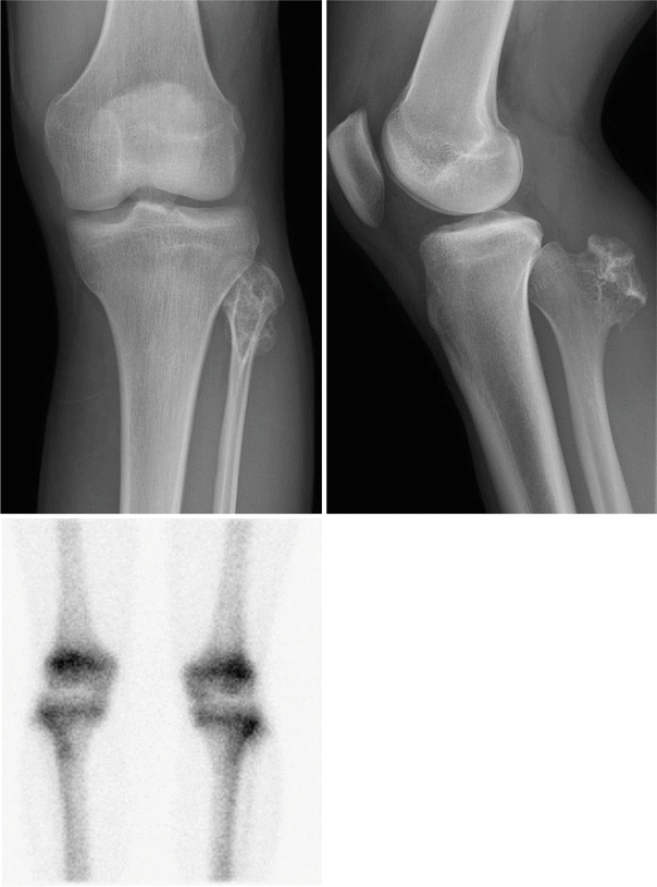
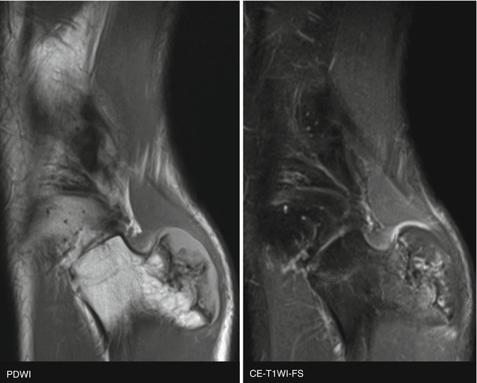
Fig. 12.17
- 1.
What are some characteristic findings of this lesion?
- 2.
List the differential diagnoses.
- 3.
What is the most likely diagnosis?
- 4.
When does this lesion stop growing?
Case 18
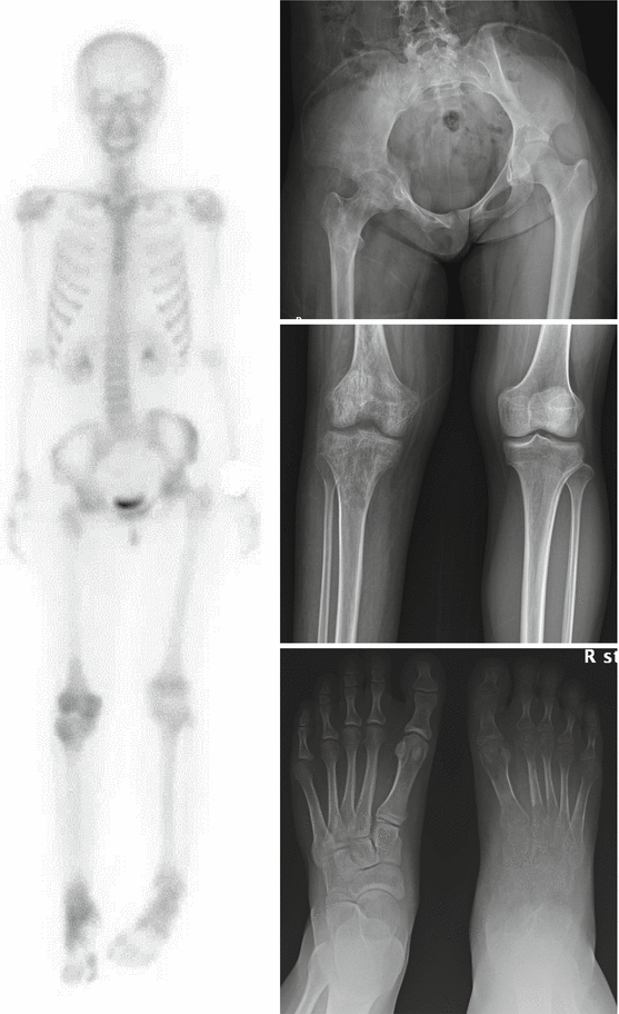
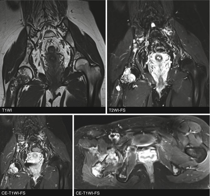
Fig. 12.18
- 1.
What observations did you make on radiographs?
- 2.
What observations did you make on MR images?
- 3.
List the differential diagnoses.
- 4.
What is the likely diagnosis?
Case 19

