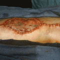
(1)
State University of New York at Buffalo Kaleida Health, Buffalo, NY, USA
Tumors on this side can grow to prodigious size. Therefore, they become palpable or otherwise cause some mild symptoms before the diagnosis is made. They may involve the diaphragm, the right lobe of the liver, the right adrenal gland and kidney, and the inferior vena cava. For large tumor masses in the right side of the abdomen, a left lateral position is preferred for surgery. A midline incision is carried out from the xiphoid to the right of the umbilicus and for a few centimeters below the umbilicus in the midline. The abdomen is opened; the surgeon then can palpate the area of the tumor mass and perhaps perform some initial maneuvers to expose and free this mass from surrounding structures. In the case illustrated, the retroperitoneal tumor is found between the liver and the hepatic flexure of the colon (Fig. 37.1). An incision in the right parietal peritoneum is made along the white line adjacent to the ascending colon and then around the lateral border of the palpable mass (Fig. 37.2). Inferiorly, the dissection of the tumor mass allows one to free the lower pole of this mass, including the kidney, which appears to be involved. The ureter is exposed at a sufficient distance from the tumor mass. The lower part of the inferior vena cava below the renal veins is also exposed (Fig. 37.3). The duodenum, with the head of the pancreas, is dissected off the area of the tumor. In this patient, the duodenum and head of the pancreas are free of involvement (Fig. 37.4). The parietal peritoneum between the right lobe of the liver and the tumor mass is incised (Fig. 37.5). The right triangular ligament is divided (Fig. 37.6). It is found that the posterior portion of the right hemidiaphragm is involved by the tumor, so the incision is extended to include the right chest. This oblique incision joins the midline incision slightly above the level of the umbilicus.
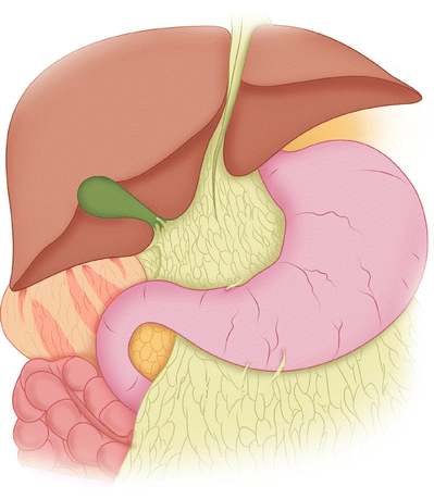
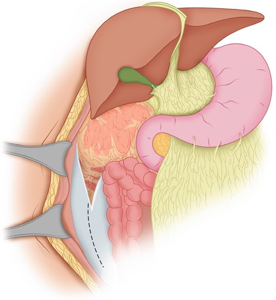
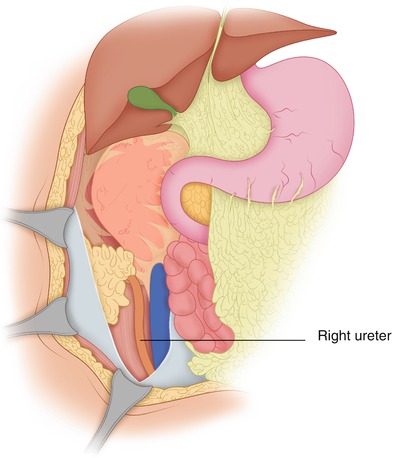
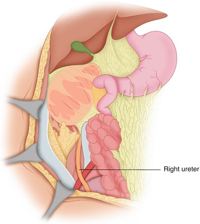
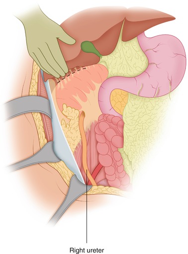

Fig. 37.1
The retroperitoneal tumor is found between the liver and the hepatic flexure of the colon

Fig. 37.2
The colon is mobilized by incising the parietal peritoneum along the white line

Fig. 37.3
The colon is further dissected off the retroperitoneal structures, exposing the right ureter and inferior vena cava

Fig. 37.4
The duodenum and head of the pancreas are mobilized off the upper medial aspect of the tumor mass

Fig. 37.5


The parietal peritoneum is incised superiorly and lateral to the tumor
Stay updated, free articles. Join our Telegram channel

Full access? Get Clinical Tree


