(1)
State University of New York at Buffalo Kaleida Health, Buffalo, NY, USA
The posterior group of the thigh includes the semimembranosus, semitendinosus, and biceps femoris muscles, the last consisting of the long and short heads. The patient is placed in a prone position, and a vertical longitudinal incision is made from the gluteal crease to the popliteal fossa, using an ellipse to circumscribe any previous biopsy incision (Fig. 49.1). When the tumor is not close to the skin, flaps are developed around the previous biopsy site, with the thickness of the flaps depending on the preoperative CT or MRI scan and the physical examination. For tumors located deep to the investing fascia, of course, the flaps do not have to be thin and one may carry the incision down to almost the fascia level and then around the fascia, leaving the fascia to be removed with the specimen. In the midline, the posterior femoral cutaneous nerve, sensory to the posterior thigh, courses directly underneath the investing fascia and is often sacrificed, because it can interfere with the exposure and en bloc resection of large tumors in this area.

Fig. 49.1
A longitudinal incision is made, extending an ellipse around the previous biopsy incision
Superiorly, below the edge of the gluteus maximus, the sciatic nerve may be exposed between the origin of the hamstring muscles from the ischial tuberosity and the greater trochanter. If the tumor comes close to the gluteal crease, one may need to incise the edge of the gluteus maximus in a superior direction to provide exposure for the identification of the sciatic nerve proximal to the tumor (Fig. 49.2). As the flaps are developed around the tumor and the sciatic nerve is exposed superiorly or inferiorly, depending on the location of the tumor, one then decides on the extent of resection and whether all hamstring muscles need to be removed (Fig. 49.3). The origin of the hamstring muscles may be divided off the ischial tuberosity. As the sciatic nerve continues to be exposed and dissected, it is surrounded with vessel loops. At the middle thigh, the sciatic nerve courses between the long head of the biceps laterally and the medial hamstrings (semitendinosus, semimembranosus) medially (Fig. 49.4). At the level of the ischial tuberosity, the sciatic nerve lies between the origin of the hamstrings and the greater trochanter. To expose the nerve at this level, as mentioned above, one must divide the gluteus maximus from its lower edge cephalad for several centimeters (Fig. 49.4). The short head of the biceps usually is not close to the tumor and therefore can be separated from the long head (Fig. 49.5). The sciatic nerve initially in the space between the origin of the hamstrings, at the ischial tuberocity and the greater trochanter, is crossed posteriorly by the laterally deviating long head of the biceps at the mid thigh, while in the lower thigh assumes a position between the biceps femoris and the medial hamstrings. The sheath of the sciatic nerve is entered on the side opposite to the tumor and incised along the length of the nerve, leaving the sheath attached to the tumor to be removed en bloc with the tumor. In the lower half of the thigh, close to the popliteal space, the sciatic nerve divides into the tibial nerve, proceeding in a straight downward direction, and the common peroneal nerve, proceeding to occupy a course along the edge of the biceps femoris and then down to the level of the neck of the fibula. From that point, it goes around the neck of the fibula, beneath the origin of the peroneus longus, where it splits, entering the anterior compartment of the leg to supply its muscles as the deep peroneal nerve, whereas the other terminal branch, the superficial peritoneal, supplies the peronei muscles and terminates as a sensory nerve for the lateral aspect of the leg and foot.
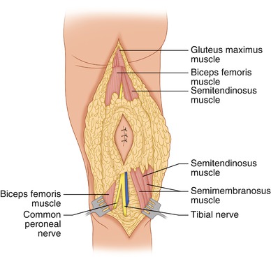
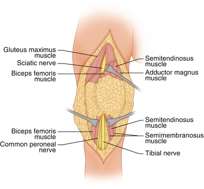
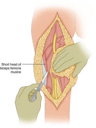
Fig. 49.2
Medial and lateral flaps have been developed. The hamstring muscles have been exposed above and below the tumor
Fig. 49.3
The sciatic nerve is found proximally between the ischial tuberosity and the greater trochanter. To expose the nerve, the gluteus maximus may be divided for several centimeters from its inferior edge in a cephalad direction. Distally, the sciatic nerve has already divided into the tibial and common peroneal nerves
Fig. 49.4


The sciatic nerve is further exposed by mobilizing the tumor and hamstrings, as the latter’s origin from the ischial tuberosity is divided. The long head of the biceps is separated from the short head, which often is not close to the tumor. This is a posterior dissection on the left leg
Related posts:

Stay updated, free articles. Join our Telegram channel
Full access? Get Clinical Tree
