(1)
State University of New York at Buffalo Kaleida Health, Buffalo, NY, USA
The adductor group of muscles consists of three layers: the superficial layer, including the pectineus, adductor longus, and gracilis; the middle layer, consisting of the adductor brevis; and the posterior layer, consisting of the adductor magnus. These muscles are supplied mainly by the obturator nerve, which forms in the substance of the psoas muscle from L2, L3, and L4 roots, emerges from the medial border of this muscle, and courses behind the common iliac vessels. The obturator nerve then proceeds lateral to the internal iliac vessels on the side of the true pelvis. On the surface of the obturator internus muscle, the obturator nerve runs anterior to the obturator vessels and then courses through the obturator canal, anteriorly located between the anterior pubic ramus and the membrane occluding the obturator opening. It then divides into an anterior division, coursing in front of the adductor brevis, and a posterior division, coursing behind it. The pectineus muscle, however, is innervated by a branch from the femoral nerve that courses behind the common femoral vessels. This muscle occasionally is also supplied by an accessory obturator nerve. The ischiocondylar portion of the adductor magnus is supplied by the sciatic nerve (tibial division). Sensory function for a small area of the medial thigh is supplied by a branch of the obturator nerve.
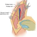
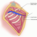
An operation for a tumor in the medial thigh is performed with the patient supine on the operating table and the affected leg prepped and draped free in a flexed and externally rotated position. This position brings out the area of the gracilis muscle, and the incision is made in the medial aspect of the thigh along the course of the gracilis muscle from the pubic bone to the medial femoral condyle. For a bulky tumor located proximally, a T extension of the proximal end of the incision may be required to provide exposure. The exact placement of the incision depends, of course, on the location of the tumor, whether it is located more in the proximal portion of the adductor muscles, and whether it is close to the femoral vessels or is more medially located. For a tumor located close to the femoral vessels, an incision starting from the anterior superior iliac spine is carried over to the site of the approximate origin of the gracilis muscle and then down along the anterior border of the gracilis to the level of the medial femoral condyle (Fig. 48.1). A flap is developed to expose the surface of the sartorius muscle, which may have to be divided proximally and distally, depending on its proximity to the area of the tumor and the need to expose the femoral vessels for their entire length (Fig. 48.2). The femoral vessels are exposed proximally as common femoral vessels, and then the profunda vessels are dissected and encircled with vessel loops (Fig. 48.3). Finally, the superficial femoral vessels are dissected down to the adductor hiatus or as far down as the location of the tumor requires. Whether the vessels need to be skeletonized or not depends on how close the tumor is to the vessel surface. Following the exposure and control of the femoral vessels proximal and distal to the tumor, the decision is made whether to proceed with a tentative dissection of the vessels off the tumor. Obviously it is best to avoid unnecessary skeletonization of the vessels, because skeletonization involves division of a significant number of lymphatics coursing along the vessels and may increase the chance of postoperative lymphedema. However, skeletonization of the femoral vessels in itself does not lead to any significant degree of lymphedema, the thinness and width of the flaps when developed, i.e. the degree of interruption of subcutaneous lymphatics being a more likely predictor of development of lymphedema. Proximal exposure of the external iliac vessels, when the tumor nears the inguinal ligament, may be obtained by extending the incision along the iliac crest and entering the retroperitoneal space as per the radical groin dissection technique, which also involves ligation and division of the inferior epigastric vessels (Figs. 48.4 and 48.5). If the tumor involves the pubic bone, however, or perhaps arises in the lesser pelvis, infiltrating the obturator foramen and the adductor muscles, then one may advisedly use the abdominoinguinal incision by making a low midline incision from the umbilicus to the pubic symphysis, then transversely along the pubic crest on the side of involvement up to the mid inguinal point, and then vertically but in a medial direction over the adductor muscles.


