(1)
State University of New York at Buffalo Kaleida Health, Buffalo, NY, USA
Retroperitoneal sarcomas located in the left upper quadrant, particularly when they are large and fill in the space between the aorta, diaphragm, and lateral abdominal wall, require placement of the patient in the right lateral position. The abdomen is opened through a midline incision, first from the xiphoid to the left of and below the umbilicus for a few centimeters. Through this incision, the initial exploration is carried out to assess the location of the tumor mass and therefore, the placement of an oblique incision, which will be connected to the midline incision but also extends to the costal margin and, if needed, into an intercostal space in order to provide the necessary exposure. The incision must be extended into the left chest if the retroperitoneal sarcoma involves the diaphragm and therefore requires resection of the diaphragm.
Left Flank Tumors
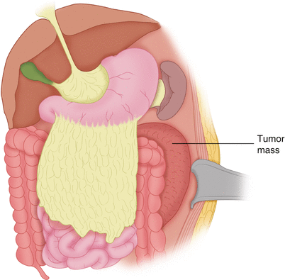
In the case illustrated, the tumor mass is located behind the splenic flexure of the colon (Fig. 38.1). The greater omentum is mobilized off the colon to allow entry into the lesser sac (Fig. 38.2). Alternatively, the greater omentum can be left attached to the transverse colon, and one may enter the lesser sac by dividing the gastrocolic ligament. The peritoneum is incised laterally so as to enter the space between the mass and the abdominal wall musculature on the lateral side (Fig. 38.2). The incision in the parietal peritoneum is continued lateral to and behind the palpable retroperitoneal mass in a caudal direction (Fig. 38.3), exposing the area of the iliac crest, the quadratus lumborum and psoas medially, and the beginning of the iliacus muscle inferiorly (Fig. 38.4). Superiorly, the incision is continued around the lienorenal ligament, which allows mobilization of the spleen and the medial and forward displacement of the spleen and the tail of the pancreas (Fig. 38.5). The surface of the aorta is exposed, and the left renal vein is visualized (Fig. 38.6). The inferior mesenteric artery issuing from the lower abdominal aorta is visualized and preserved. The plane between the lower portion of the retroperitoneal tumor and the lateral wall of the abdominal aorta is dissected, ligating and dividing any lumbar branches that may be encountered. The transverse colon is divided at about its middle and the descending colon at its junction with the sigmoid, leaving the splenic flexure of the colon attached to the area of the tumor, to which it was fairly adherent in this patient. A small bowel loop that was also adherent to the tumor mass is divided proximal and distal to the area of its attachment to the tumor, and the mesentery and the branches corresponding to this loop are serially ligated and divided (Fig. 38.5). The left renal vein may be divided between vascular clamps, and the two stumps sutured with vascular sutures. Immediately afterwards, the left renal artery is dissected, ligated, and divided (Fig. 38.7). In general, however, it is preferable to first expose and divide the ureter below the area of any involvement by the tumor of the kidney and perirenal tissues. Then the tumor may be mobilized posteriorly and laterally off the surface of the quadratus lumborum muscle and more medially off the psoas. The plane between the inferior pole of the tumor mass and the lower abdominal aorta is developed, and the tumor mass is mobilized medially and anteriorly. Superiorly, the adrenal gland is also mobilized for en bloc removal with the tumor and the kidney. The left renal artery is first found posteriorly, ligated, and divided; then one proceeds with the ligation and division of the renal vein. This method is safer not only in terms of control of the operative field, but also because ligation first of the renal artery and then of the vein prevents the engorgement of the kidney with blood, which actually results in some blood loss within the kidney if the vein is ligated and divided before the artery. The specimen is then removed (Fig. 38.8). Anastomosis is performed between the two ends of the small bowel and between the two stumps of the large bowel (Fig. 38.9). The abdominal organs, including the spleen and the tail of the pancreas, are placed again in their position. The incision is closed in layers. A Jackson-Pratt (JP) drain (or often two) is placed to drain serosanguinous fluid that is bound to accumulate for the first few postoperative days. The postoperative course is usually unremarkable. This case illustrates the possibility of dissecting and mobilizing the spleen and tail of the pancreas, bringing them to the surface of the skin without injuring their blood supply, if these organs are not involved by the tumor lying posterior to them.

