(1)
State University of New York at Buffalo Kaleida Health, Buffalo, NY, USA
Tumors occurring in the dorsal aspect of the foot preclude the possibility of limb-preserving resection unless they are small, located superficially, and do not extend into the surface of the bone. Conservative surgery is combined with neoadjuvant intra-arterial chemotherapy, systemic chemotherapy, or both, preoperative radiation or adjuvant postoperative radiation therapy. Tumors occurring in the plantar surface of the foot are more common, however, probably because of the larger amount of tissue available for the development of such tumors in the plantar surface compared with the dorsal aspect. In the sole of the foot, the superficial muscle layer consists of the abductor hallucis, flexor digitorum brevis, and abductor digiti minimi. These muscles can be removed without any obvious functional difficulty for the patient.
The following illustrations depict surgery for a soft-tissue tumor involving the superficial layer of the muscles of the foot on the plantar aspect. An incision widely circumscribing the previous biopsy site is made, and flaps are developed to include the tumor with a wide margin (Fig. 52.1). In the margins of the intended resection, the plantar fascia is incised posteriorly. The abductor hallucis, flexor digitorum brevis, and abductor digiti minimi are divided at their origin from the calcaneus and distally near their insertions (Fig. 52.2). The plane between this layer and the layer of the long flexors of the foot appeared to be free of tumor, allowing a resection of the specimen (Fig. 52.3). A skin graft was applied. In the removed specimen, there was a tumor to within 2 mm from the deep margin. The patient was given adjuvant radiation to a total of 60 Gy and remained free of tumor on long-term follow-up, but was suffering from pain in the plantar aspect of the foot on ambulation. Radiation of tumor on the plantar aspect of the foot should be given with caution, however, because the combination of surgery and radiation can be the source of chronic, disabling pain when the patient steps on this weight-bearing area.
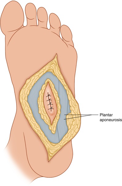
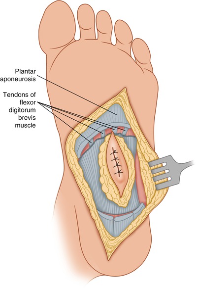
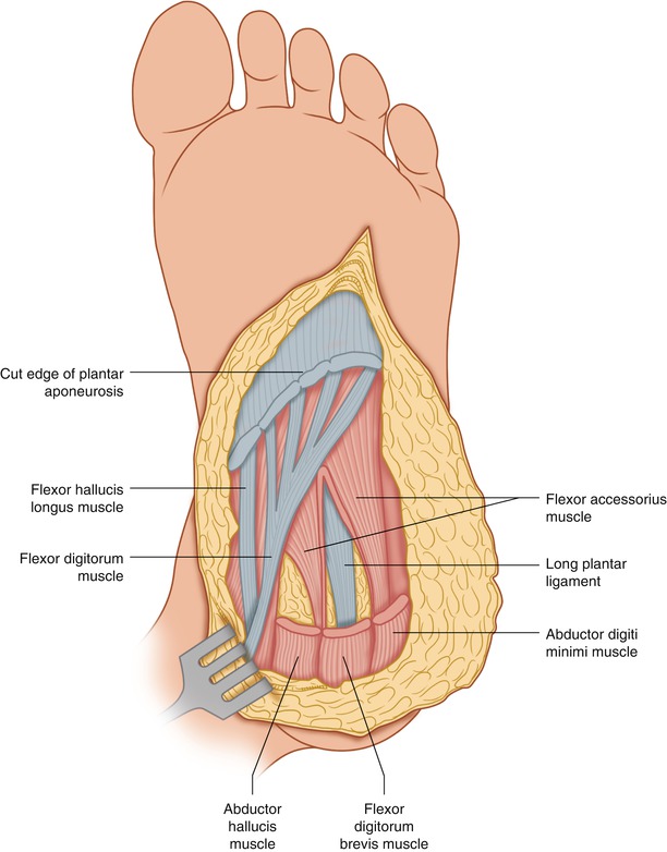
Fig. 52.1
Dissection around the previous biopsy incision exposes a large area of the plantar aponeurosis
Fig. 52.2
The plantar aponeurosis is incised distally, exposing the tendons of the flexor digitorum brevis
Fig. 52.3
The plantar aponeurosis and the underlying abductor hallucis, flexor digitorum brevis, and abductor digiti minimi have been removed en bloc with the tumor, exposing the second layer of muscles of the plantar aspect of the foot
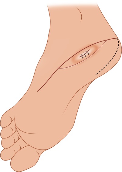
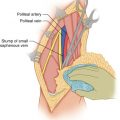
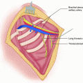
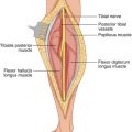
In another patient, tumor involving the lateral posterior aspect of the plantar area was managed with an elliptical incision around the previous biopsy site, which came around the heel to the medial aspect of the foot (Fig. 52.4). A flap was developed with a wide base, obtaining its blood supply from the distal and medial areas of the foot. The superficial layer of the muscles of the sole (i.e., the abductor digiti minimi, flexor digitorum brevis, and abductor hallucis) are exposed (Fig. 52.5) and divided (Fig. 52.6). On the medial aspect of the foot, the tendons of the flexor hallucis longus and the flexor digitorum longus were not involved and were preserved while the flexor accessorius muscle was exposed near its insertion to the flexor digitorum tendon (Fig. 52.7). It was divided while the peroneus longus muscle laterally was exposed (Fig. 52.8) but could also be divided and removed if adjacent to the tumor. The long plantar ligament was divided, and a wedge of the calcaneus was removed with an osteotome in order to adequately resect the gross tumor (Fig. 52.9). This particular flap, based distally, allows complete exposure of the area of the heel and has sufficient blood supply for its survival.