(1)
State University of New York at Buffalo Kaleida Health, Buffalo, NY, USA
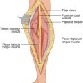
The anterior compartment of the arm consists of three muscles, the biceps brachii, brachialis, and coracobrachialis. The short head of the biceps has as its origin the coracoid process, arising lateral to the origin of the coracobrachialis and joining the long head arising from the supraglenoid tubercle to form the biceps brachii inserting in the radial tuberosity and the bicipital aponeurosis. The brachialis originates from the lower half of the anterior humeral surface and inserts in the ulnar tuberosity. The coracobrachialis originates from the coracoid process medial to the short head of the biceps and inserts in the medial aspect of the middle humerus.
The biceps is a flexor of the elbow and supinator of the forearm, the brachialis is a flexor of the elbow, and the coracobrachialis flexes the arm anteromedially (Fig. 7.1). The nerve supply is from the musculocutaneous nerve, which arises from the lateral cord in the axilla and deviates laterally from the median nerve, traversing obliquely and supplying the coracobrachialis (Fig. 7.2). It then courses in the plane between the biceps and brachialis, supplying both and emerging at the lateral border of the biceps as the lateral, antebrachial, cutaneous, terminal branch of the musculocutaneous nerve, which proceeds under the investing fascia and provides sensory supply to the lateral aspect of the forearm.
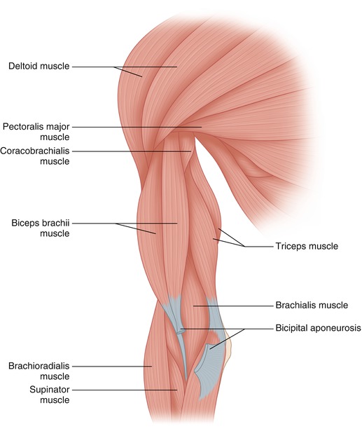
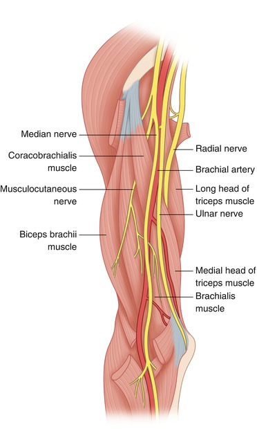
Fig. 7.1
The relationships proximally of the deltoid, the tendon of insertion of the pectoralis major at the crest of the greater tubercle of the humerus, the two heads of the biceps brachii and coracobrachialis are exposed. Distally, the relationship of biceps and brachialis is shown
Fig. 7.2
The tendon of insertion of pectoralis major to the humerus has been divided. The musculocutaneous nerve is visible in the distal axilla as it traverses and supplies the coracobrachialis, further supplying the biceps and brachialis
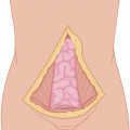
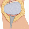
For a tumor in the anterior compartment, a longitudinal incision is used, circumscribing any previous biopsy tract with an ellipse. The length and placement of the incision depends on the location and extent of the tumor. For a tumor extending proximally, the incision may be extended to the deltopectoral groove, and the insertion of the pectoralis major to the humerus may be divided (it can be resutured at the end of the operation), exposing thus the neurovascular bundle, which courses in front of the insertion of the latissimus dorsi and teres major, leaving the axilla at the distal border of this insertion. The axillary nerve issues from the posterior cord of the brachial plexus and courses transversely at the level of the surgical neck of the humerus through the quadrangular space, providing the nerve supply to the deltoid. The quadrangular space, bound by the teres minor and major, the long head of triceps, and the surgical neck of the humerus, carries not only the axillary nerve but also the posterior humeral circumflex vessels. The radial nerve, a terminal branch of the posterior cord, proceeds obliquely, inferiorly, and laterally in the radial groove of the humerus. It supplies the triceps and emerges in the plane between the brachioradialis and brachialis, supplying the brachioradialis and the extensor carpi radialis longus and brevis; in front of the elbow, it divides into a superficial sensory branch and a deep branch (posterior interosseous nerve), which traverses the supinator and enters the posterior compartment, supplying the dorsal muscles of the forearm.
Related posts:
Stay updated, free articles. Join our Telegram channel

Full access? Get Clinical Tree


