(1)
State University of New York at Buffalo Kaleida Health, Buffalo, NY, USA
Tumors at the shoulder point arising in the subcutaneous tissue are apt to be detected when they are small because of their prominence and accessibility to self-inspection and palpation. The acromioclavicular arch and the adjacent muscles may be removed, providing some margin around the tumor, without causing significant interference with the shoulder joint and range of motion.
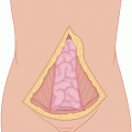
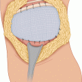
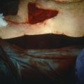
Small tumors in this area can be removed with adequate surgical margins by dividing through the free portion of the spine of the scapula posteriorly and the lateral third of the clavicle anteriorly, so that the distal portion of the clavicle can be removed with the acromioclavicular joint and the acromion. The case illustrated is that of a patient who had an incisional biopsy of a small sarcoma in the area of the shoulder (Fig. 13.1). An elliptical incision around the previous biopsy incision is made, and flaps are developed until a sufficient distance has been obtained from the area occupied by the tumor. Anteriorly, the clavicle is exposed at its lateral third; posteriorly, the acromion is exposed where it issues from the spine of the scapula (Fig. 13.2). A wide curving incision around the tumor mass in the deltoid muscle is made from the point where the clavicle has been exposed, to the spine of the scapula posteriorly, where the base of the acromion is exposed. A Gigli saw is passed around the clavicle (Fig. 13.3). The trapezius muscle is divided at a sufficient distance from the tumor along the clavicle and acromion opposite the deltoid, exposing the supraspinatus muscle between the clavicle and the spine of the scapula and the infraspinatus below the spine of the scapula. The clavicle is divided laterally (Fig. 13.4).
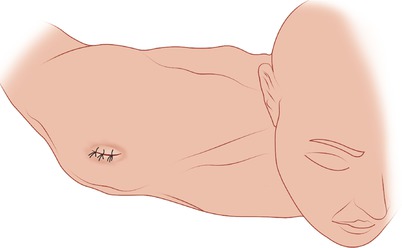
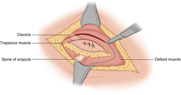

Fig. 13.1
An incisional biopsy has been obtained from a tumor at the right shoulder

Fig. 13.2


Flaps have been developed, exposing the deltoid muscle, the clavicle anteriorly, and the crest of the spine of the scapula posteriorly. The fascia covering deltoid is incised widely around the tumor mass
Stay updated, free articles. Join our Telegram channel

Full access? Get Clinical Tree


