Tuberculosis


Tuberculosis


C Scott Mahan MD

John L Johnson MD


Introduction

Tuberculosis (TB) is a chronic necrotizing granulomatous disease caused by Mycobacterium tuberculosis and, occasionally, by two closely related species, M. bovis and M. africanum. The global toll of TB is staggering. TB disproportionately affects poor, malnourished, and immunocompromised persons (Fig. 4.1, Table 4.1). Pulmonary disease is the most common manifestation of TB, but any organ can be involved including the lymph nodes, pleura, bones, joints, and central nervous system.


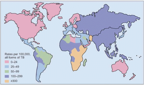
Fig. 4.1 Estimated worldwide tuberculosis incidence rate 2004. (Adapted from WHO data.)


Etiology and pathogenesis

TB is caused by the M. tuberculosis complex, which includes M. tuberculosis, M. bovis, M. africanum, M. microti, and M. canetti; M. tuberculosis is responsible for the most disease in humans. M. tuberculosis is an obligate aerobic, non-motile bacillus that stains acid fast, meaning it retains a deep red tint after staining with carbol-fuchsin followed by washing with acid-alcohol (Fig. 4.2). Humans are the only reservoir for M. tuberculosis.


Table 4.1 Persons at increased risk for developing active tuberculosis


Human immunodeficiency virus-infected


Elderly


Immunosuppressed persons and persons receiving corticosteroids or other immunosuppressive therapy or cancer chemotherapy


Comorbidities such as diabetes mellitus, silicosis, alcoholism, and chronic renal insufficiency


Institutionalized persons in jails, prisons, nursing homes, and chronic care facilities


Health care workers


Close contacts of infectious cases of tuberculosis


Malnourished persons


Prior arrested tuberculosis as evidenced by apical scarring or fibrosis on chest radiography


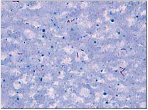
Fig. 4.2 Ziehl-Neelsen stain of sputum sample showing red acid-fast bacilli in a patient with cavitary pulmonary tuberculosis.

Tuberculosis is spread when an infected person coughs, sneezes, or speaks creating small (1-5 µm) aerosolized droplets of bacilli that can be inhaled by another person and deposited in the new host’s alveoli. Once tubercle bacilli reach the alveoli, several different events can occur: local tissue macrophages may kill the organism and clear it before infection is established; alternatively, the bacteria may enter the macrophages, multiply, and spread via the lymphatics and the bloodstream throughout the body. Over 90% of immunocompetent individuals are able to mount an effective cell-mediated immune response and contain the initial infection, leaving only small parenchymal scars, a Ghon focus (Fig. 4.3), or no evidence of infection except for a positive tuberculin skin test. Persons who fail to mount an adequate immune response may develop progressive primary TB. Illustrative cases are shown in Figs. 4.2, 4.3 and 4.4.


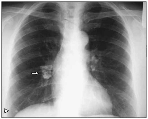
Fig. 4.3 A calcified Ghon focus is visible in the right lower lung field with associated hilar adenopathy. The combination of a Ghon focus (arrowhead) and a calcified draining lymph node is known as a primary (Ranke) complex (arrow). These radiographic findings are consistent with resolution of primary tuberculosis infection. (Courtesy of Dr Catherine Curley.)


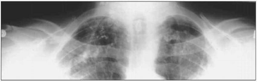
Fig. 4.4 Chest radiograph showing apical scarring due to arrested tuberculosis (Simon’s foci) in an asymptomatic 55-year-old male.


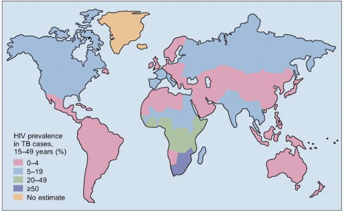
Fig. 4.5 Estimated prevalence rates of human immunodeficiency virus coinfection in patients with tuberculosis worldwide, 2004. (Adapted from WHO data.)


Global epidemiology

TB is a major global health problem that has been exacerbated by poverty, poor public health infrastructure, the acquired immunodeficiency syndrome (AIDS) epidemic and the emergence of multidrug resistant TB. Worldwide, the number of TB cases increased by 1.8% annually between 1997 and 2000. The World Health Organization (WHO) estimates that one-third of the world’s population is infected with TB. In 2005, over 9 million new cases of TB and more than 2 million deaths due to TB occurred worldwide. Ninety-five percent of all TB cases and 98% of all deaths due to TB occur in persons living in developing countries. Conditions of intense overcrowding, inadequate sanitation, malnutrition, and lack of access to medical care all contribute to the epidemic.

Human immunodeficiency virus (HIV)/AIDS and TB are closely linked. HIV coinfection is the greatest risk factor known for the progression of latent TB infection to active TB (Fig. 4.5). Increasing rates of HIV coinfection in countries in sub-Saharan Africa have led to rapid increases in the incidence of TB despite the introduction of more effective TB treatment strategies. TB is the most frequent serious opportunistic infection in adults with AIDS worldwide.


Clinical manifestations


Primary pulmonary TB

Primary TB is the development of active TB soon after infection with the tubercle bacillus in the non-immune host. Most healthy individuals are asymptomatic after TB infection, and the event is marked only by the development of a positive tuberculin skin test (TST) 4-6 weeks after infection. Infants and children are more likely than adults to develop active TB after infection. About 10% of infants and children develop symptomatic primary TB heralded by mild fever, non-productive cough, and malaise. Radiologic manifestations may include hilar or mediastinal lymphadenopathy, mid- or lower-lung infiltrates, and transient pleural effusions.

Among immunocompetent adolescents and adults infected with TB, only 5-10% will develop active disease, with roughly one-half of cases occurring during the first 2 years after infection. Illustrative cases are shown in Figs. 4.6, 4.7 and 4.8.


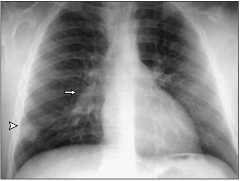
Fig. 4.6 Chest radiograph of a 4-year-old child presenting with low-grade fever and non-productive cough. There is a visible parenchymal infiltrate (Ghon focus) in the right lower lung field (arrowhead) and associated right hilar lymphadenopathy (arrow). The primary (Ranke) complex consists of the parenchymal lesion and the associated enlarged ipsilateral lymph nodes. (Courtesy of Dr Charles Daley.)


Reactivation TB

As noted earlier, most immunocompetent individuals are able to contain their initial infection with TB. Although these individuals have an effective immune response to initial infection, small numbers of viable, slow growing bacilli remain that are walled off in granulomas (Fig. 4.9). Later in life, during periods of waning immunity, these walled off lesions can break down and lead to local and disseminated disease.

The most frequent symptoms of reactivation TB in the adult include fever, anorexia, productive cough (with or without hemoptysis) for more than 3 weeks, night sweats, and malaise. Typical chest radiographic findings include upper lobe fibrocavitary lesions and infiltrates classically involving the apical and posterior segments of the upper lobes followed in frequency by involvement of the superior segments of the lower lobes. Atypical radiographic manifestations of TB in the adult include lower lobe infiltrates similar to pyogenic pneumonia and intrathoracic adenopathy. Lower lobe TB is more frequent in patients with diabetes mellitus. Illustrative cases are shown in Figs. 4.10, 4.11, 4.12, 4.13 and 4.14.


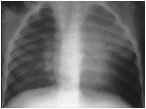
Fig. 4.7 Tuberculous pneumonia with left upper lobe infiltrate in a 4-year-old with fever and progressive primary tuberculosis. (Courtesy of Dr Charles Daley.)


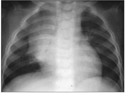
Fig. 4.8 Lobar collapse due to extrinsic bronchial compression from enlarged right hilar lymph nodes and endobronchial tuberculosis in a 5-year-old child with fever and chronic cough. (Courtesy of Dr Charles Daley.)


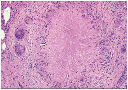
Fig. 4.9 Pulmonary granuloma (H&E, x100). Notice the area of central caseation (arrow) with a surrounding rim of lymphocytes, mononuclear cells, multinucleated giant cells, and fibrosis.


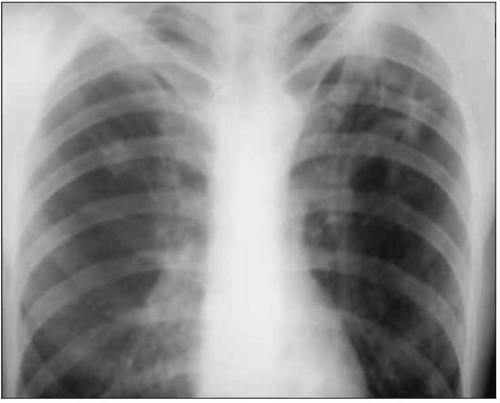
Fig. 4.10 Left upper lung cavity in patient with reactivation tuberculosis who presents with fever, productive cough, and weight loss.


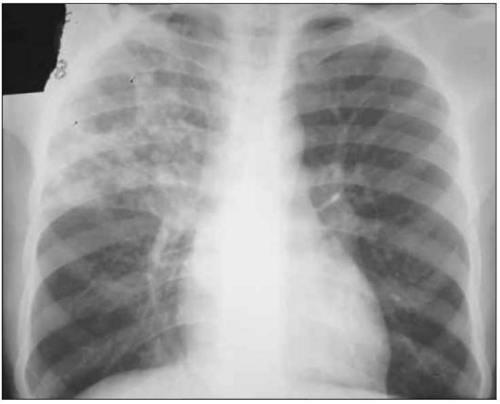
Fig. 4.11 A chest radiograph from a 38-year-old male with 1 month of fever, weight loss, and productive cough. His chest radiograph shows a right upper lobe cavitary lesion with spreading throughout the right upper lobe and into the right lower lobe. This is an example of how caseous material and tubercle bacilli from an open cavitary lesion can spread endobronchially to other areas of the lung.


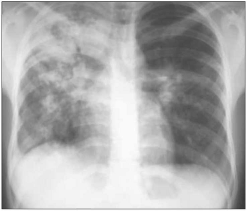
Fig. 4.12 Chest radiograph of a 48-year-old male presenting with wasting and a productive cough. It shows right upper lung involvement with a cavitary lesion and an air bronchogram.


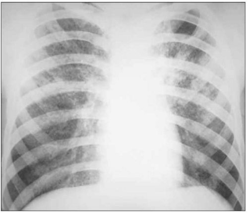
Fig. 4.13 Chest radiograph of a 34-year-old male with intermittent fever, weight loss, and no pulmonary symptoms. He was found to have miliary tuberculosis. Although the chest radiograph may be normal in a minority of cases, it usually shows classic miliary lesions, which are diffuse 1-2 mm rounded opacities (similar in size to millet seeds) scattered throughout all lung fields. Sometimes these lesions are best seen on a lateral view. Miliary tuberculosis is due to lymphatic and hematogenous seeding of tubercle bacilli to all areas of the lungs and other organs.


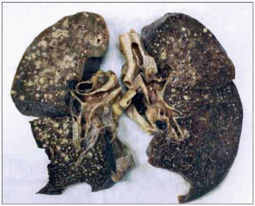
Fig. 4.14 Gross view of lungs from a fatal case of miliary tuberculosis. (Courtesy of Dr Rosana Eisenberg.)


TB in HIV/AIDS and the immunocompromised

Persons infected with HIV are at an 80-100-fold increased risk for active TB after infection by the tubercle bacillus. In the immunocompetent individual, the risk of reactivation of latent TB is about 10% over the course of their lifetime; however, in an HIV-positive individual, the risk is about 8-10% per year. Early in the course of HIV, when host defenses are less impaired, patients usually present with typical upper lung field fibrocavitary disease, similar to reactivation TB in HIV-uninfected adults. In advanced HIV/AIDS (CD4+ count <200/mm3), patients are more likely to present atypically with non-cavitary lower lobe infiltrates or extrapulmonary or disseminated TB. In addition, sputum acid-fast bacilli (AFB) smears are more likely to be negative in patients with advanced HIV/AIDS. Table 4.2 presents the clinical and laboratory manifestations of active TB in early and late infection with HIV. Illustrative cases are shown in Figs. 4.15, 4.16, 4.17 and 4.18.


Table 4.2 Clinical and laboratory manifestations of active tuberculosis in early and late infection with human immunodeficiency virus


Early (CD4+ >200/mm3)


Late (CD4+200/mm3)


TST


Usually positive (5 mm or greater induration)


Usually negative


Adenopathy


Uncommon


Common


Affected lung areas


Upper lobes


Lower and middle lung fields


Cavitary disease


Frequent


Uncommon


Extrapulmonary disease


15%


30-50%


Sputum AFB smear


60% positive


40% positive


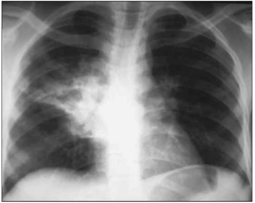
Fig. 4.15 Chest radiograph of a 28-year-old male with advanced acquired immunodeficiency syndrome (CD4+ count of 80/mm3) presenting with pleuritic right-sided chest pain and productive cough. A right mid-lung field infiltrate and associated hilar adenopathy are visible.


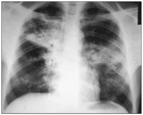
Fig. 4.16 Multilobar disease with associated hilar adenopathy in a patient with advanced human immunodeficiency virus/acquired immunodeficiency syndrome.


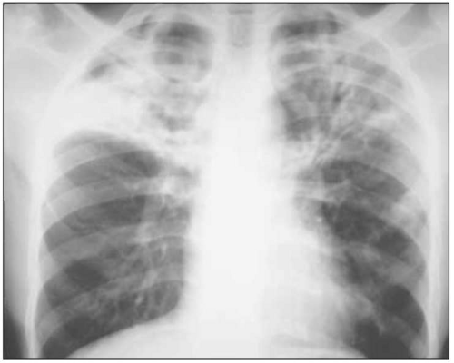
Fig. 4.17 Chest radiograph of a 35-year-old HIV-infected man with classic upper lobe fibrocavitary disease. Chest radiographic findings in patients with early HIV (CD4+ >200/mm3) are similar to findings in HIV-uninfected persons.


Immune reconstitution inflammatory reactions

Patients starting on antituberculous chemotherapy may develop acute worsening of their symptoms or existing tuberculous lesions or new lesions; these are termed paradoxical or immune reconstitution inflammatory syndrome (IRIS) reactions. IRIS reactions occur more frequently in HIV-infected patients with TB (7-36% of patients) and are more common when highly active antiretroviral therapy (HAART) and anti-TB treatment are started at the same time. The cause of IRIS reactions is unclear, but they may be due to reconstitution of the host immune response and an increased inflammatory response to mycobacterial antigens after beginning HIV and anti-TB therapy. Most occur within 4-12 weeks after starting HAART. IRIS reactions are more common in patients with extrapulmonary TB or low CD4+ lymphocyte counts. New or worsening adenopathy, fever, and new pulmonary infiltrates or pleural effusion are the most common presentations. After excluding other opportunistic infections, non-adherence with TB treatment, and drug resistant TB, management is by treatment of symptoms. Adjunctive corticosteroids may be helpful in severe cases. HAART therapy can usually be continued. Illustrative cases are shown in Figs. 4.19, 4.20 and 4.21.

Only gold members can continue reading. Log In or Register to continue

Stay updated, free articles. Join our Telegram channel

Aug 1, 2016 | Posted by in INFECTIOUS DISEASE | Comments Off on Tuberculosis

Full access? Get Clinical Tree

Get Clinical Tree app for offline access