Emerging viral respiratory illnesses


Emerging viral respiratory illnesses


Nandhitha Natesan MD

Rana B Hejal MD


Introduction

Respiratory tract infections remain a major cause of morbidity and mortality throughout the world. A number of viruses are known to infect the respiratory tract producing different clinical syndromes (Table 3.1). Recently, several previously unrecognized viruses have come to the forefront of both media and scientific attention as important causes of acute respiratory illnesses. In this chapter, we will focus on three of these newer agents; namely, severe acute respiratory
syndrome-associated coronavirus (SARS-CoV), human metapneumovirus (hMPV) and a novel strain of avian influenza A (H5N1).


Table 3.1 Respiratory viral infections and corresponding clinical syndromes in the immunocompetent host


Clinical syndromes


Virus


Common cold


Pharyngitis


Croup


Bronchiolitis


Pneumonia


Adenoviruses


Rare


Uncommon


Rare


Uncommon


Uncommon


Coronaviruses


Group I-III


Common


Rare


Rare


Rare


Uncommon


SARS-CoV


Rare


Rare


Rare


Rare


Very common


Herpesviruses


Cytomegalovirus


Rare


Uncommon


Rare


Rare


Uncommon


Epstein-Barr virus


Rare


Common


Rare


Rare


Uncommon


Herpes simplex virus


Rare


Common


Rare


Rare


Uncommon


Varicella-zoster virus


Rare


Rare


Rare


Rare


Uncommon


Orthomyxoviruses


Influenza A, B, C


Uncommon


Common


Uncommon


Uncommon


Common


Paramyxoviruses


Measles


Rare


Rare


Rare


Rare


Uncommon


Parainfluenza 1,2,3


Uncommon


Common


Very common


Common


Rare


Respiratory syncytial virus


Rare


Uncommon


Common


Common


Common


Metapneumovirus


Rare


Uncommon


Common


Very common


Common


Picornovirus


Enterovirus


Uncommon


Rare


Rare


Rare


Rare


Rhinovirus


Very common


Common


Uncommon


Uncommon


Rare


Fig. 3.1 Summary of SARS cases (A) and deaths (B) by country. (Adapted from WHO data.)


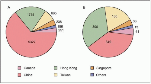
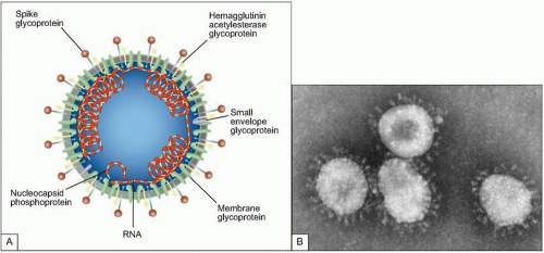
Fig. 3.2 A: Structure of coronavirus virion. (Adapted from Holmes KV. SARS-associated coronavirus. N Engl J Med 2003; 348(20):1948-1951.) B: The negative stain electron micrograph of coronavirus. Coronaviruses have a halo, or crown-like (corona) appearance when viewed under electron microscopy. (Courtesy of CDC.)


SARS coronavirus


Background

In late 2002, an outbreak of severe, atypical pneumonia began in southern China. Within months, the illness named SARS had affected more than 8,000 patients in 29 countries, leading to more than 900 fatalities worldwide (Fig. 3.1). Led by the World Health Organization (WHO), a massive global collaboration was undertaken to identify the pathogen and clinical and epidemiologic spectrum of this syndrome in order to arrest the spread of a threatened pandemic.


Table 3.2 Spectrum of coronavirus species, hosts, and corresponding clinical syndromes


Host


Clinical syndrome


Group I


Human respiratory coronavirus


HCoV-229E


Human


Colds, pneumonia


Human coronavirus-Netherlands


HCoV-NL


Human


Colds, pneumonia


Porcine transmissible gastroenteritis virus


TGEV/PRCoV


Pig


Enteric, pneumonia


Canine respiratory coronavirus


CCoV


Dog


Enteric


Feline enteric coronavirus


FECoV


Cat


Enteric


Feline infectious peritonitis virus


FICoV


Cat


Systemic, peritonitis


Rabbit coronavirus


RbCoV


Rabbit


Enteric


Group II


Human respiratory coronavirus


HCoV-OC43


Human


Colds, pneumonia


Bovine respiratory coronavirus


BCoV


Cow


Enteric, pneumonia


Hemagglutinating encephalitis virus


HEV


Pig


Respiratory, enteric, neurologic


Rat coronavirus


RCoV


Rat


Respiratory


Sialodacryoadenitis virus


SDAV


Rat


Neurologic


Murine hepatitis virus


MHV


Mouse


Hepatitis, encephalitis


Group III


Avian bronchitis virus


IBV


Chicken


Tracheitis, hepatitis, renal


Turkey respiratory coronavirus


TCoV


Turkey


Respiratory, enteric


?Group IV


Severe acute respiratory syndrome-associated coronavirus


SARS-CoV


Human


Pneumonia, enteric


Virology

Coronaviruses are large enveloped, single stranded, positivesense ribonucleic acid (RNA) viruses that cause the ‘common cold’ in humans as well as a variety of illnesses in other species (Table 3.2). They have been divided into three groups based on serologic cross-reactivity, but more recently on genomic sequence homology. When the SARS-CoV genome was identified, it appeared similar to other coronaviruses in its organization, but quite different in phylogenic analysis and sequence comparisons, making it
likely the representative of a new fourth group of coronaviruses (Fig. 3.2). In fact the sequence analysis suggested that this new virus is an animal virus that has gained the ability to cross the species barrier. The SARS-CoV has four major proteins: the envelope (E), membrane (M), spike (S), and nucleocapsid protein (N). It is the S protein that binds to a species-specific host cell and along with the host’s immune responses determines the virulence of the organism. Several antigenically diverse strains were identified in China; however, only one went on to be responsible for the major outbreak and global spread of SARS.


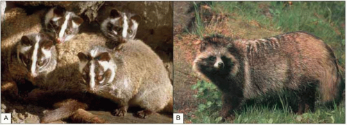
Fig. 3.3 Exotic animals regarded as a delicacy in Guangdong, China believed to harbor SARS-CoV. A: Masked palm civets (Paguma larvata). B: Raccoon dog (Nyctereutes procyonoides).


Epidemiology

The SARS-CoV is believed to be of animal origin, and to have spread from animals to humans in crowded, open air, live animal markets, which are common in east Asia. Precursors of this virus with 99% homology have been identified in several species of exotic animals sold at these markets, including civets and raccoon dogs (Fig. 3.3). It remains unclear whether these animals represent the natural reservoirs of this virus, or serve as amplifiers. The principle mode of transmission is thought to be close person-to-person contact with exposure to infected body secretions, including respiratory droplets, infected fomites, feces, and other bodily fluids.

SARS emerged in the Guangdong province of China in November 2002. By January 2003, multiple independent outbreaks had occurred within this province. A physician who had been in the Guangdong province, caring for SARS patients, returned to Hong Kong where he seeded at least 17 guests at a hotel (Fig. 3.4). Many of these guests traveled out of the country in the following days, and were responsible for the spread of this virus to Vietnam, Singapore, and Canada.

The unexplained respiratory illness was first reported to the WHO in February of 2003. Standard public health measures, along with advanced screening and communication measures, were quickly implemented. A virtual network was established, with daily teleconferences enabling real time sharing and application of information. In addition, global health alerts and travel advisories were issued and updated regularly. Many countries implemented screening for elevated body temperatures and quarantines in airports. In late March, three independent labs identified a novel coronavirus from patients with SARS from different countries. The identified virus fulfilled the six Koch’s postulates, as modified by Rivers for pathogenic viruses (Table 3.3). On April 16, 2003 WHO announced this new virus to be the definitive cause of SARS. By July of 2003, human-to-human spread of this virus was under control leading to the successful averting of the threatened pandemic.

SARS-CoV largely affects adults. A disproportionate
number of cases were in health care workers (greater than 40% of cases in China and Canada). The global average case fatality rate was just over 10% (Fig. 3.5); however, for patients requiring intensive care and/or mechanical ventilation, fatality rates greater than 50% were reached. Predictors of mortality include increasing age, presence of comorbidities, atypical initial symptoms, and some abnormal laboratory findings (Table 3.4).


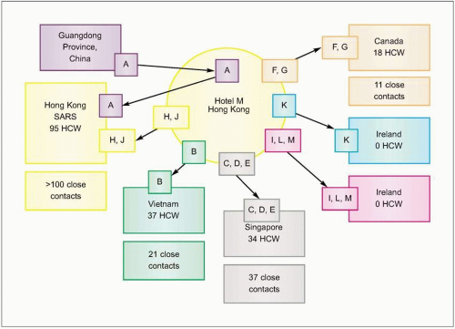
Fig. 3.4 Effect of travel and missed cases on the SARS epidemic. Spread from Hotel M, Hong Kong. Case A from Guangdong Province, China and two hotel guests who became ill, cases H and J, started outbreaks of SARS in three Hong Kong hospitals involving at least 95 health care workers (HCW) and more than 100 contacts. (Adapted from CDC data.)


Table 3.3 Koch’s postulates as modified by Rivers for viruses


Isolation of the virus from each of the suspected cases


Cultivation of the virus in host cell


Proof of filterability


Production of a comparable disease in related species


Re-isolation of the same organism


Detection of specific host immune response to the virus


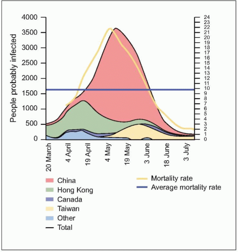
Fig. 3.5 The evolution of the people probably infected, by main countries (moving average of 7 days) and the mortality rates for the last 2 weeks. People probably infected = cumulative case x number of deaths x number of people discharged. Mortality rate = deaths / (deaths + discharged). (Adapted from WHO data.)


Table 3.4 Risk factors for death or admission to an Intensive Care Unit


Risk factor


Relative risk
or odds ratio


95% CI


Author


Age >60 years


1.8


1.16-2.81


Lee N, et al., 2003


28


3.1-253.3


Peiris J, et al. (A), 2003


3.5


1.2-10.2


Chan JWM, et al., 2003


1.57


1.26-1.95


Tsui PT, et al., 2003


19.9


11.7-33.8


Leung GM, et al., 2004


Diabetes mellitus


3.1


1.4-7.2


Booth CM, et al., 2003


9.1


2.8-29.1


Chan JWM, et al., 2003


Comorbid illness


2.5


1.1-5.8


Booth CM, et al., 2003


5.2


1.4-19.7


Chan JWM, et al., 2003


Increased neutrophil count


1.6


1.03-2.50


Lee N, et al., 2003


1.28


1.13-1.46


Tsui PT, et al., 2003


Increased lactate dehydrogenase


2.09


1.28-3.42


Booth CM, et al., 2003


1.35


1.11-1.64


Tsui PT, et al., 2003


2.3


1.4-3.8


Leung GM, et al., 2004


Clinical features

The vast majority of patients pass through three phases in this illness: exposure, early prodrome, and late respiratory
phase (Fig. 3.6). The incubation period varies between 2 and 10 days. Unlike many of the common viral respiratory pathogens, evidence suggests that there are rather few asymptomatic or mild illnesses associated with SARS-CoV, except for children, in whom the disease is uncommon, generally mild, and self limited. Typical adult illness begins with a prodrome of non-specific symptoms including high-grade fever, chills/rigors, myalgias, headache, and diarrhea (Fig. 3.7). Upper respiratory tract symptoms are less common. The clinical course tends to be insidious, and patients frequently improve transiently prior to developing lower respiratory symptoms in the second week of illness, with non-productive cough, dyspnea, and hypoxia. In 10-20% of hospitalized patients, symptoms progress to respiratory failure requiring intubation and mechanical ventilation. It is unclear whether this clinical deterioration is due to ongoing viral replication or uncontrolled immune response mediated by host defenses.


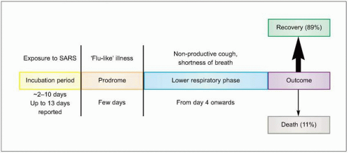
Fig. 3.6 The clinical phases of SARS.

Lymphocytopenia at presentation is common. Patients often go on to develop leukopenia and thrombocytopenia with the onset of respiratory symptoms. Elevated amino-transferases, creatinine kinase, and lactate dehydrogenase are also reported (Fig. 3.8). Moreover, these variables tend to worsen at different time points over the disease course (Fig. 3.9).


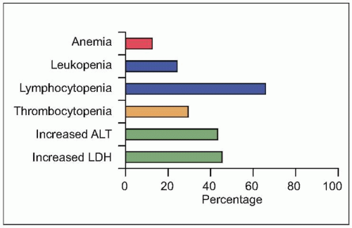
Fig. 3.8 Initial laboratory abnormalities in patients with SARS. (Data from close to 500 patients are compiled from several series, including Booth CM, et al., 2003; Hsu LY, et al., 2003; Lee N, et al., 2003; Peiris JS, et al. (B), 2003; Poutanen SM, et al., 2003; Tsang KW, et al., 2003; Zhao, Z, et al., 2003.) (LDH: serum lactate dehydrogenase level; ALT: serum alanine aminotransferase.)


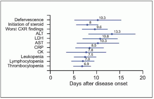
Fig. 3.9 The time relationships between the time points of defervescence, initiation of steroid, and when chest radiographic finding, as well as various laboratory parameters became most severe. Mean and standard deviation (days) are presented. (CXR: chest radiography; ALT: alanine aminotransferase; LDH: lactate dehydrogenase; AST: aspartate aminotransferase; CRP: C-reactive protein; CK: creatine kinase.) (Adapted from CDC data.)

Chest radiographs in SARS are usually abnormal (Table 3.5). Approximately 50% of patients present with unilateral focal consolidation. Lower lobe predominance is rather common. While patients presenting early can have a normal film initially, they uniformly develop some abnormality by the seventh day of their illness. In the setting of high clinical suspicion and normal chest X-ray, high resolution chest computed tomography (HRCT) may identify early parenchymal abnormalities and should be considered. Illustrative cases are shown in Figs. 3.10, 3.11 and 3.12.


Table 3.5 Radiographic features of severe acute respiratory syndrome


Chest radiograph


High resolution computed tomography


Normal, only early in course


Ill-defined ground-glass opacification


Peripheral alveolar infiltrates (most common):


Reticulation


Basilar predilection


Irregular interlobular septal thickening


Often multifocal


Subpleural reticulation


Nodular infiltrate (early)


Late manifestations (patients surviving respiratory failure):


Non-cardiogenic pulmonary edema


Pleural effusion rarely


Bronchiectasis


Pneumomediastinum


Honey-combing and fibrosis


Absence of:


Adenopathy


Cavitation

Only gold members can continue reading. Log In or Register to continue

Stay updated, free articles. Join our Telegram channel

Aug 1, 2016 | Posted by in INFECTIOUS DISEASE | Comments Off on Emerging viral respiratory illnesses

Full access? Get Clinical Tree

Get Clinical Tree app for offline access