HIV and AIDS


HIV and AIDS


Benigno Rodriguez MD

Robert A Salata MD, FACP, FIDSA


Introduction

The acquired immunodeficiency syndrome (AIDS) was first recognized in 1981, when a cluster of cases of uncommon opportunistic infections and malignancies was reported among otherwise healthy men who had sex with men in San Francisco, Los Angeles, and New York. Alert clinicians and immunologists recognized the unusual infections as indicative of a profound cellular immunodeficiency, a notion promptly confirmed by a diversity of laboratory assays. Alternative routes of acquisition, including parenteral, perinatal, and transfusion-associated were quickly identified, and further reports that an indistinguishable illness had been known for decades in sub-Saharan Africa began to emerge. Subsequent developments occurred at a remarkably fast pace, unprecedented for a novel infectious disease: the retrovirus now known as human immunodeficiency virus (HIV) was identified as the causative agent within 2 years of the first case reports by independent groups in France, Bethesda, and San Francisco; a serological test became available shortly thereafter; the genome was fully sequenced in 1985; and the first clinically usable therapeutic compound, zidovudine, became commercially available in 1987. Since then, combinations of drugs that act at different stages of the virus’ life cycle (see below), known as highly active antiretroviral therapy (HAART) have proven capable of suppressing viral replication to extremely low levels, and to restore, at least partially, the impaired cellular immune function that is ultimately responsible for the increased susceptibility to opportunistic infections in AIDS patients.

The HIV pandemic, however, continues virtually unabated, having spread to every continent, and to all demographic groups throughout the world. Moreover, no curative treatment is available, and predictions for the time to development of an effective, widely available, preventive vaccine are measured in decades. Thus, HIV infection and AIDS remain major health problems that concern virtually every practicing clinician, and the complexity of their management can only be expected to increase in coming years. This chapter focuses mostly on the clinically relevant aspects of HIV infection and AIDS. Excellent reviews of the biology, immunology, and virology of HIV have been published elsewhere.


Etiology and pathogenesis

HIV-1 is the etiologic agent of the majority of AIDS cases worldwide. A closely related agent, HIV-2, also causes AIDS in parts of West Africa; sporadic cases occur elsewhere. Throughout the remainder of this chapter, ‘HIV’ is used to refer to HIV-1, unless otherwise indicated. HIV is a member of the lentiviridae family with a plus-stranded ribonucleic acid (RNA) genome that encodes structural, regulatory and accessory proteins, as well as the enzymatic activities; the genomic organization of HIV is shown in Fig. 1.1, and the structure of the infective viral particle is shown in Fig. 1.2.

The hallmark of HIV infection is depletion of CD4+ helper T lymphocytes, with ensuing loss of immune competence. Many other immune defects are evident as HIV disease progresses, however, and not all of them can be readily accounted for by the loss of help associated with
CD4+ T cell destruction. Among these, defects in B cell proliferation and antibody production, impaired cytotoxic lymphocytic responses, decreased dendritic cell number and function, and profound perturbations of the cytokine milieu have all been shown, particularly in advanced stages of HIV infection. The precise mechanism by which HIV infection leads to these wide-ranging defects is incompletely understood, although they are related to HIV replication, and can be partially corrected by effective antiretroviral therapy that suppresses plasma viremia to very low levels. The vital cycle of HIV is complex and includes multiple steps that can be targeted for therapeutic purposes. These steps are summarized in Fig. 1.3.


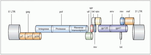
Fig. 1.1 The genomic organization of HIV. The complete genome is approximately 10 kb in size, and is similar to the general structure of other retroviruses. In the figure, the most relevant genes are represented in different colors, and the most important proteins they encode for are shown inside the corresponding symbols. Not all gene products are shown.


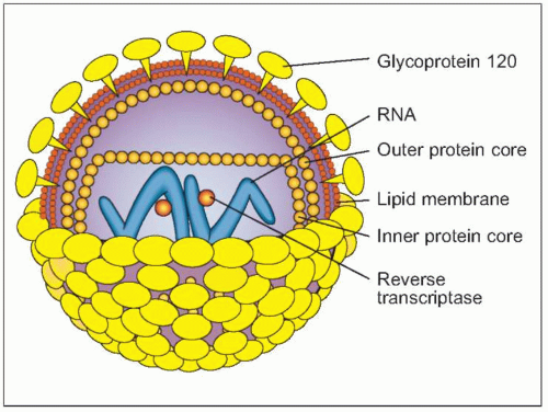
Fig. 1.2 Schematic view of HIV structure.

Active HIV replication is lytic to some, but not all infected cells. Because the predominant target of HIV is the CD4+ T cell, it has been proposed that direct destruction of these cells by HIV is the predominant mechanism of immunodeficiency in progressive HIV infection. More recent evidence, however, shows that the number and distribution of infected cells, the rate of CD4+ T cell turnover and the loss of large numbers of uninfected cells through indirect, or ‘bystander’, mechanisms do not support this model as the sole explanation for HIV-related immune deficiency. Moreover, studies in HIV-infected persons receiving clinical care show that the level of HIV viremia predicts poorly the subsequent rate of CD4+ T cell loss at the individual level, further highlighting that other, indirect mechanisms in effect lead to immunodeficiency in HIV infection. Uncontrolled immune activation is an additional feature of HIV infection that may underlie the CD4+ T cell loss and other immune derangements that eventually culminate in full-blown AIDS. Similarly, advanced HIV infection is associated with depletion of thymocytes and loss of thymic function, as well as impaired bone marrow activity, all of which limit the ability to restore the accelerated CD4+ T cell losses induced by HIV. The net result is a progressively increased susceptibility to a diversity of opportunistic complications that, in the era before the introduction of HAART, were almost invariably fatal within a short period after the initial diagnosis of AIDS.


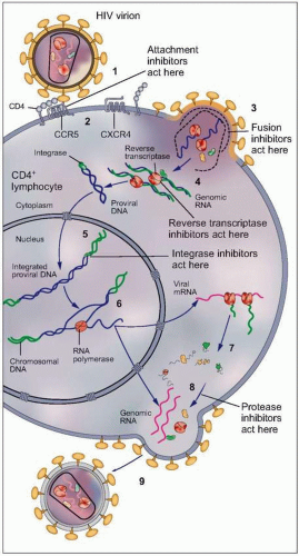
Fig. 1.3 Vital cycle of HIV and sites targeted by current anti-HIV medications. After HIV binds to its primary receptor, CD4 (1), the viral envelope undergoes a conformational change that facilitates binding to another cellular coreceptor, the most important of which are the chemokine receptors CCR5 and CXCR4 (2). Interaction with the coreceptor triggers further conformational changes in the envelope that bring the viral and cellular membranes into close proximity, thereby permitting their fusion (3) through insertion of the newly exposed fusion domain of the envelope protein gp41 into the host cell membrane. The HIV nucleocapsid then enters the cytoplasm, where the RNA genomic material of the virus is reverse transcribed into DNA (4) by the virally encoded reverse transcriptase. Next, the double-stranded viral DNA enters the nucleus, where it integrates into the host genome with the aid of the HIV-encoded enzyme integrase (5). The integrated proviral DNA is then transcribed into messenger RNA (6), which serves as the template for assembly of the main viral structural proteins (7). The protein complex is cleaved by a protease into functional segments (8), thus allowing assembly and budding of the new viral particles (9) to proceed.


Global epidemiology

Few human infections fit the description of an emerging disease better than HIV infection and AIDS. In the 25 years since its initial description, 75 million individuals worldwide have been infected with HIV, and the epidemic is now present throughout the world. The World Health Organization (WHO) estimates that, by the end of 2006, there were 39.5 million persons living with HIV/AIDS in the world, and 4.3 million acquired HIV in the previous year alone. Over 90% of these persons live in the developing world (62.5% in sub-Saharan Africa alone) and heterosexual intercourse is the route of acquisition in the vast majority of cases. Current estimates of the extent of the HIV epidemic worldwide are shown in Fig. 1.4.

In addition to the sheer number of cases, the HIV epidemic has changed dramatically over the past several years, leading to a truly re-emerging epidemiological pattern worldwide. Large epidemics are expanding rapidly in eastern Europe, Asia, and India, which is now the single country with the largest number of cases worldwide. Moreover, the proportion of cases occurring in women is increasing at an alarming pace. Close to 50% of all adults living with HIV/AIDS worldwide are women, and the proportion approaches 66% in parts of sub-Saharan Africa.

In the United States and western Europe, the introduction of HAART has produced dramatic reductions in HIV-related morbidity and mortality (Fig. 1.5), but trends in sex distribution are similar to those observed worldwide (Fig. 1.6). Furthermore, new cases are occurring disproportionately more often among minorities and disadvantaged populations, further changing the face of the epidemic.


Fig. 1.4 Global estimates of the HIV/AIDS epidemic at the end of 2006. (Adapted from UNAIDS and WHO, 2006 Report on the Global HIV epidemic, Geneva, UNAIDS, 2006.)


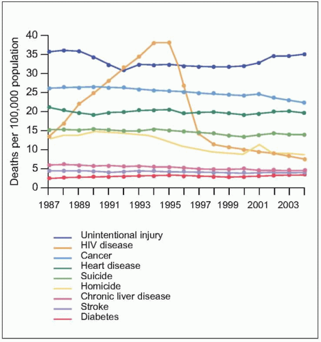
Fig. 1.5 Trends in annual rates of death due to the nine leading causes among persons 25-44 years old, USA, 1987-2004. HIV disease was the leading cause of death among person 25-44 years old in 1994 and 1995. With the introduction of HAART in 1995, the rank of HIV disease fell to 5th place from 1997 through 2000, and to 6th place in 2001 and 2002. The spike in the death rate due to homicide in 2001 resulted from the terrorist attack on September 11. (Adapted from CDC data.)


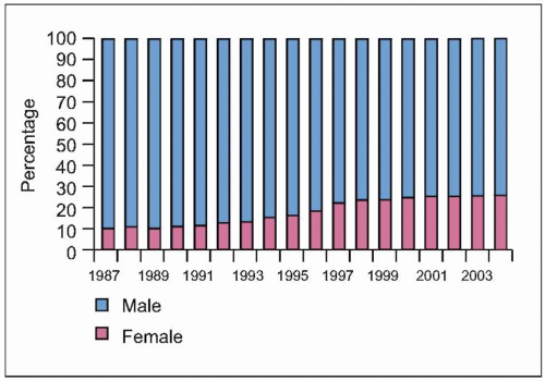
Fig. 1.6 Trends in the percentage distribution of deaths due to HIV disease by sex, USA, 1987-2004. The proportion of females among persons who died of HIV disease increased from 10% to 26% during this period, highlighting the increasing burden of disease among females in the US, as is the case in other countries. Because heterosexual transmission is emerging as the predominant mode of transmission, this trend can be expected to grow in the coming years. (Adapted from CDC data.)


Clinical manifestations

The clinical manifestations of HIV infection and AIDS are diverse and can affect virtually any organ system. The time to development of specific symptoms or syndromes varies considerably from person to person, and many cases remain asymptomatic for very prolonged periods. Nevertheless, HIV-related immunodeficiency develops in most cases as a predictable sequence of events, in which the massive depletion of CD4+ T cells that characterizes AIDS occurs only after a clinically silent interval. During this period, few clinical indications of HIV infection exist, despite vigorous viral replication in the lymphoid tissues and ongoing plasma viremia. This sequence of events is summarized in Fig. 1.7.

The first clinical manifestation of HIV infection may be a mononucleosis-like syndrome, termed acute retroviral syndrome, which occurs in over 50% of cases within 2-6 weeks of initial infection. Symptoms are nonspecific and may include fever, sore throat, lymph node enlargement, arthralgias, and headache and usually persist for several days to 3 weeks; in a significant proportion of symptomatic cases, the manifestations are severe enough to warrant medical attention. A maculopapular rash is common, as is nonspecific lymphadenopathy (Fig. 1.8), and some patients may present with self-limited aseptic meningitis, which manifests as cerebrospinal fluid (CSF) pleocytosis and isolation of HIV from CSF. In some cases, the acute retroviral syndrome may be accompanied by thrush or even opportunistic infections during the transient CD4+ T cell decline seen early in the disease course. Table 1.1 summarizes the most common clinical manifestations of acute retroviral syndrome. From the laboratory standpoint, the acute retroviral syndrome can be diagnosed on the basis of a negative HIV enzyme-linked immunosorbent assay (ELISA) and a positive antigen-based or HIV RNA test in a patient with risk factors.


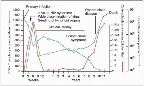
Fig. 1.7 Natural history of untreated HIV disease. Shortly after infection, viremia reaches extremely high levels, while CD4+ count decreases to levels that may be sufficient for the development of certain opportunistic complications. During this period, patients may experience the manifestations of the acute retroviral syndrome, and are highly infectious, thus making a high index of suspicion of paramount importance. After this initial phase, viremia decreases to a level (referred to as ‘set point’) at which it will remain, relatively constant, during the subsequent phase. At the same time, CD4+ T cell count rebounds, but does not return to pre-infection levels. A relatively asymptomatic period follows, during which there is ongoing viral replication and a slow but demonstrable decline in CD4+ T cell count. This phase ends years after infection with a precipitous fall in CD4+ T cell count and an exponential rise in plasma HIV RNA level, which heralds the beginning of AIDS. (Adapted from Fauci AS, Pantaleo G, Stanley S, et al. Immunopathic mechanisms of HIV infection. Ann Intern Med 1996;124:654.)

After the acute retroviral syndrome, the majority of subsequent clinical manifestations are due to complications emerging from progressive immunodeficiency. These infections will be discussed according to the stage at which they characteristically present. It should be kept in mind, however, that while diseases that are typical of profound immunodeficiency rarely appear at earlier stages, those that occur with high CD4+ T cell counts can obviously also
occur at later stages. A useful classification that includes both clinical and laboratory markers is the US Centers for Disease Control (CDC) staging system, shown in Table 1.2.


Table 1.1 Clinical manifestations of acute retroviral syndrome


Sign or symptom


%


Fever


96


Lymphadenopathy


74


Pharyngitis


70


Rash


70


Myalgia/arthralgia


54


Diarrhea


32


Headache


32


Nausea/vomiting


27


Hepatosplenomegaly


14


Thrush


12


Neurologic symptoms


12


(Data from Kahn JO, Walker BD. Human immunodeficiency Virus Type 1 infection. N Engl J Med 1998;339:33.)


Table 1.2 US Centers for Disease Control 1993 revised classification system for HIV infection in adults and adolescents


CD4+ T cell category


Clinical category


Category A: asymptomatic, PGL or acute viral syndrome


Category B: symptomatic conditions, non-AIDS-defining1


Category C: AIDS-defining conditions


Category 1: ≥500 cells/mm3


A1


B1


C1


Category 2: 200-499 cells/mm3


A2


B2


C2


Category 3: <200 cells/mm3


A3


B3


C3


Patients can be classified according to CD4+ T cell count and spectrum of clinical manifestations; increasing categories represent increasing degrees of immunodeficiency, although this classification was primarily created for surveillance purposes.


PGL: persistent generalized lymphadenopathy.


1 Conditions in this category include, but are not limited to, bacillary angiomatosis; thrush; persistent or refractory vaginal candidiasis; moderate to severe cervical dysplasia and cervical carcinoma in situ; constitutional symptoms including chronic diarrhea; oral hairy leukoplakia; recurrent or multidermatomal herpes zoster; idiopathic thrombocytopenic purpura; listeriosis; pelvic inflammatory disease; peripheral neuropathy.


Complications occurring with CD4+ T cell counts >500/mm3

Persistent generalized lymphadenopathy (PGL) can begin with the acute retroviral syndrome (Fig. 1.8), and is defined as two or more extrainguinal regions with lymphadenopathy persisting for at least 3-6 months in the absence of an alternative explanation. Up to 50-70% of HIV-infected patients may develop PGL, but PGL is not associated with adverse consequences in those individuals. Excluding a treatable etiology is critical, particularly with more localized lymphadenopathy. Some patients will experience constitutional symptoms (low-grade fevers, fatigue, and night sweats), diarrhea, or unexplained weight loss during the early stages of HIV infection; again, excluding other etiologies is imperative. Oral and vaginal candidiasis (Fig. 1.9) can also appear at all stages of HIV disease, although they increase in frequency as the CD4+ T cell count falls below 500 cells/mm3. Thrush presents as adherent, nonpainful, off-white exudates that can be scraped off with a tongue depressor, leaving a denuded area of mucosa. Dermatomal herpes zoster (Fig. 1.10) is another frequent early manifestation; in more advanced stages, the presentation may involve multiple dermatomes, prolonged persistence of lesions, or systemic dissemination.


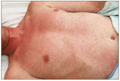
Fig. 1.8 Maculopapular rash consistent with acute HIV infection.


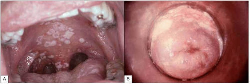
Fig. 1.9 Mucosal candidiasis in moderately advanced HIV infection. A:Thrush (oropharyngeal candidiasis). Typical white exudates that can be removed with a tongue depressor are seen covering the oral mucosa. B: Vaginal candidiasis. The similarity of the exudates covering the vaginal wall is obvious. (A: courtesy of Dr S Silverman, CDC; B: courtesy of CDC.)


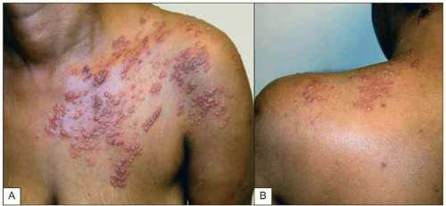
Fig. 1.10A, B Multidermatomal herpes zoster (shingles) in a patient with advanced HIV infection and incomplete CD4+ T cell replenishment after highly active antiretroviral therapy. Note the characteristic erythematous-vesicular rash that does not cross the midline and involves several dermatomes. Some of the lesions have begun to crust, while others (e.g. on the upper arm) are in a pustular phase.


Complications occurring with CD4+ T cell counts 200-500/mm3

Oral hairy leukoplakia (OHL) is the main differential diagnosis for oral candidiasis, as it also presents as a white, raised lesion of the oral mucosa (Fig. 1.11). OHL, however, typically presents on the lateral margin of the tongue and cannot be scraped off easily. Worldwide, the most common complication at this stage is tuberculosis (TB), both in pulmonary and extrapulmonary forms. TB can occur at any stage of HIV disease, however, and its clinical presentation relates to the CD4+ T cell count; thus, ‘typical’ cavitary forms are more commonly seen at early stages (Fig. 1.12), whereas more diffuse pulmonary and extrapulmonary forms become more frequent as the immunodeficiency progresses (Fig. 1.13). Kaposi’s sarcoma (KS) is a vascular neoplastic disorder characteristically seen in homosexual men that presents as red-purple nodules involving the skin and/or mucous membranes (Fig. 1.14); visceral involvement also occurs (Fig. 1.15), particularly with more advanced stages of disease.


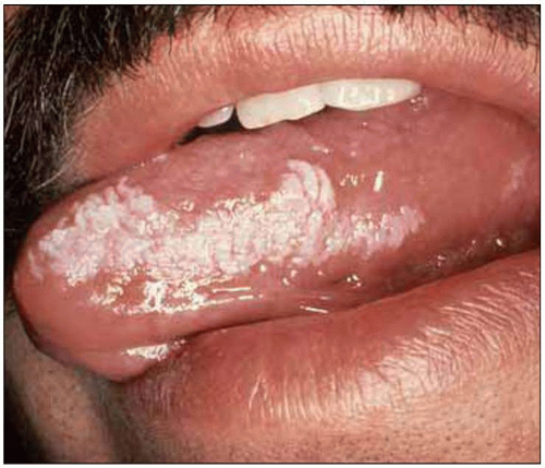
Fig. 1.11 Oral hairy leukoplakia. This lesion is associated with Epstein-Barr infection of the keratinized epithelium of the tongue and the buccal mucosa, and can be distinguished from oral candidiasis by the characteristic location on the lateral surface of the tongue, its resistance to scraping with a tongue depressor and its failure to respond to antifungal therapy. OHL can be seen at relatively early stages of HIV disease, but its prevalence increases with decreasing CD4+ T cell counts. (Courtesy of Dr S Silverman, DDS.)


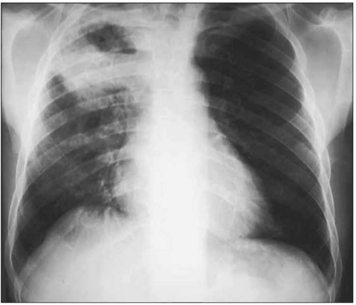
Fig. 1.12 Cavitary pulmonary tuberculosis in a Ugandan patient with AIDS. This person was not known to be HIV infected when he presented to the TB clinic complaining of subacute respiratory symptoms, fevers, and diaphoresis. Note the characteristic right upper lobe location of the lesion, and the air-fluid level, indicative of a cavitating lesion in that location. At the time of this visit, the patient’s CD4+ T cell count was approximately 400 cell/mm3.

Recurrent bacterial pneumonia (most often due to Streptococcus pneumoniae) and other serious bacterial infections are common during this stage, and their
frequency increases with progressive immunodeficiency. The risk of bacterial pneumonia is up to 100-fold greater in HIV infection. Other encapsulated organisms such as Hemophilus influenzae and highly virulent pathogens such as Pseudomonas aeruginosa are also more common in HIV-infected patients. Pneumococcal pneumonia typically presents with a similar clinical picture to that in HIV-negative patients (Figs. 1.16, 1.17), but mortality is increased in HIV-infected patients, particularly with advanced disease, when atypical presentations including subtle interstitial infiltrates and a protracted course reminiscent of Pneumocystis jirovecii can be seen. Concomitant bacteremia is especially common in this setting, making blood cultures an important part of the diagnostic evaluation. Nodular and cavitary lesions, in addition to mycobacterial and fungal disease, should raise suspicion for less common bacterial pulmonary pathogens including Nocardia species (Fig. 1.18) and Rhodococcus equi, particularly in the setting of clinical manifestations in other organ systems (Fig. 1.19).


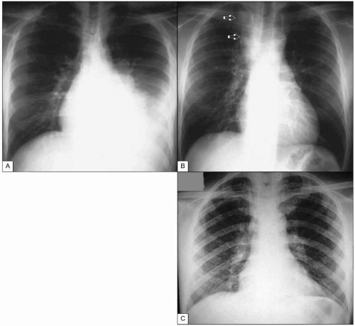
Fig. 1.13 Tuberculosis in advanced AIDS. A: Pleural tuberculosis. This massive pleural effusion resolved completely after anti-TB treatment. B: Tuberculous adenitis. This mass (arrows) can easily be mistaken for a neoplasia, leading to unnecessary interventions. C: Miliary tuberculosis in a patient with advanced AIDS. Note the finely micronodular infiltrate involving virtually all the pulmonary parenchyma. CD4+ T cell count at the time of diagnosis was <5 cells/mm3. (A and B courtesy of Dr R Kalayjian.)

Only gold members can continue reading. Log In or Register to continue

Stay updated, free articles. Join our Telegram channel

Aug 1, 2016 | Posted by in INFECTIOUS DISEASE | Comments Off on HIV and AIDS

Full access? Get Clinical Tree

Get Clinical Tree app for offline access