Treatment of Locally Recurrent Prostate Cancer


Treatment of Locally Recurrent Prostate Cancer


▪ 13A Recurrence Following Radiotherapy: Definitions, Prognosis, and Management

Eric M. Horwitz

Prostate cancer recurrence following primary radiation therapy (RT) can be defined in several ways including a biochemical (PSA) or clinical failure (CF). Most common in the PSA era is a recurrence in the asymptomatic patient defined entirely by a rise in posttreatment PSA (1,2). Current definitions of a PSA recurrence following surgery include any detectable PSA after a prostatectomy or a PSA above a particular threshold, most commonly >0.2 ng/mL (3,4,5). Following radiation, defining a PSA recurrence can be more problematic. The current definition of a PSA or biochemical recurrence in a population, commonly referred to as the Phoenix definition, was adopted in 2006 and is defined as a posttreatment PSA > nadir + 2 ng/mL (6). However, this definition is to be used for defining recurrence in a population only. For an individual, a PSA recurrence can be defined as a steadily rising PSA over time following a posttreatment nadir. Because posttreatment PSA levels can fluctuate, multiple rising PSA levels are required to establish a definitive trend. A clinical recurrence can be defined as local, disease confined to the prostate (a change in DRE and confirmed by biopsy) or distant, most often observed in radiographic imaging first before the development of symptomatic disease.

Salvage treatment depends upon what the primary treatment was, and this chapter focuses on how a recurrence is defined and what the predictors of a recurrence are following definitive RT. Treatment options for a local recurrence (LR) depend upon the original treatment, and choices following external beam RT include prostatectomy, cryosurgery, high intensity focused ultrasound (HIFU), or brachytherapy (7,8,9,10). One of the largest series of patients describing patterns of failure for men following primary treatment for prostate cancer was reported utilizing data from the Cancer of the Prostate Strategic Urological Research Endeavor (CapSURE) database. Agarwal et al. examined the risk of, patterns of care of, and outcome of men who recur after primary surgical or radiation management of prostate cancer. More than 5,000 men were identified who were treated with either radical prostatectomy (RP) (4,342 patients) or RT (935 patients). Of these patients, 1,590 men developed recurrent disease (1,003 RP and 587 RT). Patients in this series who developed recurrent disease had statistically significant increased rates of overall death (15% vs. 1%, p = 0.01). Overall, 420 men in the RP group and 319 men in the RT group failed salvage treatment, and these patients had significantly greater rates of death compared to the patients who did not fail salvage treatment (24.8% vs. 6.9%, p < 0.001). Androgen deprivation therapy (ADT) was the most common salvage treatment for both groups, 60% for the RP patients and >90% for the RT patients. It is worth noting that of the 430 patients, only 13 (3%) were treated with salvage cryosurgery, 4 men received a salvage prostatectomy (0.9%), and one patient had a salvage implant (0.2%); the overwhelming majority of patients in the CapSURE database did not receive potentially curative salvage treatment (11).

Before salvage treatment options can be addressed, it is important to understand the myriad of issues surrounding posttreatment PSA and how failure is defined following treatment with definitive RT. In an ideal world, comparisons to assess efficacy of various treatment options for localized prostate cancer would be standardized. However, inherent differences exist between surgery and radiation that must be reflected in how we assess success or failure. Since the prostate is not removed when radiation is used as treatment, a detectable PSA following treatment is expected. However, the presence of a detectable PSA does not mean that active cancer remains, as attested to by large multi-institutional data evaluating local and distant control rates over many years (12,13). Because it may never be possible to completely compare “apples to apples” when evaluating the results of surgery or radiation for prostate cancer, it is probable that two “gold standards” exist for determining outcomes based on PSA.

The need for a standard definition to define PSA failure after radiation treatment was first recognized in the late 1990s when several groups began publishing their results with increasingly large numbers of patients utilizing different definitions of PSA failure. Outcomes were different, but it was not recognized that at first some of these different results were based solely on the definition of PSA failure used. Horwitz et al. first demonstrated that the different definitions of biochemical failure (BF) could, by themselves, significantly alter the perceived efficacy of external beam RT for prostate cancer. In this original study, three different definitions of biochemical (bNED) control taken from the literature at the time were used: (1) PSA nadir < 1 ng/mL within 1 year of treatment completion. After achieving nadir, if two consecutive increases of PSA were noted, the patient was scored a failure at the time of the first increase. (2) PSA nadir < 1.5 ng/mL within 1 year of treatment completion. After achieving nadir, if two consecutive increases of PSA were noted, the patient was scored a failure at the time of the first increase. (3) Posttreatment PSA nadir < 4 ng/mL without a time limit. Once the nadir was achieved, if it did not rise above normal, the patient was considered to be biochemically controlled. Pretreatment PSA values were correlated with treatment outcome using the three definitions of biochemical control. Pretreatment PSA values were stratified into five groups (Group 1: PSA < 4; Group 2: PSA 4 to 10; Group 3: PSA 10 to 15; Group 4: PSA 15 to 20; and Group 5: PSA > 20), and 5-year actuarial rates of biochemical control were calculated using the three biochemical control definitions. As an example, for Group 2, 5-year actuarial control rates were 45%, 54%, 74%, and 92% for the four definitions, respectively. Differences between all three definitions for all pairwise comparisons ranged from 5 to 53% (p < 0.001) (14).
In 1997, the American Society of Therapeutic Radiology and Oncology (ASTRO) convened a Consensus Panel to develop a unified definition of BF, and the original definition included the following guidelines:


  • BF is not justification per se to initiate additional treatment and is not equivalent to CF. BF is an appropriate early end point for clinical trials.


  • Three consecutive increases in PSA is a reasonable definition of BF after RT. For clinical trials, the date of failure should be backdated to the midpoint between the post-RT nadir PSA and the first of the three consecutive rises.


  • No definition of PSA failure has, as yet, been shown to be a surrogate for clinical progression or survival.


  • Nadir PSA (nPSA) is a strong prognostic factor, but no absolute level is a valid cut point for separating successful and unsuccessful treatments.

The Panel also recommended minimum follow-up of 24 months for published data (15). With time, experience, and clinical data using the consensus definition, several weaknesses in this original ASTRO definition were identified. In 1999, Vicini et al. reported data demonstrating that treatment success could be overestimated with short follow-up (16). The Fox Chase group also identified problems with the original ASTRO definition because backdating the date of failure with short follow-up could also alter outcome (17). Other issues for which the original ASTRO definition could not account for included the PSA bounce phenomena, differences between current and absolute posttreatment PSA nadir and decreased sensitivity and specificity compared with newer alternate definitions of BF. The original definition was developed for patients treated with external beam RT only and was not validated for patients receiving ADT or brachytherapy; but it was applied to series using these treatment modalities. Using data from the largest multi-institutional dataset, the ASTRO definition was compared with alternate PSA failure definitions against the “gold standard” of CF. Nine participating institutions agreed to submit follow-up results for all patients with clinically localized prostatic cancer (Stage T1b, T1c, T2, N0M0) treated between 1986 and 1995 by external beam RT only, to doses of ≥60 Gy, with no ADT before treatment. A total of 4,839 men met the study criteria, with a median follow-up time of 6.3 years. The prediction of CF by 102 definitions of BF was assessed using various quantitative measures (Table 13A.1). Four definitions of BF were superior to the original ASTRO definition
as measured by the sensitivity, specificity, positive and negative predictive values, and hazard of CF after BF: two rises of at least 0.5 ng/mL backdated, PSA level at or greater than the absolute nadir + 2 ng/mL at the call date, and PSA level at or greater than the current nadir + 2 or 3 ng/mL at the call date. Backdating the failure time introduced bias into the estimate of freedom from BF, which was increasingly overestimated at shorter median follow-up times. The authors concluded that alternate definitions of BF were superior as assessed by various quantitative measures of concordance of biochemical and ultimate CF (18,19). Using this same dataset, Horwitz et al. determined the sensitivity and specificity of several BF definitions using distant failure (DF) alone or CF, defined as local failure and/or DF. The sensitivity and specificity of the ASTRO definition to predict DF alone was 55% and 68%, respectively. The sensitivity and specificity of the ASTRO definition to predict CF was 60% and 72%, respectively. Three definitions had higher sensitivity and specificity: (a) PSA > current nadir + 3 ng/mL (sensitivity 66% and specificity 77%), dated at the call; (b) PSA > absolute nadir + 2 ng/mL (sensitivity 64% and specificity 74%), dated at the call; and (c) two consecutive increases of at least 0.5 ng/mL, back dated (sensitivity 67% and specificity 78%) (20).


TABLE 13A.1 SENSITIVITY AND SPECIFICITY OF MULTIPLE BF DEFINITIONS COMPARED TO THE ORIGINAL ASTRO DEFINITION


Definition


Sensitivity


Specificity


Consecutive rises in PSA


2 rises backdated


0.81


0.59


2 rises call date


0.72


0.59


3 rises backdated (original ASTRO definition)


0.61


0.80


3 rises call date


0.51


0.80


4 rises backdated


0.41


0.89


4 rises call date


0.32


0.89


2 rises backdated


Final PSA > 1.5 ng/mL


0.79


0.73


2 rises call date


0.69


0.73


3 rises backdated


0.61


0.83


3 rises call date


0.50


0.83


4 rises backdated


0.41


0.90


4 rises call date


0.32


0.90


2 rises backdated


Each rise > 0.5 ng/mL


0.68a


0.87a


2 rises call date


0.57


0.87


3 rises backdated


0.41


0.94


3 rises call date


0.31


0.94


4 rises backdated


0.22


0.97


4 rises call date


0.15


0.97


PSA ≥ absolute nadir PSA + X ng/mL at call date


X =


1


0.74


0.74


2


0.67a


0.84a


3


0.59


0.88


4


0.52


0.91


5


0.46


0.93


PSA ≥ current nadir PSA + X ng/mL at call date


X =


1


0.83


0.70


2


0.74a


0.82a


3


0.66a


0.86a


4


0.57


0.90


5


0.50


0.94


PSA > 0.2 ng/mL


0.91


0.09


PSA > 0.5 ng/mL


0.90


0.26


a Both sensitivity and specificity are significantly higher than the original ASTRO definition (15) for three rises backdated (p < 0.01)


One of the criticisms of the original ASTRO definition was that it was supposed to only apply to patients treated with external beam RT alone although it was ultimately used for other patients (those treated with ADT or brachytherapy). As the radiation oncology community recognized the need to modify this definition grew, groups tested some of these proposed modifications on these patients treated with ADT or brachytherapy. As with the external beam RT group, a second multi-institutional project was organized to examine outcomes of patients treated with brachytherapy alone (13). Alternate PSA failure definitions were tested with this dataset. Two thousand six hundred ninety three patients treated with permanent prostate implants for T1-2 prostate cancer were studied. All patients had a pretreatment PSA, were treated at least 5 years before analysis, 1988 to 1998, and did not receive ADT before recurrence. As with the external beam RT group, multiple PSA failure definitions were tested for their ability to predict CF. The sensitivity and specificity for the nadir + 2 definition were 72% and 83%, vs. 51% and 81% for 3 PSA rises. Conclusions from this large dataset included that for prostate cancer patients treated with a permanent implant, the definition nadir + 2 ng/mL provides the best surrogate for CF, results similar to patients treated with external beam RT (21). With evidence provided by these studies, a second Consensus Conference was held in Phoenix, Arizona in January 2005, to formerly modify or replace the ASTRO definition. Following presentations and discussion, the Panel recommended the following as the current standard definition of BF after RT with or without a short course of ADT—a rise by 2 ng/mL or more above the nadir PSA (defined as the lowest posttreatment PSA level achieved). The date of failure was not to be backdated (6).

Prognostic factors for recurrence include pretreatment and posttreatment PSA, Gleason score (GS), T stage, PSA nadir, and PSA doubling time (PSADT). This last factor has consistently been shown to be most predictive for the development of clinical distant metastases (1,22,23,24). Analysis of patients from the multi-institutional group correlated PSA failure patients with the development of clinical metastases. Out of the entire group, 1,421 patients had a BF, as the first event, by the original ASTRO definition; 895 patients had no ADT for the entire duration of follow-up or until the development of distant metastasis (DM) and 526 patients were treated with ADT for PSA recurrence. However, for those in the worst prognostic group, PSADT ≤ 4 months, distant metastasesfree survival (DMFS) was improved with ADT, 78% versus 35% at 5 years, p = 0.001, with corresponding cause-specific survival (CSS) of 93% versus 81%, p = 0.02. In classification and regression tree (CART) analysis in patients who did not receive ADT, those with a PSADT of ≤4 months and an interval from treatment to BF (iPSAF) of ≤2 years had a risk of DM 47 times higher than patients with a PSADT > 9 months. Outcome after BF following external beam RT varies significantly and is dependent most on PSADT and the interval from BF. Patients with short PSADTs (≤4 months) and interval from BF (≤2 years) are those who would benefit most from ADT and not an alternative local therapy (25).

At Fox Chase, we had shown that PSADT of <12 months is an important predictor for the development of DM following 3DCRT using the original ASTRO definition of BF. With the introduction of the Phoenix definition, we sought to determine if this approach was still valid using intensity-modulated radiation therapy (IMRT) in a larger dataset with longer follow-up. Four hundred and thirty two men with T1-3N0M0 prostate cancer who demonstrated PSA failure according to the Phoenix definition after completing definitive 3DCRT or IMRT alone or with ADT treated at Fox Chase Cancer Center from 1989 to 2005 were tested. PSADT was stratified by 0 to 6, 6 to 12, 12 to 18, 18 to 24, and >24 months. Using both RPA and the log-rank test, PSADT ≤6 months was the significant breakpoint in the FDM for patients who were PSA failures. Seven-year FDM was 50% for PSADT < 6 months versus 83% for PSADT > 6 (p = 0.0001). Seven-year CSS was 61% for PSADT < 6 and 85% for PSADT > 6 (p = 0.0001). Seven-year overall survival (OS) was 47% for PSADT < 6 and 53% for PSADT > 6 (p = 0.04). The use of ADT was associated with a significant improvement in the 7-year CSS (68% vs. 46%, p = 0.015). In the group of men with PSADT > 6 months, there was no improvement in the 7-year CSS with the use of ADT, (87% vs. 79%, respectively; p = 0.758). With this expanded dataset, we concluded that PSADT remained a significant predictor of CF and CSS for men treated with 3DCRT or IMRT who fail according to the Phoenix definition. The immediate use of ADT in patients with PSADT < 6 months demonstrated significant improvements in CSS; the benefit was less apparent in patients with longer PSADT and these are the patients who should consider a local treatment for their recurrence (26).

Patients with a short PSADT as defined as <6 months likely have occult micrometastatic disease that will present clinically in short order. For these patients, ADT is most often the treatment of choice and early, rather than late, ADT should be considered for these patients. Other systemic agents including chemotherapy or vaccines can be considered on a clinical trial (27). For patients with a PSADT > 1 year, the long time interval between the first rise in PSA following treatment and the development of clinical metastatic disease suggests that the presence of micrometastatic disease is much less likely. In these patients, the rise in PSA is most likely a marker/harbinger of a LR in the prostate. In this situation, treatment options are quite different and more varied as compared to patients with short PSADTs. Once a metastatic workup has ruled out the presence of metastatic disease, treatment options include salvage prostatectomy, cryosurgery, brachytherapy, HIFU, or continued close surveillance.

Because patients who are treated definitively with external beam RT have a prostate after treatment, some measurable baseline PSA is expected. In an ideal situation, a patient’s PSA level would decrease after treatment and remain stable over time, providing confidence that the treatment was successful and there was no evidence of disease. As our clinical experience has grown and follow-up after treatment increase, investigators have recognized that patient’s posttreatment PSA levels are not stable. A transient rise, or bounce, in PSA was first identified in 1997 by Wallner et al. (28) and in 2000 by Critz et al. In that report, the PSA bounce phenomenon was described in a series of 779 early stage patients treated with permanent prostate
implants and external beam RT. In this series, a transient rise in PSA was found in 35% of the patients. The bounce was defined as an increase of 0.1 ng/mL or greater above the preceding level followed by a subsequent decrease below that level. The median time to the bounce was 18 months and more than 90% occurred within 3 years. The median bounce height was 0.4 ng/mL (0.1-15.8 ng/mL). No independent predictors of bounce were identified and no clinical or biochemical correlation was made with the bounce in this early series (29).

At first, the PSA bounce was thought to be unique to these patients treated with a prostate implant; however, Hanlon et al. reported that 30% of patients treated with 3DCRT alone also experienced PSA bounces sometime following treatment (30). In this Fox Chase series, 306 patients received a median radiation dose of 74 Gy. The PSA bounce was defined as a minimum rise of 0.4 ng/mL over 6 months, followed by a drop of any magnitude. As with the implant series, approximately one third of patients bounced at some point after treatment. Lower radiation doses (73 Gy vs. 75 Gy) and higher pretreatment PSA levels were independent predictors of bounce. This was the first series to suggest that patients who bounced had lower rates of bNED control, 69% versus 52% at 5 years (p = 0.0024) although no clinical correlation was observed with a median follow-up of 79 months. The fact that decreased bNED control rates did not translate into inferior clinical control may have been a function of the ASTRO definition requirement of several consecutive rises (30). Additional data from Cavanagh and Rosser supported the general recognition that the PSA bounce phenomenon is common in patients treated with radiation and is not modality specific (31,32).

While these series described the frequency, timing, and magnitude of the bounce, the available data on its clinical significance were scarce. Were there different biochemical and clinical control rates between patients with a bounce and those without? Could physiological and interassay variations of the PSA test itself be separated from a true bounce? To try and answer these questions, data from the multi-institutional group were reviewed, this time with attention to the PSA bounce. The power of large patient numbers and long followup of this multi-institutional pooled dataset was to determine the biochemical and clinical significance of the PSA bounce in patients treated with EBRT alone. In the first analysis, data from the 4,839 patients treated with external beam RT were used to determine if there was a difference in biochemical and clinical control between the bounce and nonbounce (NB) patients. Twenty percent (978) of the patients experienced at least 1 posttreatment PSA bounce. Patients <70 years had a 72% chance of remaining bounce-free at 5 years compared with 75% for older patients (p = 0.04). The NB patients had 72% bNED control at 10 years compared with 58% for the bounce patients. However, this did not translate into a difference in CF (DF, CSF, or OS) (33).

In the second report, data on 7,532 patients from the 2 multi-institutional pooled datasets- 4,839 patients with T1-2 prostate cancer treated with RT alone and 2,693 patients treated with BT alone and identify any predictors of a bounce was used to determine if there was a difference in biochemical and clinical control between the bounce and NB patients. No neoadjuvant ADT was allowed. A posttreatment PSA bounce was defined as an increase of at least 0.2 ng/mL over a previous PSA measurement followed by a decline. The median follow-up for the RT and BT patients were 75 and 63 months, respectively. Endpoints included bNED failure (Phoenix definition), DMFS, CSS and OS. At 10 years, there were no statistically significant differences in DMFS, CSS, and OS between the NB and bounce RT patients (DMFS—93.9% vs. 94.5% [p = 0.16]; CSS—91.7% vs. 93.2% [p = 0.21]; OS—64.5% vs. 67.5% [p = 0.10]) or BT patients (DMFS—94.5% vs. 96.9% [p = 0.74]; CSS—94.3% vs. 96.3% [p = 0.56]; OS—62.5% vs. 66.3% [p = 0.22]). In this multi-institutional analysis with large patient numbers and long follow-up, patients treated with either RT alone or BT alone who experience a posttreatment PSA bounce do not have increased risk of BF or CF compared to men who do not bounce at 10 years. Interestingly, BT patients treated with 125I bounce more often than patients who receive 103Pd. Younger men (≤70 years) treated with either modality bounce more frequently, but there is no difference in outcome (34).

Once the PSA bounce phenomenon was recognized as a relatively common event, various groups focused on both identifying independent predictors of the PSA bounce and determining its effect on outcome. Merrick et al. identified younger age, T stage, first implant PSA and V150 (the volume of prostate receiving 150% of the prescribed dose) as predictors of PSA bounce although there was no significant difference in bNED control. Fewer patients were in this series (218) and all received either 125I or 103Pd implants (120 patients also received supplemental EBRT). One quarter of these patients experienced a bounce, defined as a transient increase ≥0.2 ng/mL (35).

Stock et al. summarized the risk of experiencing a PSA bounce in their dataset using three different definitions of bounce in 373 patients who received 125I or 103Pd implants (337 125I and 36 103Pd implants) alone. These authors defined bounce as a PSA rise ≥0.1 ng/mL, a PSA rise ≥0.4 ng/mL, and finally >35% over the previous value. The likelihood of developing a bounce at 5 years was 31%, 17%, and 20% for the three definitions, respectively. The median time to developing a bounce was 19.5 months for the first two definitions and 20.5 months for the third definition. Only D90 > 160 Gy (dose that 90% of the prostate receives) was predictive of bounce using the first definition (36). Czieki et al. reported similar results in their Cleveland Clinic experience of 162 patients treated with an implant. BF was also defined using two definitions—the original ASTRO definition and the Phoenix definition. At 5 years, bNED control was 87% using the ASTRO definition and 96% for the Phoenix definition (37). Here we see evidence that the Phoenix definition was not as sensitive to the bounce phenomenon and was likely a better definition of PSA failure for these patients.

With time and clinical experience, other authors have come to similar conclusions (37,38,39,40). As with other series, Pickles observed that the PSA bounce was common and that the Phoenix definition had a lower false call rate of BF compared to the original ASTRO definition (41). In addition to testing the validity of the Phoenix definition with brachytherapy patients, Zietman et al. tested the definition, along with several others, using patients treated with external beam RT neoadjuvant ADT. At Massachusetts General Hospital, characteristics of the PSA bounce were calculated for all patients treated by EBRT with neoadjuvant ADT between 1992 and 1998. The percentage of bounces scoring as false positives according to the ASTRO definition of BF was compared with those for three alternative definitions (Vancouver, nadir + 2, and nadir + 3). The authors concluded that a substantial proportion of patients treated by external beam RT with neoadjuvant ADT experienced a PSA bounce. A large percentage of these bounces scored as BF according to the ASTRO definition. The alternate definitions, including the Phoenix definition, were less vulnerable to this bias (42).

Many prognostic factors which were first thought to be important early in the PSA era have become even more significant predictors of recurrence with greater patient numbers and longer follow-up. Understanding the significance of the posttreatment PSA nadir as a predictor of failure is as important as understanding PSADT and the PSA bounce. The clinical significance of the PSA nadir has been studied by many authors in an effort to better assess treatment efficacy and help predict which
patients may need additional treatment (43,44). Zelefsky et al. detailed the influence of posttreatment PSA nadir response at 2 years after external beam RT on distant metastases (DM) and cause-specific mortality (CSM) using data from 844 patients with localized prostate cancer treated with 3DCRT with long follow-up. Multivariate analysis demonstrated that nadir PSA ≤ 1.5 ng/mL was an independent predictor of progression-free survival after adjusting for T stage, GS, pre-RT PSA value, and RT dose (p = 0.03). The 5- and 10-year cumulative incidences of DM were 2.4% and 7.9%, respectively in those with nadir PSA levels ≤1.5 ng/mL, and were 10.3% and 17.5%, respectively, in patients with higher nadir values. Multivariate analysis showed that the higher nadir PSA value (p = 0.002), higher GSs (p < 0.001), and increasing T stage (p = 0.03) were predictors of DM after adjusting for pre-RT PSA values and RT dose. Multivariate analysis also showed that higher GSs (p = 0.002), and higher nadir PSA values (p = 0.03) were risk factors associated with CSM after adjusting for T stage and pre-RT PSA value. The authors concluded that nadir PSA values of ≤1.5 ng/mL at 2 years after RT for prostate cancer predict for long-term DM and CSM outcomes. The authors recommended that patients with higher absolute nadir levels at 2 years after treatment should be evaluated for the presence of nonresponding disease in the context of considering earlier salvage treatment interventions (45).

Alcantera et al. presented data from another single institution series where the nadir PSA at 1 year (nPSA12) was investigated as an early estimate of biochemical and clinical outcome after RT alone. One thousand men were treated with 3DCRT alone (median dose 76 Gy) with minimum and median follow-up of 26 and 58 months, respectively. Multivariate analyses were used to determine the association of nPSA12 to BF (original ASTRO definition), DM, CSM, and overall mortality (OM). In MVA, nPSA12 as a continuous variable was independent of RT dose, T-stage, GS, pretreatment initial PSA, age, and PSADT in predicting for BF, DM, CSM, and OM. The authors concluded that nPSA12 is a strong independent predictor of outcome after RT alone for prostate cancer and should be useful in identifying patients at high risk for progression to metastasis and death (46).

Although these first two series had large patient numbers, they were still limited by the fact that they originated from single institutions. Ray et al. used data from the multi- institutional pooled analysis to test the prognostic significance of PSA nadir in the largest dataset in predicting biochemical or clinical disease-free survival (PSA-DFS) and DMFS in patients treated with definitive RT. bNED control and DMFS were the two endpoints correlated with nPSA and time to nPSA. A greater nPSA level and shorter T(nPSA) were associated with decreased PSA-DFS and DMFS in all patients and in all risk categories, regardless of RT dose. The 8-year PSA-DFS and DMFS rate for patients with nPSA < 0.5 ng/mL was 75% and 97%; nPSA 0.5 to 1.0 ng/mL, 52% and 96%; nPSA 1 to 2 ng/mL, 40% and 91%; and nPSA ≥ 2 ng/mL, 17% and 73%, respectively. The 8-year PSA-DFS and DMFS rate for patients with T(nPSA) < 6 months was 27% and 66%; T(nPSA) 6 to 12 months, 31% and 85%; T(nPSA) 12 to 24 months, 42% and 94%; and T(nPSA) ≥ 24 months, 75% and 99%, respectively. A shorter T(nPSA) was associated with decreased PSA-DFS and DMFS, regardless of the nPSA. Both nPSA and T(nPSA) were significant predictors of PSA-DFS and DMFS in multivariate models incorporating clinical stage, GS, initial PSA level, and RT dose (47). Both the individual series and pooled analyses have demonstrated that nPSA and time to nPSA after external beam RT are not only predictive of a predominantly PSA endpoint (PSA-DFS), but are also predictive of DM in all clinical risk categories.

Before choosing an appropriate salvage treatment, it is important to understand the patient’s risk of recurrence based on their pretreatment prognostic factors, their post-primary treatment PSA kinetics, and the ability to predict a local or distant recurrence. It is important to understand the proper use of the BF definitions. Current data suggests that the Phoenix definition, is less dependent on time, PSA bounce, ADT, and treatment modality when assessing efficacy. There are several prognostic factors that are predictive of outcome across treatment techniques for patients who undergo local salvage therapy. The most common factors include presalvage PSA, pretreatment and presalvage GS, and pre-RT and presalvage T stage. Other factors which have been shown to be predictive of outcome include surgical margin status, tumor ploidy, and androgen independence (7). Although obvious, patient selection before therapy can have one of the biggest outcomes for successful salvage. Consistently, patients with presalvage PSA ≤ 10 ng/mL, T stage ≤ T2, and pretreatment GS ≤ 7 do better than patients with higher PSA, GS, or T stage. For individual patients, PSADT and nadir PSA can aid in determining a local versus distant recurrence in an asymptomatic patient to guide the treatment decision for recurrent disease. Clinicians need to understand that the PSA bounce after RT is a relatively common phenomena that is not predictive for CF and should not be a trigger to diagnose treatment failure and initiate salvage treatment. PSADT is an extremely important factor to determine treatment options. If a PSADT is <6 months, distant disease should be suspected and local salvage treatment is probably not an appropriate option. If PSADT is >1 year, prostate confined disease is much more likely and local salvage treatment should be considered after biopsy confirmation.


References

1. Lee WR, Hanks GE, Hanlon A. Increasing prostate-specific antigen profile following definitive radiation therapy for localized prostate cancer: clinical observations. J Clin Oncol 1997;15(1):230-238.

2. D’Amico AV, Moul JW, Carroll PR, et al. Surrogate end point for prostate cancerspecific mortality after radical prostatectomy or radiation therapy. J Natl Cancer Inst 2003;95(18):1376-1383.

3. Pound CR, Partin AW, Eisenberger MA, et al. Natural history of progression after PSA elevation following radical prostatectomy. JAMA 1999;281(17):1591-1597.

4. Grossfeld GD, Li YP, Lubeck DP, et al. Patterns of failure after primary local therapy for prostate cancer and rationale for secondary therapy. Urology 2002;60(3 Suppl 1):57-62; discussion 62-53.

5. Ward JF, Blute ML, Slezak J, et al. The longterm clinical impact of biochemical recurrence of prostate cancer 5 or more years after radical prostatectomy. J Urol 2003;170(5):1872-1876.

6. Roach M III, Hanks G, Thames H Jr, et al. Defining biochemical failure following radiotherapy with or without hormonal therapy in men with clinically localized prostate cancer: recommendations of the RTOG-ASTRO Phoenix Consensus Conference. Int J Radiat Oncol Biol Phys 2006;65(4):965-974.

7. Ward JF, Sebo TJ, Blute ML, et al. Salvage surgery for radiorecurrent prostate cancer: contemporary outcomes. J Urol 2005;173(4):1156-1160.

8. Izawa JI, Madsen LT, Scott SM, et al. Salvage cryotherapy for recurrent prostate cancer after radiotherapy: variables affecting patient outcome. J Clin Oncol 2002;20(11):2664-2671.

9. Grado GL, Collins JM, Kriegshauser JS, et al. Salvage brachytherapy for localized prostate cancer after radiotherapy failure. Urology 1999;53(1):2-10.

10. Gelet A, Chapelon JY, Poissonnier L, et al. Local recurrence of prostate cancer after external beam radiotherapy: early experience of salvage therapy using high-intensity focused ultrasonography. Urology 2004;63(4):625-629.

11. Agarwal PK, Sadetsky N, Konety BR, et al. Treatment failure after primary and salvage therapy for prostate cancer: likelihood, patterns of care, and outcomes. Cancer 2008;112(2):307-314.

12. Kuban DA, Thames HD, Levy LB, et al. Longterm multi-institutional analysis of stage T1-T2 prostate cancer treated with radiotherapy in the PSA era. Int J Radiat Oncol Biol Phys 2003;57(4):915-928.


13. Zelefsky MJ, Kuban DA, Levy LB, et al. Multi-institutional analysis of long-term outcome for stages T1-T2 prostate cancer treated with permanent seed implantation. Int J Radiat Oncol Biol Phys 2007;67(2):327-333.

14. Horwitz EM, Vicini FA, Ziaja EL, et al. Assessing the variability of outcome for patients treated with localized prostate irradiation using different definitions of biochemical control. Int J Radiat Oncol Biol Phys 1996;36(3):565-571.

15. Consensus statement: guidelines for PSA following radiation therapy. American Society for Therapeutic Radiology and Oncology Consensus Panel. Int J Radiat Oncol Biol Phys 1997;37(5):1035-1041.

16. Vicini FA, Kestin LL, Martinez AA. The importance of adequate follow-up in defining treatment success after external beam irradiation for prostate cancer. Int J Radiat Oncol Biol Phys 1999;45(3):553-561.

17. Horwitz EM, Uzzo RG, Hanlon AL, et al. Modifying the American Society for Therapeutic Radiology and Oncology definition of biochemical failure to minimize the influence of backdating in patients with prostate cancer treated with 3-dimensional conformal radiation therapy alone. J Urol 2003;169(6):2153-2157; discussion 2157-2159.

18. Kuban D, Thames H, Levy L, et al. Failure definition-dependent differences in outcome following radiation for localized prostate cancer: Can one size fit all? Int J Radiat Oncol Biol Phys 2005;61(2):409-414.

19. Thames H, Kuban D, Levy L, et al. Comparison of alternative biochemical failure definitions based on clinical outcome in 4839 prostate cancer patients treated by external beam radiotherapy between 1986 and 1995. Int J Radiat Oncol Biol Phys 2003;57(4):929-943.

20. Horwitz EM, Thames HD, Kuban DA, et al. Definitions of biochemical failure that best predict clinical failure in patients with prostate cancer treated with external beam radiation alone: a multi-institutional pooled analysis. J Urol 2005;173(3):797-802.

21. Kuban DA, Levy LB, Potters L, et al. Comparison of biochemical failure definitions for permanent prostate brachytherapy. Int J Radiat Oncol Biol Phys 2006;65(5):1487-1493.

22. Patel A, Dorey F, Franklin J, et al. Recurrence patterns after radical retropubic prostatectomy: clinical usefulness of prostate specific antigen doubling times and log slope prostate specific antigen. J Urol 1997;158(4):1441-1445.

23. Sartor CI, Strawderman MH, Lin XH, et al. Rate of PSA rise predicts metastatic versus local recurrence after definitive radiotherapy. Int J Radiat Oncol Biol Phys 1997;38(5):941-947.

24. Lee AK, Levy LB, Cheung R, et al. Prostatespecific antigen doubling time predicts clinical outcome and survival in prostate cancer patients treated with combined radiation and hormone therapy. Int J Radiat Oncol Biol Phys 2005;63(2):456-462.

25. Kuban D, Thames H, Levy L, et al. Predicting outcome after PSA failure in prostate cancer patients treated by radiation. Who needs salvage therapy? Int J Radiat Oncol Biol Phys 2004;60:167-168.

26. Horwitz E, Ruth K, Uzzo R, et al. PSA doubling time predicts for the development of distant metastases for patients who fail 3DCRT or IMRT using the Phoenix definition. Int J Radiat Oncol Biol Phys 2008;72(1):135-136.

27. Eisenberger MA, Blumenstein BA, Crawford ED, et al. Bilateral orchiectomy with or without flutamide for metastatic prostate cancer. N Engl J Med 1998;339(15):1036-1042.

28. Wallner K, Blasko JC, Dattoli MJ. Prostate brachytherapy made complicated: a practical guide to iodine-125 and palladium-103 implants, 1st ed. Canaan, NY: Smart Medicine Press, 1997.

29. Critz FA, Williams WH, Benton JB, et al. Prostate specific antigen bounce after radioactive seed implantation followed by external beam radiation for prostate cancer. J Urol 2000;163:1085-1089.

30. Hanlon AL, Pinover WH, Horwitz EM, et al. Patterns and fate of PSA bouncing following 3D-CRT. Int J Radiat Oncol Biol Phys 2001;50(4):845-849.

31. Cavanagh W, Blasko JC, Grimm PD, et al. Transient elevation of serum prostate-specific antigen following 125I/103Pd brachytherapy for localized prostate cancer. Sem Urol Oncol 2000;18:160-165.

32. Rosser CJ, Kuban DA, Levy LB, et al. Prostate specific antigen bounce phenomenon after external beam radiation for clinically localized prostate cancer. J Urol 2002;168(5):2001-2005.

33. Horwitz EM, Levy LB, Thames HD, et al. Biochemical and clinical significance of the posttreatment prostate-specific antigen bounce for prostate cancer patients treated with external beam radiation therapy alone: a multiinstitutional pooled analysis. Cancer 2006;107(7):1496-1502.

34. Horwitz E, Levy L, Martinez A, et al. The biochemical and clinical significance of the post-treatment PSA bounce for prostate cancer patients treated with external beam RT or permanent brachytherapy alone: a multiinstitutional pooled analysis of more than 7500 patients. Int J Radiat Oncol Biol Phys 2006;66(3):205.

35. Merrick GS, Butler WM, Wallner KE, et al. Prostate-specific antigen spikes after permanent prostate brachytherapy. Int J Radiat Oncol Biol Phys 2002;54(2):450-456.

36. Stock RG, Stone NN, Cesaretti JA. Prostatespecific antigen bounce after prostate seed implantation for localized prostate cancer: descriptions and implications. Int J Radiat Oncol Biol Phys 2003;56(2):448-453.

37. Ciezki JP, Reddy CA, Garcia J, et al. PSA kinetics after prostate brachytherapy: PSA bounce phenomenon and its implications for PSA doubling time. Int J Radiat Oncol Biol Phys 2006;64(2):512-517.

38. Sheinbein C, Teh BS, Mai WY, et al. Prostatespecific antigen bounce after intensity-modulated radiotherapy for prostate cancer. Urology 2009;76(3):728-723.

39. Mitchell DM, Swindell R, Elliott T, et al. Analysis of prostate-specific antigen bounce after I(125) permanent seed implant for localised prostate cancer. Radiother Oncol 2008;88(1):102-107.

40. Crook J, Gillan C, Yeung I, et al. PSA kinetics and PSA bounce following permanent seed prostate brachytherapy. Int J Radiat Oncol Biol Phys 2007;69(2):426-433.

41. Pickles T. Prostate-specific antigen (PSA) bounce and other fluctuations: which biochemical relapse definition is least prone to PSA false calls? An analysis of 2030 men treated for prostate cancer with external beam or brachytherapy with or without adjuvant androgen deprivation therapy. Int J Radiat Oncol Biol Phys 2006;64(5):1355-1359.

42. Zietman AL, Christodouleas JP, Shipley WU. PSA bounces after neoadjuvant androgen deprivation and external beam radiation: impact on definitions of failure. Int J Radiat Oncol Biol Phys 2005;62(3):714-718.

43. Buyyounouski MK. Radiotherapy: PSA nadir predicts long-term mortality. Nat Rev Clin Oncol 2010;7(4):188-190.

44. Hussain M, Goldman B, Tangen C, et al. Prostate-specific antigen progression predicts overall survival in patients with metastatic prostate cancer: data from Southwest Oncology Group Trials 9346 (Intergroup Study 0162) and 9916. J Clin Oncol 2009;27(15):2450-2456.

45. Zelefsky MJ, Shi W, Yamada Y, et al. Postradiotherapy 2-year prostate-specific antigen nadir as a predictor of long-term prostate cancer mortality. Int J Radiat Oncol Biol Phys 2009;75(5):1350-1356.

46. Alcantara P, Hanlon A, Buyyounouski MK, et al. Prostate-specific antigen nadir within 12 months of prostate cancer radiotherapy predicts metastasis and death. Cancer 2007;109(1):41-47.

47. Ray ME, Thames HD, Levy LB, et al. PSA nadir predicts biochemical and distant failures after external beam radiotherapy for prostate cancer: a multi-institutional analysis. Int J Radiat Oncol Biol Phys 2006;64(4):1140-1150.


▪ 13B Salvage Radical Prostatectomy for Recurrence Prostate Cancer After Radiation Therapy

James A. Eastham


INTRODUCTION

One of the most perplexing problems faced by urologists and oncologists is the management of patients with a rising serum prostate-specific antigen (PSA) level after definitive local therapy (1). Specifically, the interpretation of early PSA changes after radiation therapy (RT) is problematic, because RT spares some PSA-producing nonneoplastic prostate epithelial cells. The initial challenge is to determine whether the PSA elevation is temporary and benign, or whether it originates from local recurrence of cancer or from distant metastases or both. If the recurrence is local, there remains an opportunity for cure by additional treatment to the primary site. While patients with relapsing disease after RT vary in their risk of death from prostate cancer, many will develop local progression or metastasis, or die of their disease (2,3). The recognition that local recurrence after RT is associated with a poor prognosis has led to the development of improved methods for early detection of recurrence, as well as the development of alternative treatment strategies for radiation-resistant cancers; however, the fact remains that by the time relapse becomes clinically evident, the cancer has usually progressed beyond the point where salvage therapy might be beneficial. The challenge to the clinician, therefore, is to detect local recurrence while the cancer is still curable with salvage therapy. To accomplish this, diagnostic tests of subclinical local recurrence must be both sensitive and specific; that is, they need to identify clinically threatening localized cancers while excluding metastatic disease and evidence of relapse that is not likely to be life threatening.

Treatment options for men with local recurrence of prostate cancer after RT include expectant management, hormonal therapy, whether continuous or intermittent, or further local (salvage) therapy with high-intensity focused ultrasound (HIFU), brachytherapy, cryoablation, or radical prostatectomy (RP).

Salvage RP, while technically challenging, provides excellent local control of radiation-resistant cancer. It can eradicate the disease in a high proportion of patients treated when the cancer is confined to the prostate or immediate periprostatic tissue. Currently, the major hurdle for salvage RP is that the cancer is already advanced by the time most patients and their physicians will consider surgery—and the associated morbidities. However, while the majority of patients undergoing salvage RP have a pathologically advanced cancer (seminal vesicle invasion and/or lymph node metastases), comparisons of similar pathological stages show that outcomes after salvage RP resemble results of standard RP. As with standard RP, patient selection is of utmost importance in planning appropriate treatment.


POSTRADIATION PROSTATE BIOPSY

Local recurrence after RT is defined as a rising PSA level in conjunction with a positive needle biopsy of the prostate at least 18 to 24 months after completion of RT. A biopsy taken earlier is not reliable, as the cancer may be regressing (4). Interestingly, the location of the recurrent cancer is likely to be at the site of the original cancer, suggesting that radiation resistance—rather than development of a new cancer—is the most likely cause of local failure after RT (5).

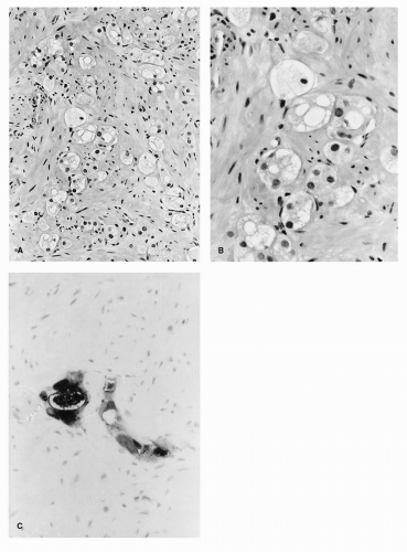
Care must be taken in evaluating post-RT prostate biopsies because radiation-induced atypia may be difficult to distinguish from residual cancer exhibiting severe radiation changes (6,7). Gaudin et al. (7) reviewed prostate sextant needle biopsies from 137 patients obtained at a median of 35.7 months after three-dimensional conformal RT (3DCRT) to investigate histologic changes within the prostate as a result of RT. After 3DCRT, histopathologic changes in benign prostate glands consisted of glandular atrophy, cytologic atypia, and basal cell prominence. The benign glands showed intensely positive reactions with antibodies to high-molecular-weight cytokeratin (34[beta]E12), and either negative or weakly positive reactions to PSA. The changes in benign prostate tissues were similar between patients treated with neoadjuvant androgen deprivation therapy and 3DCRT and those treated with 3DCRT alone. In contrast, two histologic patterns of prostate cancer after 3DCRT were identified: (a) prostate cancer showing no apparent RT effect and (b) prostate cancer showing RT-related changes characterized by poorly formed, PSA-positive/34[beta] E12-negative glands, and residual neoplastic cells containing abundant clear to finely granular cytoplasm (Figs. 13B.1A-C). The investigators concluded that, while the effect of 3DCRT on prostate cancer is variable, with some cases showing profound therapy-related changes and others showing no apparent therapy effect, post-3DCRT benign prostate glands show profound histopathologic changes that may be confused with prostate cancer. This distinction is critical because there is mounting evidence that a positive post-RT biopsy showing only prostate cancer with profound therapy-related changes identifies a subset of tumors with little or no biological activity. Crook et al. (8) studied irradiated prostate cancer and correlated the degree of RT effect to proliferating cell nuclear antigen (PCNA) immunohistochemistry. They found that prostate cancer with marked therapy effect showed significantly less PCNA immunoreactivity than prostate cancer with little or no therapy effect (17% vs. 61%). No local progression was observed in patients whose biopsies showed marked RT effect. Postradiation prostate biopsy showing only “prostate cancer with marked radiation effect” indicated a low risk for local failure, leading the investigators to suggest that such patients did not require additional local (i.e., salvage) therapy.


SALVAGE RADICAL PROSTATECTOMY

The ultimate goal of early detection of prostate cancer recurrence is to improve the efficacy of salvage therapy, the hypothesis being that early recurrences will more likely be organ confined and therefore amenable to salvage strategies. Salvage RP has been used successfully to eradicate locally recurrent prostate cancer after definitive radiotherapy, but complications are common (9,10,11,12,13,14,15,16,17,18,19,20,21). From these studies, several
generalizations can be made. First, the procedure should be reserved for patients in excellent health with a life expectancy of at least 10 years. Second, patients must have no evidence of metastatic disease and no evidence of lymph node involvement before RT if an initial pelvic lymph node dissection was performed. Salvage surgery should be offered only to those whose cancer (both initial and recurrent) is clinically organ confined and potentially curable. Third, patients should have no evidence of severe radiation cystitis or proctitis. In all of the studies referenced above, candidates for salvage surgery were advised of, and willing to accept, the higher morbidity associated with this approach.


FIGURE 13B.1. (A) RT-related changes in a prostate needle biopsy are characterized by a diminution in the number of neoplastic glands, which are often poorly formed and haphazardly arranged within the prostatic stroma. (B) The residual neoplastic cells had abundant clear, vacuolated, or reticulated cytoplasm and contained nuclei with only minimal degrees of pleomorphism. Nucleoli were often inconspicuous in prostate cancer with therapy effect. (C) The residual neoplastic cells with RT-related changes were intensely immunoreactive with antibodies to PSA. (Reproduced from Gaudin PB, Zelefsky MJ, Leibel SA, et al. Histopathologic effects of three-dimensional conformal external beam radiation therapy on benign and malignant prostate tissues. Am J Surg Pathol 1999;23:1021-1031, with permission.)

Salvage RP is technically feasible, with acceptable immediate intraoperative and postoperative outcomes, using current
surgical techniques (21). Salvage RP can be safely performed after failed external beam RT, brachytherapy (open or ultrasound-guided), or combinations of these techniques. The majority of patients can be treated via a retropubic approach, which is familiar to most urologic surgeons. Rarely is a combined abdominoperineal approach required.


TABLE 13B.1 COMPLICATIONS OF SALVAGE RADICAL PROSTATECTOMY


Author


Year


No. of Patients


EBL (mL)


Strictures (%)


Rectal Injuries (%)


Other (%)a


Incontinence (%)b


Mild


Severe


Thompson et al. (13)


1988


5


0


0


20


20


60


Link and Freiha (14)


1991


14


1,000


7


0


3.6


Moul and Paulson (15)


1991


4


800


100


0


Ahlering et al. (16)


1992


11


0


64


Pontes et al. (17)


1992


35


11.5


6


9


28


17


Stein et al. (18)


1992


11


1,100


18


0


27


64


Zincke (19)


1992


32


19


6.3


25


26.7


Brenner et al. (20)


1994


10


1,650


10


0


20


10


10


Rogers et al. (9)


1995


40


910


27.5


15


20


58


Gheiler et al. (12)


1998


30


1,100


13


7


17


23


26


Stephenson et al. (21)


2004


100


1,000


30


7


38


23


a Other major operative complications include postoperative hemorrhage, ureteral injury, and prolonged anastomotic leakage.

b Mild incontinence indicates stress incontinence requiring fewer than two pads per day, and severe incontinence implies greater than two pads per day.


EBL, estimated blood loss.


Overall, mean estimated blood loss and operative time do not differ significantly from estimated blood loss and operative time for standard RP. However, short-term and long-term complication rates exceed those of standard RP, due in part to the loss of the normal anatomic planes as a consequence of radiation. Table 13B.1 lists the rate of complications in several salvage RP series. Approximately 5% to 15% of patients in these series had rectal injuries, and as many as 25% had some other early complication of surgery, such as ureteral transection, prolonged anastomotic leakage, and/or pulmonary embolism. Rectal and other intraoperative injuries are especially common in patients who had prior staging pelvic lymphadenectomy and/or open seed implantation, both of which are associated with extensive fibrosis between the bladder, iliac vessels, prostate, and rectum; the former procedure is now infrequently performed and the latter has been abandoned. In the series from Baylor Medical Center (Rogers et al. [9]), 31% of patients who had previously undergone pelvic lymphadenectomy had a surgical complication, compared with only 9% of patients who initially received radiation alone.

In our series, 100 consecutive patients with biopsy confirmed, locally recurrent prostate cancer were treated with salvage RP with curative intent after failed RT (21). Forty patients were treated between 1984 and 1992, and 60 were treated between 1993 and 2003. Early in our experience (prior to 1993), the mean estimated blood loss, transfusion requirements, and average hospital stay were greater for salvage RP than for standard RP, but over time the morbidity of the operation has changed substantially. Rectal injuries during salvage RP occur less commonly today but are particularly concerning because of impaired healing of radiated tissues. With full bowel preparation before the operation, however, rectal injuries can usually be repaired primarily without altering postoperative recovery. In our series, the reoperation rate was significantly lower for patients treated since 1993 (3% vs. 15%, p = 0.05), and there were no perioperative deaths during that time.

The development of an anastomotic stricture and persistent urinary incontinence continue to be problematic after salvage RP. The overall anastomotic stricture rate is as high as 30%. Most patients will require multiple interventions for management of these strictures. For our entire series of 100 patients undergoing salvage RP, the overall recovery of urinary continence was 62% (95% CI, 49%-74%) (21). For the period prior to 1993, 45% (95% CI, 26%-64%) recovered urinary continence, while after 1993 the rate of recovery improved to 66% (95% CI, 49%-84%). This likely reflects not only an improvement in surgical technique but also better targeted radiation therapies leading to better preservation of the sphincteric mechanism. Twenty-three patients with persistent severe urinary incontinence were dry after insertion of an artificial urinary sphincter and only one patient has required sphincter revision. The artificial sphincter insertion rate, however, did not improve over time.

Erectile dysfunction has been considered almost inevitable after salvage RP, but in selected cases one or both neurovascular bundles may be preserved. While overall postsurgical potency in our series was low (16% [95% CI, 4%-28%]), many men had erectile dysfunction prior to salvage RP. The 5-year recovery was 45% (95% CI, 16%-75%) for men who were potent preoperatively. Of seven patients who underwent bilateral nerve-sparing procedures, five (71%) recovered functional erections. Importantly, in men undergoing neurovascular bundle preservation, we have not had a positive surgical margin in the area where the nerve bundle was preserved.

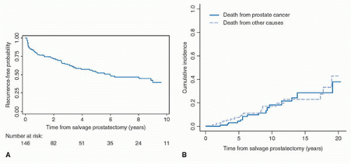
Data for cancer control outcomes from several salvage RP series are summarized in Table 13B.2. Most series demonstrate excellent cancer-specific survival at 5 years (>90%). We have recently updated our experience with oncologic outcomes after salvage RP (22). Of the 146 patients treated with salvage RP, the 5-year recurrence-free probability was 54% (95% CI, 44%-63%) (Fig. 13B.2A). Clinical local recurrence occurred in only one patient, who also had bone metastases. Overall, there were 16 prostate cancer-specific deaths and 19 deaths from other causes (Fig. 13.2B). The 5-year cumulative incidence of death from prostate cancer was 4% (95% CI, 2%-11%). Presalvage RP serum PSA level and biopsy Gleason score were significantly associated with death due to prostate cancer (p < 0.0005 and p = 0.002, respectively). These data confirm that salvage RP provides excellent local cancer control.


TABLE 13B.2 OUTCOMES AFTER SALVAGE RADICAL PROSTATECTOMY IN SELECTED SERIES OF PATIENTS


Clinical Nonprogression Rate (%)


Clinical Nonprogression Rate (%)


Cancer-Specific Survival Rate (%)


Authors


N


Clinical Stage


5 yr


10 yr


5 yr


10 yr


5 yr


10 yr


Rogers et al. (9)


38


T1-3 N0NX


55


33


83


67


95


87


Amling et al. (23)


108


T1b-N+


70


44


42


90


60


Gheiler et al. (12)


40


T2-3N0


47.4


87.5


Paparel et al. (22)


146


T1-3 N0NX


54


96


FIGURE 13B.2. (A) Kaplan-Meier probability of freedom from biochemical recurrence following salvage prostatectomy. (B) Cumulative incidence of death from prostate cancer following salvage prostatectomy. (Reproduced from Paparel P, Cronin AM, Savage C, et al. Oncologic outcome and patterns of recurrence after salvage radical prostatectomy. Eur Urol 2009;55:404-410, with permission.)


SALVAGE LAPAROSCOPIC RADICAL PROSTATECTOMY

Salvage RP has been described using standard and robotic-assisted laparoscopic approaches (24,25,26). Nuñez-Mora et al. (24) reported their results from nine patients treated with salvage laparoscopic RP between 2005 and 2007 after failure of either external beam RT (four patients) or brachytherapy (five patients). The average operating time was 170 minutes (range, 120-240). There were no conversions to open surgery, no cases of rectal injuries, and no patient required a blood transfusion. Four cases were pT2c, one was pT3a, three were pT3b, and one was pT4a. The Gleason score was 7 in three cases, 8 in two cases, and 9 in another four. Two patients had nodal metastasis. Postoperative PSA was undetectable in seven of the nine patients. Two patients experienced biochemical recurrence 16 and 13 months after the surgery. After a minimum follow-up period of 15 months, they were free from recurrence. There were no cases of anastomotic stricture. Three patients manifested severe incontinence (more than two pads per day), which was corrected in two cases by implanting an artificial sphincter. The other six patients required zero to one pads per day. The investigators concluded that there was less morbidity in their series in comparison with the rates of anastomotic stricture and rectal injury published for open salvage RP, but further study with larger numbers of patients is needed to confirm their findings.

Boris et al. (26) reported their initial results for salvage robotic-assisted RP in 11 patients. Six patients had brachytherapy, three had external beam RT, one had intensity-modulated RT, and one received brachytherapy with an external beam radiation therapy (EBRT) boost. The mean preoperative PSA level was 5.2 ng/mL. Average duration of surgery was 183 minutes, and the estimated blood loss was 113 mL. One patient had prolonged lymphatic drainage, one had an anastomotic leak, and one had an anastomotic stricture requiring direct vision internal urethrotomy at 3 months. Three patients had a biochemical recurrence at 1, 2, and 43 months, respectively. In patients with a minimum follow-up of 2 months, eight of ten were continent (defined as zero to one pad per day) and two had erections adequate for intercourse with the use of phosphodi-esterase-5 inhibitors. The investigators concluded that salvage robotic-assisted laparoscopic RP is feasible, with minimal perioperative morbidity and early functional outcomes appearing to be at least equivalent to historical salvage RP series.


ALTERNATIVE SALVAGE THERAPIES

Concerns regarding the risk of anastomotic stricture and urinary incontinence after salvage RP have prompted the search for less invasive salvage therapies. Although these treatments
are generally well tolerated, there are concerns that locally ablative therapies provide incomplete and inadequate treatment. The primary goal of any salvage local therapy for radiationrecurrent prostate cancer is to provide a durable cure; the prevention of symptomatic local and systemic progression is a secondary objective. As such, the oncologic efficacy of any treatment is judged by the ability of that treatment to accomplish these goals. It is difficult to assess the outcomes of most currently available salvage therapies because of a lack of long-term outcome data and/or the small sample sizes reported in published series (27). While each of these ablative salvage therapies (HIFU [28,29], brachytherapy [30-32], cryotherapy [33,34]) can be safely delivered, information regarding the benefit they provide in terms of cancer control is limited.


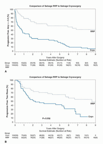
FIGURE 13B.3. (A) Biochemical disease-free survival defined as PSA 0.4 ng/mL or greater in all patients undergoing salvage local therapy. (B) Biochemical disease-free survival defined as 2 increases in PSA above nadir in all patients undergoing salvage local therapy. (Reproduced from Pisters LL, Perrotte P Scott SM, et al., Patient selection for salvage cryotherapy for locally recurrent prostate cancer after radiation therapy. J Clin Oncol 1999;17:2514-2520, with permission.)


Salvage HIFU

Zacharakis et al. (28) reviewed 31 patients treated with post-RT salvage HIFU between 2005 and 2007. Side effects included stricture or necrotic tissue in 11 of the 31 patients (36%), urinary tract infection or dysuria syndrome in eight (26%), and urinary incontinence in two (7%). Prostate-rectal fistula occurred in two (7%). After a mean follow-up of 7.4 months (range, 3-24), half of the patients had PSA levels of <0.2 ng/mL and 71% had no evidence of disease The investigators concluded that salvage HIFU is a minimally invasive outpatient procedure that can achieve low PSA nadirs and good cancer control in the short term, with morbidity comparable to other forms of salvage treatment. Gelet et al. (29) reported their experience treating 71 patients with salvage HIFU after EBRT. Mean follow-up was 14.8 months (range, 6-86). Efficacy was assessed by posttreatment biopsy; 57 patients (80%) had a negative biopsy. A nadir PSA level of <0.5 ng/mL was achieved in 43 of 71 patients (61%) within 3 months. At the last follow-up, 31 patients (44%) had no evidence of disease progression. Whether the results of salvage HIFU are durable awaits further follow-up, but these short-term oncologic outcomes seem inferior to salvage RP.


Salvage Cryoablation

Criteria to select men for salvage cryotherapy are similar to those used to select men for any local salvage treatment: an increasing PSA value after radiation, a positive postradiation biopsy, and a negative metastatic workup (33,34,35). While any man identified with local-only failure after radiotherapy is a potential candidate for salvage treatment, a variety of clinical factors have been suggested to predict the likelihood of success. Pisters et al. (36) examined outcomes after salvage cryotherapy in 145 men failing radiation for clinically localized prostate cancer. A PSA > 10 ng/mL prior to salvage cryotherapy and postradiation prostate biopsy Gleason score ≥9 were associated with failure. Additional recent studies have suggested that a postradiation PSA level >10 ng/mL or a PSA doubling time of 16 months or less was associated with increased likelihood of failure after salvage failure cryotherapy (33,37).

Longer-term outcomes with salvage cryotherapy have recently been reported. Ng et al. (33) assessed the efficacy of salvage cryotherapy in 187 patients with locally recurrent prostate cancer after RT. Approximately 70% of these patients also received ≥3 months of neoadjuvant androgen deprivation therapy. The mean follow-up period was 39 months. Biochemical recurrence was defined as nadir PSA level plus 2 ng/mL (Phoenix definition). Patients with precryotherapy PSA levels of <4 ng/mL had 5-year and 8-year biochemical recurrencefree survival rates of 56% and 37%, respectively. In contrast, patients with precryotherapy PSA levels of ≥10 ng/mL had 5-year and 8-year biochemical recurrence-free survival rates of only 1% and 7%, respectively. The four-quadrant positive prostate biopsy rate after salvage cryotherapy was 17%. Serum PSA level immediately prior to salvage cryotherapy was a predictive factor for biochemical recurrence, according to multivariate analyses. The investigators recommended that salvage cryotherapy should be performed when serum PSA level is still relatively low because the procedure may potentially be curative in these patients.

Pisters et al. (34) retrospectively compared treatment outcomes between 42 patients undergoing salvage RP and 56 patients receiving salvage cryotherapy at two large cancer centers. Eligibility criteria were locally recurrent prostate cancer after RT, PSA < 10 ng/mL, post-RT biopsy Gleason score ≤8 or less, and prior RT alone (i.e., no presalvage or postsalvage hormonal therapy). Biochemical failure was assessed using two criteria: (a) PSA > 0.4 ng/mL and (b) two increases above the postsalvage therapy nadir PSA. Mean follow-up was 7.8 years for the salvage RP group and 5.5 years for the salvage cryotherapy group. Compared to salvage cryotherapy, salvage RP resulted in superior biochemical disease-free survival at 5 years by both definitions of biochemical failure (PSA > 0.4 ng/mL: salvage cryotherapy 21% vs. salvage RP 61%, p < 0.001; two increases above nadir: salvage cryotherapy 42% vs. salvage RP 66%, p = 0.002) (Fig. 13B.3A and B). Salvage RP also resulted in superior overall survival at 5 years (salvage cryotherapy 85% vs. salvage RP 95%, p = 0.001). After adjusting for post-RT biopsy Gleason sum and presalvage treatment PSA, on multivariate analysis, salvage RP remained superior to salvage cryotherapy for the endpoints of any increase in PSA > 0.4 ng/mL (hazard ratio [HR] 0.24, p < 0.0001), two increases in PSA (HR 0.47, p = 0.02) and overall survival (HR 0.21, p = 0.01). The authors suggest that young, healthy patients with recurrent prostate cancer after RT should consider salvage RP because it offers superior biochemical diseasefree survival and may potentially offer the best chance of cure. Further studies are required to appropriately select candidates for salvage ablative therapies and to determine the long-term oncologic efficacy of these treatments.


CONCLUSION

Salvage RP, while technically challenging, provides excellent local control of radiation-recurrent prostate cancer, and it can eradicate the disease in a high proportion of patients treated when the cancer is confined to the prostate or immediate periprostatic tissue. As with standard RP, patient selection is of utmost importance. Continuing surgical challenges include the high rate of urinary incontinence and anastomotic strictures. Because the presalvage therapy PSA and biopsy Gleason score markedly predict outcome, improved methods of identifying radiationrecurrent prostate cancer while it is still confined to the prostate are needed to enhance patient outcomes. Improving our ability to better identify patients with radiation-recurrent prostate cancer earlier in the course of their disease is paramount.


References

1. Djavan B, Moul JW, Zlotta A, et al. PSA progression following radical prostatectomy and radiation therapy: new standards in the new Millennium. Eur Urol 2003;43:12-27.

2. Kaplan ID, Prestidge BR, Bagshaw MA, et al. The importance of local control in the treatment of prostatic cancer. J Urol 1992;147:917-921.

3. ZelefskyMJ,ShiW,YamadaY,etal.Postradiotherapy 2-year prostate-specific antigen nadir as a predictor of long-term prostate cancer mortality. Int J Radiat Oncol Biol Phys 2009;75(5):1350-1356.

4. Crook JM, Perry GA, Robertson S, et al. Routine prostate biopsies following radiotherapy for prostate cancer: results for 226 patients. Urology 1995;45:624-631; discussion 631-622.

5. Pucar D, Hricak H, Shukla-Dave A, et al. Clinically significant prostate cancer local recurrence after radiation therapy occurs at the site of primary tumor: magnetic resonance imaging and step-section pathology evidence. Int J Radiat Oncol Biol Phys 2007;69:62-69.

6. Bostwick DG, Egbert BM, Fajardo LF. Radiation injury of the normal and neoplastic prostate. Am J Surg Pathol 1982;6:541-551.

7. Gaudin PB, Zelefsky MJ, Leibel SA, et al. Histopathologic effects of three-dimensional conformal external beam radiation therapy on benign and malignant prostate tissues. Am J Surg Pathol 1999;23:1021-1031.


8. Crook J, Robertson S, Collin G, et al. Clinical relevance of trans-rectal ultrasound, biopsy, and serum prostate-specific antigen following external beam radiotherapy for carcinoma of the prostate. Int J Radiat Oncol Biol Phys 1993;27:31-37.

9. Rogers E, Ohori M, Kassabian VS, et al. Salvage radical prostatectomy: outcome measured by serum prostate specific antigen levels. J Urol 1995;153:104-110.

10. Cheng L, Sebo TJ, Slezak J, et al. Predictors of survival for prostate carcinoma patients treated with salvage radical prostatectomy after radiation therapy. Cancer 1998;83:2164-2171.

11. Tefilli MV, Gheiler EL, Tiguert R, et al. Salvage surgery or salvage radiotherapy for locally recurrent prostate cancer. Urology 1998;52:224-229.

12. Gheiler EL, Tefilli MV, Tiguert R, et al. Predictors for maximal outcome in patients undergoing salvage surgery for radio-recurrent prostate cancer. Urology 1998;51:789-795.

13. Thompson IM, Rounder JB, Spence CR, et al. Salvage radical prostatectomy for adenocarcinoma of the prostate. Cancer 1988;61:1464-1466.

14. Link P, Freiha FS. Radical prostatectomy after definitive radiation therapy for prostate cancer. Urology 1991;37:189-192.

15. Moul JW, Paulson DF. The role of radical surgery in the management of radiation recurrent and large volume prostate cancer. Cancer 1991;68:1265-1271.

16. Ahlering TE, Lieskovsky G, Skinner DG. Salvage surgery plus androgen deprivation for radioresistant prostatic adenocarcinoma. J Urol 1992;147:900-902.

17. Pontes JE, Montie J, Klein E, et al. Salvage surgery for radiation failure in prostate cancer. Cancer 1993;71:976-980.

18. Stein A, Smith RB, deKernion JB. Salvage radical prostatectomy after failure of curative radiotherapy for adenocarcinoma of prostate. Urology 1992;40:197-200.

19. Zincke H. Radical prostatectomy and exenterative procedures for local failure after radiotherapy with curative intent: comparison of outcomes. J Urol 1992;147:894-899.

20. Brenner PC, Russo P, Wood DP, et al. Salvage radical prostatectomy in the management of locally recurrent prostate cancer after 125I implantation. Br J Urol 1995;75:44-47.

21. Stephenson AJ, Scardino PT, Bianco FJ Jr, et al. Morbidity and functional outcomes of salvage radical prostatectomy for locally recurrent prostate cancer after radiation therapy. J Urol 2004;172:2239-2243.

22. Paparel P, Cronin AM, Savage C, et al. Oncologic outcome and patterns of recurrence after salvage radical prostatectomy. Eur Urol 2009;55:404-410.

23. Amling CL, Blute ML, Lerner SE, et al. Influence of prostate-specific antigen testing on the spectrum of patients with prostate cancer undergoing radical prostatectomy at a large referral practice [see comments]. Mayo Clin Proc 1998;73:401-406.

24. Nunez-Mora C, Garcia-Mediero JM, Cabrera-Castillo PM. Radical laparoscopic salvage prostatectomy: medium-term functional and oncological results. J Endourol 2009;23:1301-1305.

25. Vallancien G, Gupta R, Cathelineau X, et al. Initial results of salvage laparoscopic radical prostatectomy after radiation failure. J Urol 2003;170:1838-1840.

26. Boris RS, Bhandari A, Krane LS, et al. Salvage robotic-assisted radical prostatectomy: initial results and early report of outcomes. BJU Int 2009;103:952-956.

27. Nguyen PL, D’Amico AV, Lee AK, et al. Patient selection, cancer control, and complications after salvage local therapy for postradiation prostate-specific antigen failure: a systematic review of the literature. Cancer 2007;110:1417-1428.

28. Zacharakis E, Ahmed HU, Ishaq A, et al. The feasibility and safety of high-intensity focused ultrasound as salvage therapy for recurrent prostate cancer following external beam radiotherapy. BJU Int 2008;102:786-792.

29. Gelet A, Chapelon JY, Poissonnier L, et al. Local recurrence of prostate cancer after external beam radiotherapy: early experience of salvage therapy using high-intensity focused ultrasonography. Urology 2004;63:625-629.

30. Grado GL, Collins JM, Kriegshauser JS, et al. Salvage brachytherapy for localized prostate cancer after radiotherapy failure. Urology 1999;53:2-10.

31. Lo K, Stock RG, Stone NN. Salvage prostate brachytherapy following radiotherapy failure. Int J Radiat Oncol Biol Phys 2005;63:S290-S291.

32. Lee B, Shinohara K, Weinberg V, et al. Feasibility of high-dose-rate brachytherapy salvage for local prostate cancer recurrence after radiotherapy: the University of California-San Francisco experience. Int J Radiat Oncol Biol Phys 2007;67:1106-1112.

33. Ng CK, Moussa M, Downey DB, et al. Salvage cryoablation of the prostate: followup and analysis of predictive factors for outcome. J Urol 2007;178:1253-1257; discussion 1257.

34. Pisters LL, Leibovici D, Blute M, et al. Locally recurrent prostate cancer after initial radiation therapy: a comparison of salvage radical prostatectomy versus cryotherapy. J Urol 2009;182:517-525; discussion 525-517.

35. Finley DS, Pouliot F, Miller DC, et al. Primary and salvage cryotherapy for prostate cancer. Urol Clin North Am 2010;37(1):67-82.

36. Pisters LL, Perrotte P, Scott SM, et al. Patient selection for salvage cryotherapy for locally recurrent prostate cancer after radiation therapy. J Clin Oncol 1999;17: 2514-2520.

37. Spiess PE, Lee AK, Leibovici D, et al. Presalvage prostate-specific antigen (PSA) and PSA doubling time as predictors of biochemical failure of salvage cryotherapy in patients with locally recurrent prostate cancer after radiotherapy. Cancer 2006;107:275-280.


▪ 13C Salvage Brachytherapy for Prostate Cancer

Ryan J. Burri

Richard G. Stock


INTRODUCTION

Local failure following external beam radiation therapy (EBRT) for early prostate cancer has been noted to occur in up to one third of patients and has been associated with an increased risk of distant metastases and death due to prostate cancer (1,2). The ideal management of the patient who experiences a biopsy-proven local-only failure (i.e., distant disease is ruled out using computed tomography [CT] or magnetic resonance imaging [MRI] of the abdomen and pelvis and whole body bone scan) after definitive radiotherapy is controversial. Observation and androgen deprivation therapy (ADT) alone are not considered curative alternatives. Options for potentially curative local treatment include radical prostatectomy, cryotherapy, brachytherapy (BT), highintensity focused ultrasound (HIFU), and photodynamic therapy (PDT) (3).

Several institutions have published short- and intermediateterm biochemical control using these potentially curative local approaches, but long-term biochemical outcomes and toxicity data are limited. Toxicities that may potentially result from all five local therapeutic options include rectal injury, incontinence, and impotence. Anastomotic leak and bladder neck contracture are unique to surgery, and obstructive uropathy requiring transurethral resection of the prostate (TURP) may occur after HIFU, PDT, cryotherapy, or BT. This chapter outlines the oncologic and quality of life outcomes in patients with locally recurrent prostate cancer treated with salvage BT.


PATIENT SELECTION

The typical presentation of a potential candidate for salvage BT is a prior history of external beam irradiation delivered 2 to 8 years in the past and a slow rising prostate-specific antigen (PSA). In a series from Mount Sinai, the median patient age was 70 years and the median time to salvage BT was 63 months (range 27-171 months) (4). For RTOG 0526, an open trial testing salvage BT, patients are eligible if they have a duration from external radiation to salvage of >30 months (www.RTOG.org).

Prior to proceeding with salvage local therapy, the patient must undergo a postirradiation prostate biopsy. Six to twentyfour core samples can be taken via a transrectal or transperineal
prostate biopsy. The pathology slides should be reviewed by a pathologist with experience interpreting postradiation prostate biopsies. Biopsy specimens showing persistent prostate cancer with no radiation effect or those with persistent cancer in the setting of radiation effect should be considered positive. Following pathologic confirmation of local failure, the extent of disease workup includes a thorough history and physical examination followed by routine laboratory studies, pelvic CT scan, whole body bone scan, and PSA determination. In addition, seminal vesicle biopsies may be performed to rule out more extensive local spread of disease.

At the time of salvage, most patients have a Gleason score of 7 or 8 to 10, which is higher than the average Gleason score at the time of the initial radiation therapy. In the Mount Sinai series, the Gleason score at post radiation biopsy was ≤6 in 19%, 7 in 46%, 8 to 10 in 32%, and unknown Gleason score in 3% (4). In this series, the PSA levels of patients prior to BT were ≤10 ng/mL in 76%, >10 to 20 ng/mL in 22%, and >20 ng/mL in 3% (4). Tumor staging with digital rectal examination is performed but is not considered prognostic following an initial course of radiation therapy.

In RTOG 0526, a phase II trial testing the efficacy and safety of salvage BT in the treatment of recurrent cancer following external beam irradiation, the selection criteria included the following conditions, initial presentation of T1-2 disease, Gleason score 2 to 7, PSA < 20 ng/mL, post radiation and pre-BT PSA < 10 ng/mL, and AUA urinary symptom score <15 with no history of TURP or other surgical procedures to the prostate (www.RTOG.org).

There are little published data on contraindications for salvage prostate BT. There are two types of patients who present with relative contraindications for salvage BT. The first groups are those who have experienced morbidity related to their original external beam treatment. These include patients with radiation proctitis and rectal bleeding as well as those who went into urinary retention following their external beam treatment. These patients are at too great a risk for further complications following seed implantation. The other group represents those who present with a very high risk of having microscopic systemic disease. These include patients with PSA > 20 ng/mL, PSA doubling times ≤3 months and those with positive seminal vesicle biopsies.


SALVAGE BRACHYTHERAPY PROCEDURE

Salvage prostate low dose rate prostate BT is performed in a similar manner to upfront BT treatment. It is always important to obtain the prior records from the initial external beam radiation treatment to make sure that critical normal adjacent structures were not overdosed. The procedure itself is usually performed under spinal or general anesthesia in much the same way as primary prostate BT. This most commonly involves the placement of low-dose-rate (LDR) permanent radioactive seeds (typically iodine-125 [125I], or palladium-103 [103Pd]) via needles inserted into the prostate though a transperineal template under endorectal ultrasound guidance (4). The lower energy and shorter half-life of 103Pd makes it an ideal choice for salvage BT on a theoretical basis. There are a few reports of high-dose-rate (HDR) salvage prostate BT, with comparable results to those of LDR procedures but with shorter follow-up (5,6). Radiation dose plans are generated either prior to the procedure or intraoperatively, and final dosimetric analysis of the implant is performed using CT scans, typically a month post procedure. With few exceptions (7,8), the entire prostate is treated as the high-dose target volume.

The appropriate dose to use in the salvage setting is unknown. In upfront BT procedures, the prescription dose for a full 103Pd implant is 124 Gy and for an implant to be combined with 45 Gy of external beam it is 100 Gy. At Mount Sinai, we have chosen a dose in between these two doses of 110 Gy. This takes into account the prior full dose of external beam as well as the need to control gross recurrent disease. Since the dose is less than an upfront dose, we have added both neoadjuvant and adjuvant hormonal therapy to the implant. This typically takes the form of an LHRH agonist given 3 months before the implant and 3 months after the implant for a total duration of 6 months (4).

In RTOG 0526, the prescription dose for an 125I implant is 140 Gy to cover the planning target volume (PTV) and for 103Pd it is 120 Gy (www.RTOG.org).


SALVAGE BRACHYTHERAPY SERIES

The following series have in common a group of men with biopsy-proven, locally recurrent prostatic adenocarcinoma following definitive EBRT with distant disease ruled out, typically by whole body bone scan and CT or MRI of the abdomen and pelvis. In addition, the number of patients in each is relatively small compared to upfront treatment outcome reports. Biochemical control rates and toxicity data, where available, are presented.

In a retrospective series of 49 patients treated with salvage BT by Grado et al. (9) from the Mayo Clinic Scottsdale, the 5-year actuarial biochemical disease-free survival was 34%. The median follow-up was 64 months, and failure was based on the definition of two successive rising PSA values above the posttreatment PSA nadir value. Postsalvage PSA nadir <0.5 ng/mL was significantly associated with improved biochemical disease-free survival. Serious complications (Grade ≥ 3) requiring surgical intervention developed in eight patients (16%).

Only gold members can continue reading. Log In or Register to continue

Stay updated, free articles. Join our Telegram channel

Jul 15, 2016 | Posted by in ONCOLOGY | Comments Off on Treatment of Locally Recurrent Prostate Cancer

Full access? Get Clinical Tree

Get Clinical Tree app for offline access