Adrenal Neoplasms


Adrenal Neoplasms


Mhd. Yaser Al-Marrawi

Inderbir S. Gill

Ronald M. Bukowski

Brian I. Rini


HISTORICAL PERSPECTIVE

The adrenal glands play a prominent role in maintaining normal homeostasis. Bartholomeo Eustachi first recognized them as organs that are distinct from the kidneys in 1563 (1). Knowledge of structure and function of the adrenal glands originated in 1805 when Currier recognized the presence of a cortex and medulla within the gland. In 1855, almost 300 years after the original description of these glands, Thomas Addison described the clinical effects of adrenal insufficiency (2). The demonstration by Brown-Sequard that death occurred following adrenalectomy firmly established the importance of the adrenal gland (3).

Sir William Osler reported the beneficial effects of adrenal extracts for the treatment of Addison disease and thus began attempts at studying and using crude adrenal extracts. Isolation of cortisol from the adrenal cortex and adrenaline from the medulla was accomplished in the first half of the 20th century, and the modern era of adrenal physiology can be dated from the time of these significant research efforts. In view of the central and complex role of this gland in normal homeostasis, a discussion of adrenal neoplasms, their diagnosis, and treatment requires an introduction to normal as well as pathologic adrenal physiology.


ADRENAL PHYSIOLOGY


Adrenal Cortex

The adrenal cortex comprises the bulk of the adrenal gland and is the site of synthesis of many types of steroid hormones. The steroid synthesis pathway is a complex sequence of enzymatically mediated biochemical steps by which adrenal steroids are derived from cholesterol. Clinical manifestations of steroid excess result from autonomous production of various steroid compounds and precursors. The terms functional and nonfunctional have been applied to cortical neoplasms based on their clinical manifestations and/or excess hormone production. This may be misleading as adrenocortical tumor steroidogenesis differs from normal steroidogenesis in several aspects. Adrenal tumors often synthesize steroids that are not normally major adrenal products; instead these steroids may be early precursors such as Δ5-pregnenolone, dehydroepiandrosterone (DHEA), or similar compounds (4,5,6). These steroids may not have glucocorticoid/androgenic activity or may have only weak activity and, therefore, clinical syndromes of excess may not be seen. Many tumors and probably most malignant tumors do not respond to adrenocorticotropin (ACTH), another cardinal characteristic of constitutive steroidogenesis in adrenal neoplasms (5).


Adrenal Medulla

The adrenal medulla consists of chromaffin tissue originating from neuroectoderm. This portion of the adrenal gland is the major source of catecholamines, including epinephrine. Synthesis of these compounds proceeds through a series of enzymatic reactions involving tyrosine to epinephrine. Catecholamine synthesis occurs predominantly in chromaffin cells but also occurs in parts of the central nervous system (CNS) and postganglionic nerves (7).

The major portion of plasma catecholamine is composed of norepinephrine, with dopamine and epinephrine accounting for <30% (8). The metabolic products of these catecholamines include homovanillic acid (dopamine), metanephrines, and vanillylmandelic acid (epinephrine, norepinephrine). Only small amounts of epinephrine, norepinephrine, and dopamine are excreted unchanged in the urine.

The metabolic and physiologic effects of catecholamines include promotion of hepatic glycogenolysis and inhibition of hepatic glyconeogenesis plus insulin release (9). Cardiovascular effects include positive inotropy of the heart and both vasoconstriction and vasodilation. The overproduction of various catecholamines by tumors of chromaffin tissue (e.g., pheochromocytoma) is responsible for syndromes typically associated with these neoplasms.


ADRENOCORTICAL TUMORS—PATHOLOGY

In autopsy series, the prevalence of clinically silent adrenal masses is about 2.1%, which increases to about 7% in patients older than 70 years (10,11). In contrast, the prevalence of adrenocortical carcinoma (ACC) is rare. The National Cancer Institute’s Surveillance, Epidemiology, and End Results database suggests an incidence of 0.5 to 2.0 per million populations per year, forming 3.6% of all endocrine malignancies (12). The age-adjusted incidence rates were reported to be 0.1 to 0.2 per 100,000 over this period (13).

The relation between adrenal adenoma and carcinoma is unclear. Progression from a benign adenoma to carcinoma was felt to be unlikely in view of the wide disparity in incidence. On the basis of clonal analysis and many studies with comparative genomic hybridization, data have been emerging that suggest that adrenal tumorigenesis is a multistep process and that sequential progression from normal to adenomatous cells and then to malignant cells occurs (14,15).


TABLE 50.1 HISTOLOGIC CLASSIFICATIONS OF NEOPLASMS OR TUMOR-LIKE LESIONS OF ADRENAL CORTEX AND MEDULLA


Adrenal cortical tumors


Adenoma


Carcinoma


Myelolipoma


Tumor-like adrenocortical nodules


Other


Adrenal medullary tumors


Pheochromocytoma


Neuroblastoma, ganglioneuroblastoma


Other


Miscellaneous neoplasms and tumor-like lesions


Adrenal cyst


Primary mesenchymal or neural tumors


Metastatic tumors


Other


Recommendations for reporting of tumors of the adrenal cortex and medulla. Association of Directors of Anatomic and Surgical Pathology.
Adapted from Hum Pathol. 1999 Aug;30(8):887-890.


The Association of Directors of Anatomic and Surgical Pathology recommended guidelines (Table 50.1) for the histologic classification of neoplasms or tumor-like lesions of the adrenal cortex and medulla (16).


Adrenocortical Adenoma Pathology

Adrenocortical adenomas are benign tumors of the adrenal cortex and, on cross-section, are often well defined with apparent encapsulation. Occasionally, on cut section, there may be areas of hemorrhage, but confluent tumor necrosis is unusual (17). Microscopically, adenomas typically contain nests and cords of cohesive vacuolated cells with abundant cytoplasm (Fig. 50.1A). They are composed of lipid-rich spongiocytes or of compact cells that are devoid of lipid. Generally, mitoses are lacking, but focal nuclear atypism and hyperchromasia may be seen (18). Vascular and capsular invasion are also absent. The histologic and anatomic differences distinguishing benign and malignant adrenal tumors are still controversial (19,20).


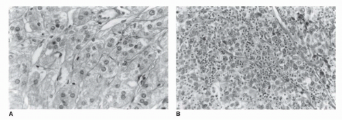
FIGURE 50.1. Histologic comparison of a benign adrenocortical adenoma and a carcinoma. (A) Benign adenoma. Adenomas are characterized by nests and cords of clear cells with abundant cytoplasm separated by fibrovascular trabeculae. Mitoses are absent or rare, and cells have an orderly, monotonous appearance. (B) Adrenocortical carcinoma (ACC). This cancer retains the fibrovascular trabeculae, but the cells demonstrate marked pleomorphism, enlarged and hyperchromatic nuclei, and obvious nucleoli.


Adrenocortical Carcinoma Pathology

Grossly, ACC has a coarse nodular structure with areas of necrosis, hemorrhage, and, occasionally, cystic degeneration. Microscopically (Fig. 50.1B), the architecture can be trabecular, alveolar (nested), diffuse (solid), or a combination of these patterns (17,21).

Reliable differentiation of ACC from adenoma or benign lesions may be difficult. Hough et al. (22) evaluated 41 patients with ACC for the presence of 15 histologic and 5 nonhistologic features that had been associated with malignant behavior. The most significant criteria were evidence of weight loss, broad fibrous bands, diffuse pattern, vascular invasion, necrosis, and tumor mass. Van Slooten et al. (23) assessed the discriminatory value of seven histologic parameters. In their series of 45 patients with more than 10 years of follow-up, tumor weight and mitotic activity as single parameters had the highest discriminating value. The calculated histologic index (based on the sum of the numeric value of each parameter) had a critical value of 8, such that tumors with a histologic index <8 were smaller and less likely to be malignant. However, absolute differentiation between adenoma and carcinoma was not possible.

A third set of criteria proposed by Weiss et al. (19) on evaluation of 43 adrenocortical tumors is listed in Table 50.2. In a review by Medeiros and Weiss (20), it is noted that the following characteristics are seen only in malignant adrenocortical tumors: mitotic rate of 5 or more per 50 high-power field, atypical mitotic figures, and venous invasion. It appears that all three sets of criteria are useful in predicting the behavior of adrenocortical neoplasms.

There does not appear to be a diagnostic immunoprofile for ACC, but immunohistochemistry still plays some role in its evaluation (18,24,25). Keratin and vimentin expression have been studied; keratin positivity, however, varies with the methods of tissue fixation and preparation. Vimentin expression appears to have an inverse relation with cytokeratin. Most carcinomas express this protein intensely. Normal cortical cells are negative and adenomas demonstrate intermediate expression. Another study has reported positive staining for neuron-specific enolase, synaptophysin, and neurofilament protein (26). The oncogene p53 and MIB-1
(a marker of cell proliferation) may be overexpressed in carcinomas compared to normal adrenal tissue, and benign adenomas (27). Immunostaining for chromogranin is positive in pheochromocytomas and negative in ACC. Similarly, the monoclonal antibody D11 has utility in distinguishing adrenocortical tumors from other tumors, but not benign from malignant. It therefore appears that immunohistochemical studies are useful in distinguishing adrenal neoplasms from other tumors but are of limited value in differentiating benign and malignant adrenal tumors.


TABLE 50.2 HISTOLOGIC CRITERIA OF MALIGNANCY IN ACC


1.


High nuclear grade


2.


Mitotic rate of 5 or more per 50 high-power field


3.


Atypical mitoses


4.


Eosinophilic tumor cell cytoplasm (≥75% cells)


5.


Diffuse pattern/architecture


6.


Presence of necrosis


7.


Vascular invasion


8.


Sinusoidal invasion


9.


Invasion of capsule


Adapted from Medeiros LJ, Weiss LM. New developments in the pathologic diagnosis of adrenal cortical neoplasms: a review. Am J Clin Pathol 1992;97:73.


Early studies suggested that carcinomas are aneuploid (28); in one series, however, 6 of 30 (20%) adenomas also contained aneuploid cell lines (29). Similarly, studies of telomerase, which is an enzyme that adds repeated telomere sequences to the ends of chromosomes arms maintaining cell proliferation, have shown conflicting results (30,31).


Myelolipoma

Myelolipomas are benign endocrine-inactive tumors (Fig. 50.2) composed of mature adipose tissue and fatty elements (32). The hematopoietic cells include granulocytic, erythroid, and, rarely, megakaryocytic precursors. Sparse adrenocortical tissue may also be found. These tumors are thought to arise from metaplasia of undifferentiated stromal tissue (33). Myelolipomas are usually small, nonfunctional, and discovered incidentally (33). They may occasionally reach an extremely large size and cause symptoms. Asymptomatic individuals can be clinically monitored, with periodic radiographic assessment every few years.


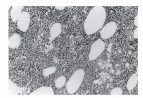
FIGURE 50.2. Histologic appearance of a myelolipoma showing fat (white areas) intermixed with myeloid tissue.


Adrenal Cysts

Adrenal cysts are rare, usually asymptomatic lesions occurring more frequently in women. Endothelial-lined cysts of lymphatic origin account for almost one half of all adrenal cysts. The next most common cysts are pseudocysts, which result from old adrenal hemorrhages. These are usually thick walled and unilocular and contain red-brown fluid. Least common are epithelial cysts derived from cystic cortical adenomas (Fig. 50.3). Adrenal cysts are benign and generally treated by simple excision. Because adrenal cancers and pheochromocytomas may bleed and give rise to pseudocysts, frozen section examination of the cyst wall should be obtained at the time of excision.


Evaluation of the Incidental Adrenal Mass

Given the increased use of radiologic imaging, incidental adrenal masses are detected in 0.6% to 1.3% of abdominal computed tomography (CT) scans and in 2% to 9% of autopsy series (34,35). Histologically, most of these tumors are benign and nonfunctional adenomas. The clinical challenge is to differentiate between clinically important entities that require therapy versus the nonfunctioning adenomas that can be safely observed. Table 50.3 outlines the differential diagnosis of an adrenal mass lesion. Figure 50.4 illustrates a useful algorithm in the workup and treatment of incidentally discovered adrenal masses. The history and physical examination should include a careful survey for subtle manifestations of hyperfunctioning tumors. The syndromes associated with functional adrenal masses include hypercortisolism or Cushing syndrome (Tables 50.3 and 50.4; Fig. 50.5), hyperaldosteronism or Conn syndrome, pheochromocytomas (see subsequent text and Fig. 50.6), and feminization or virilization syndromes. A careful search for occult primary tumors of other sites should be made, with special attention to the lung and breast cancers as the most common source of adrenal metastases. Hypertension is nonspecific and may be associated with Cushing syndrome, aldosterone-secreting tumors, and pheochromocytoma. A history of upper abdominal or flank trauma may suggest adrenal hemorrhage.


Biochemical Assessment of the Incidental Adrenal Mass

The initial biochemical assessment of an incidental adrenal mass is straightforward, with more detailed confirmatory tests reserved for equivocal cases or those in which the initial functional evaluation is normal but the clinical suspicion of a functional tumor is high. An initial biochemical screen should include measurement of serum levels of electrolytes and of 24-hour urine metanephrines, catecholamines, and free cortisol. A positive result from any screening test or high clinical suspicion based on a patient’s history/physical examination should prompt a full functional screening. This tailored style of screening helps lessen costs without negatively affecting accuracy (36). In settings where initial tests are normal, the tumor is <3 cm, the patient is asymptomatic, no evidence of occult primary tumor exists, and the radiographic appearance is compatible with a benign nonfunctioning adrenocortical adenoma, the patient may be safely followed radiographically with serial CT scanning.

An elevated urinary cortisol level suggests the presence of Cushing syndrome. The rationale for recommending the dexamethasone suppression test is the detection of subclinical glucocorticoid excess (Table 50.5). This subclinical syndrome
is not well characterized and its impact on long-term morbidity of patients with benign adrenal masses is unclear. The reader is referred to an article by Boscaro et al. (37) for a detailed review of the diagnosis and management of Cushing syndrome. A finding of hypokalemia suggests an aldosterone-secreting tumor. This suspicion should be confirmed by a plasma aldosterone concentration to plasma renin activity (PAC:PRA) ratio. A ratio >30 and a PAC of >0.5 nmol/L are highly suggestive of autonomous aldosterone production. For a detailed review of the diagnosis and management of adrenal aldosterone-secreting tumors, the reader is referred to an outstanding review (38). Elevated 24-hour urinary catecholamines are indicative of pheochromocytoma. For diagnosis of pheochromocytoma, plasma-free metanephrines (normetanephrine and metanephrine) should be measured. These combined tests have a sensitivity of 99% and specificity of 89%.


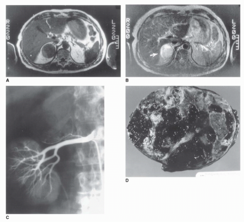
FIGURE 50.3. Radiographic and gross pathologic characteristics of a cystic benign adrenal adenoma. (A) T1-weighted magnetic resonance image in a 57-year-old woman performed after incidental binding of an adrenal mass on CT scan obtained because of vague abdominal discomfort. A 5-cm homogeneous mass isodense with the liver replaces the right adrenal gland. (B) T2-weighted MRI in the same patient. The tumor (arrow) appears hyperintense compared with the liver, a finding suggestive of malignancy. (C) Selective right renal angiogram in the same patient. The tumor is hypovascular and displaces branches of the right adrenal artery. The upper pole of the right kidney is displaced interiorly and laterally. (D) Gross specimen revealing multiple hemorrhagic pseudocysts arising from a benign cortical adenoma.


Radiologic Assessment of Incidental Adrenal Masses


Ultrasound

The diagnosis of adrenal tumors using ultrasonography is nonspecific because both benign and malignant tumors can be solid or contain cystic areas due to hemorrhage or necrosis. Ultrasound has a low sensitivity for detection of small adrenal lesions and has poor characterization of extension to adjacent structures for larger lesions.


TABLE 50.3 DIFFERENTIAL DIAGNOSIS OF AN ADRENAL MASS


Benign Masses


Malignant Masses


Adenoma


ACC


Amyloidosis


Angiosarcoma


Adrenal cyst


Leiomyosarcoma


Congenital hyperplasia


Malignant scwannoma


Ganglioneuroma


Malignant melanoma (primary)


Granuloma


Metastatic carcinoma


Hamartoma


Malignant pheochromocytoma


Hemorrhage


Neuroblastoma


Hemangioma


Infection


Leiomyoma


Lipoma


Myelolipoma


Neurofibroma


Nodular hyperplasia


Pheochromocytoma


ACC, adrenocortical carcinoma.


Computed Tomography Scan

CT imaging of adrenal incidentalomas has been used to assess the risk of malignancy. Adrenal cysts, myelolipomas, and adrenal hemorrhage have CT features that are diagnostic and seldom require further evaluation. Myelolipomas have large amounts of fat, which produces low-signal-intensity on CT scans (Fig. 50.7). CT scans also define adrenal cysts better than ultrasound studies, especially with contrast enhancement. In a review of 37 benign cysts, 19 had mural and 7 had central calcifications, 28 were unilocular and 7 had high attenuation values (39). The wall thickness was <3 mm in 31 lesions. The authors concluded that the finding of a nonenhancing mass with or without calcifications could distinguish a cyst from an adenoma (Fig. 50.8). The high attenuation value of a hemorrhage is also readily apparent on a nonenhanced CT scan. Benign adenomas are usually homogeneous lesions with smooth, regular, encapsulated margins that do not increase in size over time.

Using data from multiple published studies, the optimal sensitivity (71%) and specificity (98%) for the diagnosis of adenoma results from choosing a threshold attenuation of <10 Hounsfield units (HU) on a nonenhanced CT scan (40). Several studies have also reported high sensitivity and specificity of density readings on a delayed postcontrast CT scan. In a study of 166 adrenal masses on nonenhanced CT scans, those with attenuation values >10 HU were prospectively evaluated with contrast-enhanced CT scans and contrast-enhanced CT scans with 15-minute delays (41). The sensitivity and specificity of diagnosing an adrenal adenoma versus metastasis were 98% (124/127) and 97% (33/34), respectively. Primary ACCs usually have a more aggressive appearance on CT scan, characterized by irregular contour, large size, invasion of surrounding structures, and heterogeneous internal architecture (Fig. 50.9). Central necrosis is common, and calcification is seen in 20% to 30% of cases (42).


Magnetic Resonance Imaging

Magnetic resonance imaging (MRI) has the advantage of avoiding ionizing radiation and has been used in several studies in an attempt to differentiate benign from malignant adrenal masses (43). Adenomas typically show low signal intensity on T2-image sequence, whereas both pheochromocytomas and adrenal metastases have bright signal intensity on T2-weighted images (Figs. 50.8, 50.9 and 50.10). Carcinomas are typically heterogeneously hyperintense on both T1- and T2-weighted images, reflecting frequent internal hemorrhage and necrosis. Intravenous extension is very well depicted on MRIs, and they can be helpful in defining the cephalad extension of the tumor (44). Chemical-shift MRI imaging results in a decrease in signal intensity of tissue containing both lipid and water as compared to lipid alone. Benign adenoma exhibits a signal drop on T2-weighted images and can be distinguished from metastases. There are no conclusive data using this technique with ACC, and, therefore, its use cannot be justified in the differentiation of adenoma from ACC (44).


TABLE 50.4 CLINICAL FEATURES OF HYPERCORTISOLISM


General


Neuropsychiatric


Weight gaina


Emotional liabilitya


Hypertensiona


Psychosis


General skin disorders


Metabolic changes


Plethoraa


Glucose intolerancea


Hirsutisma


Hyperlipidemiaa


Striae


Polyuria


Acne


Hypokalemia


Bruising


Kidney stones


Musculoskeletal


Gonadal dysfunction


Osteopeniaa


Menstrual disordersa


Weakness/fatiguea


Impotence/decreased libido


a Noted in more than 50% of patients.


Adrenocortical Scintigraphy

Adrenocortical scintigraphy with 131I-labeled 6β-iodomethylnorcholesterol (NP59) and adrenal medullary imaging with 131I-labeled or 123I-labeled metaiodobenzylguanidine (MIBG) provide functional metabolic information for characterization of adrenal lesions (45,46). NP59 is a cholesterol analog and as a radiotracer is taken up by functional adrenocortical cells. If the uptake corresponds to a lesion on a CT or MRI, the diagnosis of adenoma is specific (44). Nonfunctioning malignancies or other lesions demonstrate decreased or absent (discordant) uptake. MIBG has high specificity for pheochromocytomas. These techniques, however, are expensive, time consuming, and not readily available. Therefore, data on their clinical usefulness are insufficient.


Positron Emission Tomography Scan

Data are emerging on the use of 18FDG positron emission tomography (PET) in characterization of adrenal lesions. In one reported series of 50 adrenal lesions in 41 patients, PET identified 18/18 malignant lesions with 100% sensitivity and 94% specificity (46). However, as more data mature, it is becoming evident that adenomas can also have increased FDG uptake and give false-positive results (47). The value of PET scans for differentiating various adrenal lesions remains uncertain.

A prospective study was conducted to evaluate the usefulness of [18]F-FDG PET to predict malignancy in patients without prior history of cancer. The study patient underwent surgery because of hypersecretory, growing benign lesions, obvious ACCs, or radiologically indeterminate lesions. The degree of PET max SUV was compared with that of CT scan for each lesion. This study showed that PET scan can predict the benignity in most suspicious CT scan lesions by using an adrenal to liver max SUV ratio <1.45 (222).


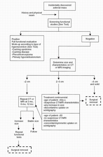
FIGURE 50.4. Algorithm illustrating the stepwise clinical evaluation and treatment of a patient with an incidentally discovered adrenal mass. (Modified from Moinzadeh A, Libertino JA. Asymptomatic adrenal mass. In: Resnick MI, Elder JS, Spirnak JP, eds. Critical decisions in urology, 3rd ed. Hamilton, ON: BC Decker Inc., 2004:381.)


Treatment of the Incidental Adrenal Mass

The major treatment issue is whether an incidentally discovered adrenal lesion is functional or nonfunctional and whether it is benign or malignant. If the history, physical examination, and radiologic and biologic assessment confirm the presence of a unilateral functioning lesion, the treatment of choice even for smaller lesions is surgical excision. The long-term effects of mild biochemical derangement in the absence of clinical symptoms are unknown; therefore, adrenalectomy and careful observation remain options for such patients. In patients with nonfunctioning tumors, consideration should be given to size, imaging characteristics, and growth rate to help in distinguishing benign from malignant tumors (221). Tumors larger than 6 cm have a high likelihood of malignancy. The incidence of ACC in adrenal masses <6 cm in diameter has varied from 35% to 98% in the literature (43). In a review of 630 cases of incidentalomas, 26 ACCs were found, and 85% of these were larger than 6 cm (48). In another retrospective review of 210 patients with adrenal masses, 15 adrenocortical cancers were discovered that formed 13% of their adrenal masses in patients who were operated upon. Using a cutoff of 5.0 cm yielded a specificity of 93% and sensitivity of 64% for the diagnosis of malignant lesion (49).


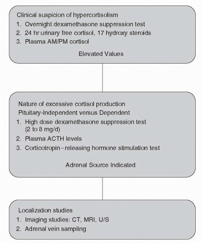
FIGURE 50.5. Nomogram illustrating the stepwise clinical evaluation of patients with suspected hypercortisolism.


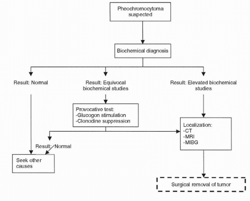
FIGURE 50.6. Algorithm illustrating the stepwise clinical evaluation and treatment of a patient with pheochromocytoma. (Modified from Moinzadeh A, Libertino JA. Pheochromocytoma. In: Resnick MI, Elder JS, Spirnak JP, eds. Critical decisions in urology, 3rd ed. Hamilton, ON: BC Decker Inc., 2004:381.)

The treatment of nonfunctioning tumors of 3 to 6 cm in diameter remains controversial because of insufficient data on their natural history in untreated patients. Management options include surgical removal, percutaneous biopsy, and observation with serial radiographs. Surgical excision offers the certainty of histologic diagnosis and the potential for cure of the occasional small primary tumor. Since the first description of laparoscopic adrenalectomy by Gagner in 1992 (50), numerous series have been reported supporting the advantages of the laparoscopic approach for the excision of the adrenal gland (51,52). Studies have documented that laparoscopy is safe, feasible, and capable of removing large tumors (53). Established advantages of this approach over the conventional “open” approach include less postoperative pain, shorter hospital stay, better cosmesis, and earlier postoperative convalescence (53,54). Laparoscopy has been applied to functional and nonfunctional adenomas, ACCs, adrenal metastases, and pheochromocytomas with acceptable morbidity (50,54). At this writing, laparoscopic adrenalectomy may be considered the standard approach for benign surgical tumors of the adrenal gland. As with any surgical procedure, proper patient selection and laparoscopic expertise are necessary for successful surgical outcomes. The laparoscopic approach may be contraindicated in patients with large tumors (>10 cm), malignant pheochromocytoma with adenopathy, and suspected ACCs with invasion of surrounding structures.

Percutaneous needle biopsy is limited by difficulties in distinguishing benign from malignant adrenocortical tumors, but it has a role in confirming the presence of metastatic tumors. In one series of 28 patients with metastatic tumors to the adrenal gland, 26 (94%) were accurately diagnosed by percutaneous biopsy (55). However, biopsy of an incidental adrenal mass without suspicion of metastatic disease is not indicated. In summary, decisions for management should be individualized based on the patient’s age, medical status, and willingness to undergo surgery versus tolerating uncertainty of diagnosis, as well as the size and functional status of the adrenal mass.


TABLE 50.5 DEXAMETHASONE SUPPRESSION TESTS: INTERPRETATION OF RESULTS


Diagnosis


Additional studies


Low-dose suppression test


Serum cortisol level decreases


Normal pituitaryadrenal axis


Recheck serum cortisol level; obtain 24-hr urine sample for free cortisol level


No change in serum cortisol level


Perform high-dose dexamethasone suppression


High-dose suppression test


Serum cortisol decreases


Cushing disease (pituitary adenoma)


Surgical/medical management


No change in serum cortisol


Check ACTH level; if low, consider functional adenoma or carcinoma; if high, consider ectopic sources of production


ACTH, adrenocorticotropic hormone.


Prognosis

Most adrenal incidentalomas are nonfunctional adenomas that do not require surgery. In a series of 27 patients with 30 incidentalomas followed up for 7 years, none of the nine deaths were attributed to the incidentaloma (56). Five of the twenty-one adrenal masses had increased in size and none had developed hormonal production on evaluation. Young (57) reported on four studies that included a total of 238 patients with adrenal incidentalomas followed for an average of
4.2 years. The adrenal masses enlarged in 8% and decreased in size in 1.3%, and none of the lesions proved to be malignant.


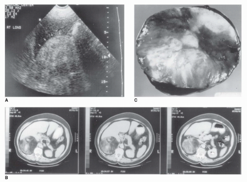
FIGURE 50.7. Radiographic and pathologic appearance of an adrenal myelolipoma. (A) Renal ultrasound in a 47-year-old woman with right flank pain. A large, solid tumor with the hyperechoic appearance typical of fat is demonstrated (arrows). (B) CT scan demonstrating a heterogeneous tumor in the right adrenal gland with areas of low attenuation also typical of fat. Myelolipoma was suspected based on these studies. The tumor measured 10 cm in diameter. (C) Gross appearance of a myelolipoma after removal, demonstrating heterogeneous nature.


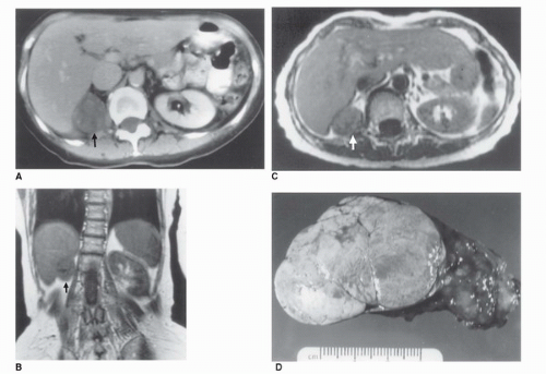
FIGURE 50.8. Radiographic and gross pathologic appearance of a benign functioning adrenocortical adenoma. (A) CT scan in a 64-year-old woman with hypercortisolism. A 4.5-cm mass replaces the right adrenal gland (arrow). (B) T1-weighted magnetic resonance image in the same patient in the coronal plane. The right upper quadrant mass (arrow) appears isodense with liver. (C) T2-weighted MRI in the same patient in a transverse plane. The tumor appears homogeneous and remains isodense with liver (arrow). (D) Gross specimen after surgical removal, with the typical smooth, lobulated, and homogeneous appearance of a benign cortical adenoma.


ADRENOCORTICAL CARCINOMAS


Epidemiology

The incidence of ACCs in the United States is approximately 0.5 to 2.0 cases per million population yearly (12). It has been estimated that 75 to 115 new cases are seen each year, and that 0.05% or less of all malignant tumors are of adrenal origin (58).

Occurrence is bimodal, with an initial peak in children younger than 5 years and the second peak in adults in the fourth and fifth decades of life (59). In a review of 1,891 cases reported in literature in the English language, Wooten and King (60) noted a similar distribution, with more than 855 tumors in children being functional. This neoplasm is more common in women (4:3) and appears to be functional more frequently in women (7:3) than in men (3:2). Several studies have shown a left-sided prevalence, while a few have shown right-sided preponderance. Approximately 2% to 10% of patients with adrenal cancer have bilateral disease (61).

Although the incidence of adrenal incidentalomas seems to be higher in some familial neoplasia syndromes—such as multiple endocrine neoplasia, type I (MEN-I), familial adenomatous polyposis (FAP), Li-Fraumeni syndrome—it is not entirely clear whether this is accompanied by a higher predisposition to adrenal cancer (62). In some areas of the world, such as southern Brazil, the incidence of adrenal cancer is high, especially in children. The role of environmental mutagens is postulated in these geographic areas (63).


Molecular Pathogenesis


Genetic Tumor Syndromes

Several hereditary tumor syndromes are associated with the formation of benign or malignant adrenocortical tumors. The discovery of genetic events involved in these syndromes, and further characterization of these candidate genes in sporadic carcinomas, has increased the understanding of adrenal tumorigenesis. Sandrini et al. (63) investigated individuals with childhood adrenal cancers from several cancer-prone families. Novel p53 mutations in exon 8 were detected. Germline
mutations in p53 are responsible for autosomal dominant Li-Fraumeni syndrome, and 5% of such patients develop ACCs (63). Germline mutations of p53 have been found in children with ACCs without classic family history of Li-Fraumeni syndrome, as well (63). The complete loss of one insulin-like growth factor type II (IGF-II) allele, which maps to 11p15.5 locus, and a duplication of the remaining allele in association with IGF-II overexpression has been demonstrated in tumors with Beckwith-Wiedemann syndrome and in sporadic adrenocortical tumors (63,64). In a report by Gicquel et al. (65), IGF-II was overexpressed in 27 of the 29 malignant adrenocortical tumors compared to 3 of 35 benign adenomas. Carney complex is another hereditary syndrome that is associated with primary pigmented nodular adrenocortical disease in 20% to 30% of patients (66). Two chromosomal loci have been identified (2p16 and 17q22-17q24); however, no deletions at these loci have been identified, implying that the responsible gene may be an oncogene rather than a tumor- suppressor gene (66,67). Approximately 35% of patients with MEN I have adrenal nodules, and the syndrome is related to germline mutations in the tumor-suppressor menin gene (11q13). In a series of 33 patients with MEN I, 12/33 had adrenocortical tumors, with loss of constitutional heterozygosity reported in 1 patient with ACC and not in 11 patients with benign adenomas (68). The genes involved in familial adenomatous polyposis coli (APC gene) and McCune Albright syndrome (activating mutations in the GNAS1 gene) have also been investigated as possible contributors to the presence of adrenal lesions in patients with these genetic syndromes.


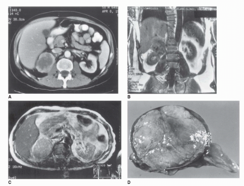
FIGURE 50.9. Radiographic and pathologic characteristics of an adrenocortical carcinoma (ACC). (A) CT scan in a 46-year-old man performed because of malaise and fever. A 7 × 5 cm heterogeneous mass with an irregular border replaces the right adrenal gland. (B) T1-weighted MRI in the same patient in a coronal plane. The tumor is isodense with liver and displaces right kidney. (C) Transverse T2-weighted MRI in the same patient demonstrating a slight increase in the tumor image intensity compared with the liver. (D) Adrenal tumor after excision, demonstrating an irregular lobulated surface and areas of hemorrhage suggestive of ACC.


Sporadic Adrenocortical Tumors

The genetic abnormalities in nonfamilial adrenal cancers are complex. Several studies have investigated the clonal composition of adrenocortical tumors using X chromosome inactivation. Gicquel et al. (69) found a monoclonal pattern in four carcinomas. Most data thus suggest that ACCs are monoclonal as a result of oncogenic mutations in single cells. Yano et al. (70) studied loss of heterozygosity (LOH) in one primary and eight recurrent ACCs and in eight sporadic benign lesions. The carcinomas showed LOH on 17p, 11p, and 13q with no changes in benign lesions. Using CGH, a high frequency of genetic aberrations, involving losses on chromosome 2, 11q, and 17p and gains on chromosome 4 and 5 have been reported (14). Subsequent studies using CGH have
shown equal distributions of chromosomal gains and losses in benign and malignant adrenocortical tumors, although the specific genetic events have been different (15). Bernard et al. (71) suggested multistep tumorigenesis. They studied a patient with malignant and benign sections in a tumor. This was confirmed by molecular analysis (expressions of IGF II and allelic status of 11p15 and 17p13 loci) and CGH. Using the “candidate gene approach,” several studies have looked at putative oncogenes and tumor-suppressor genes such as p53, IGF II, APC, MEN I, and Ras; however, a low prevalence of mutations were found (72). Mutations of ACTH receptors and constitutive activations of regulatory proteins of cAMP such as G-protein-coupled receptors have also been implicated in adrenal tumorigenesis. Mutational analysis of the coding region of the receptor in 41 adrenocortical tumors, however, did not demonstrate presence of any mutation (73).


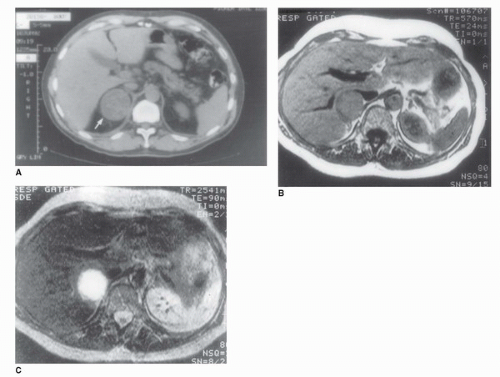
FIGURE 50.10. CT and MRI of a pheochromocytoma. (A) An abdominal CT in a 45-year-old patient with hypertension and elevated serum catecholamine level demonstrates a rounded homogeneous tumor replacing the right adrenal gland (arrow). (B) T1-weighted MRI of the 45-year-old patient illustrated in (A). The tumor is isodense with the liver. (C) T2-weighted MRI in the same patient. The tumor has a very high signal intensity and appears bright white, which is pathognomonic of a pheochromocytoma.


Staging

The staging commonly used for adrenal cancer is outlined in Table 50.6 (74). Stages I and II include tumors confined to the adrenal gland without capsular invasion; stages III include tumors confined to the adrenal gland but with involvement of the regional lymph nodes, or any tumor invading the capsule but not the adjacent organs. Stage IV includes any tumor with distant metastasis or invasion of adjacent organs even without any distant metastasis. In different series, 40% to 50% of patients have advanced disease when the diagnosis is made, and the median survival of these individuals is <1 year. In an analysis of 602 patients with adrenal carcinoma treated between 1936 and 1999 at seven institutions, Ng and Libertino (75) found that the 5-year survival by stage was as follows: stage I, 30% to 45%; stage II, 12.5% to 57%; stage III, 5% to 18%; and stage IV 0%. Median survival in unresectable tumors was 3 to 9 months, whereas after complete surgical resection it ranged from 13 to 28 months.


Clinical Presentation

The biochemical basis and the various syndromes resulting from the excessive production of corticosteroids, androgens, estrogens, or mineralocorticoids have been described. In patients with adrenal cancers, the most common syndrome is Cushing syndrome. This syndrome has been reported in 30% to 40% of patients with cancer (76). Virilization alone or in combination with excess cortisol has been found in 20% to 25% of patients.
Feminization or aldosterone excess is rarely reported (76,77). Both functional and nonfunctional adrenal cancers have been described. Reviews of various case series in patients with adrenal carcinoma demonstrate that clinical presentation varies based on whether the report comes from an oncologic or endocrine clinic. Functional carcinomas are more frequent in the endocrinologic literature. In a review of 1,480 patients in which functional status was mentioned, Wooten and King (60) noted that 60% of the cases were associated with evidence of function. In large retrospective series of patients with adrenal cancers, functional tumors have been reported in 34% to 72% (78).


TABLE 50.6 STAGING SYSTEM FOR ACC


Tumor characteristics, disease extent


TX


Primary tumor cannot be assessed


T0


No evidence of primary tumor


T1


Tumor ≤5 cm, no capsule invasion


T2


Tumor >5 cm, no capsule invasion


T3


Tumor with capsular invasion into periadrenal fat but not invading adjacent organs.


T4


Tumor invading adjacent organs (kidney, diaphragm, great vessels, pancreas, spleen, and liver).


NX


Regional lymph nodes cannot be assessed


N0


Negative lymph nodes


N1


Metastasis in regional lymph nodes


M0


No distant metastases


M1


Distant metastases


Staging categories


I


T1, N0, M0


II


T2, N0, M0


III


T1, N1, M0


T2, N1, M0


T3, N0, M0


IV


Any T, any N, M1


T3-4, N1, M0


T4, N0, M0


Adapted from American Joint Committee on Cancer (AJCC) Adrenal at-a-glance. In: AJCC cancer staging handbook, 7th ed. S.B. Edge, D.R. Byrd, C.C. Compton, A.G. Fritz, F.L. Greene, and A. Trotti, Eds. New York, NY: Springer Science + Business Media LLC, 2010:585, Chapter 47 .


In patients with nonfunctioning tumors, the diagnosis is generally made when a neoplasm produces symptoms by virtue of its size or disease spread. Nonfunctional ACCs can be more difficult to diagnose because they remain clinically silent. These tumors typically present late in their natural history and are more often of advanced stage than are functional tumors. Typical symptoms of nonfunctional tumors include abdominal mass, pain on the affected side, and constitutional symptoms of weight loss, fever, anorexia, nausea, and myalgia, owing to advanced malignancy (79). Some patients will have symptoms of metastatic disease before a primary diagnosis, whereas others present with incidental findings on abdominal CT scans performed for unrelated reasons.


Diagnostic Evaluation of Adrenal Cancer

The diagnostic evaluation of ACC is a two-step process and includes (a) biochemical assessment and (b) radiographic localization and staging.


Biochemical Assessment

Studies in patients with adrenal cancers have demonstrated the following well-documented biochemical abnormalities: elevated levels of urinary free cortisol; loss of circadian rhythm of cortisol secretion; loss of suppressibility of pituitary-adrenal axis; elevated urinary levels of steroid precursors; and low levels of ACTH in the serum due to high levels of circulating cortisol. Decisions on specific components of biochemical parameters depend to some degree on the clinical presentation of the patient.

In patients who present with Cushing syndrome, a 24-hour urinary free cortisol level determination is the most sensitive measure of excess cortisol production. Levels of cortisol metabolites in the urine, including 17-hydroxycorticosteroids and 17-ketosteroids, may also be elevated. Serum studies in a patient with suspected Cushing syndrome should include cortisol levels measured early in the morning and in the late afternoon or early evening. The normal adrenal cortex exhibits a circadian rhythm such that the morning cortisol levels are approximately two to three times higher than late afternoon/evening levels; hypersecreting adrenal tumors lose this diurnal variation and produce high levels at all times.

In patients with virilization, serum measurements of levels of androstenedione, testosterone, and DHEA and its sulfate, are appropriate. In men with feminization, estradiol levels are often elevated. In patients with hypertension and hypokalemia, the PAC:PRA ratio should be measured. Finally, in patients with an uncertain etiology, a dexamethasone suppression test should be considered, as previously outlined.

Jul 15, 2016 | Posted by in ONCOLOGY | Comments Off on Adrenal Neoplasms

Full access? Get Clinical Tree

Get Clinical Tree app for offline access