Management of Low Stage Germ Cell Tumors of the Testis


Management of Low Stage Germ Cell Tumors of the Testis


▪ 33A Management of Low-Stage Testicular Seminoma

Nathan Lawrentschuk

Neil E. Fleshner

Within oncology, early-stage seminoma represents the paradigm of a curable malignancy. No matter what treatment option is chosen, a survival of almost 100% is an expected outcome. Seminoma is managed in a truly multidisciplinary fashion with surgeons, radiation oncologists, and medical oncologists all playing an important role in disease detection, diagnosis, treatment, and follow-up. This chapter focuses on the management of men who have early-stage seminoma, which represents stage I and II (retroperitoneal spread ≤5 cm nodal disease, stage IIA/B). For patients diagnosed with early-stage testicular cancer, radical orchiectomy is the primary therapeutic intervention, and these early-stage seminomas all remain in the “good-” prognosis group under the International Germ Cell Cancer Collaborative Group (IGCCCG).

However, consensus has not been reached among urologists and oncologists regarding the optimal postoperative management in men with clinical stage I (CSI) testicular seminoma. Currently, men are offered active surveillance, adjuvant radiation therapy, or single-agent chemotherapy. Surveillance is associated with an 80% chance of no relapse, with all such events salvageable. Radiotherapy has a relapse rate of around 4% but like other therapies an almost universal cure rate if salvaged. Yet it also has the potential for long-term toxicities and an overtreatment rate of about 80%. Chemotherapy efficacy and toxicity data are still immature. Thus, a focus on long-term complications, such as cardiovascular disease, gastrointestinal disease, and secondary cancers has led to the concept of increased surveillance with therapy for those who relapse. Radiation therapy remains the mainstay of therapy for patients who have stage IIA/B disease while IIC disease is managed exclusively with chemotherapy.

Clearly, defining the clinical stage of a patient with a germ cell tumor is based on the UICC TNM classification (1). It is imperative for verification of stage I disease that markers are followed after orchiectomy until normalization is achieved, as patients without marker normalization after orchiectomy are defined as stage IS disease, although this subgroup is less relevant in seminoma. AFP elevation equates to nonseminomatous disease and should be treated as such.


STAGE 1 SEMINOMA

In stage I disease, the major controversies continue to revolve around surveillance versus adjuvant treatment (radiotherapy or more recently carboplatin-based chemotherapy). Recent guidelines recognize that the options of surveillance, chemotherapy, and radiotherapy are appropriate in the treatment of patients with CSI seminoma. However, without a “gold-standard” treatment option, but with three efficacious and acceptable therapeutic options available, informing patients is the key, so that they make an informed choice in a multidisciplinary setting (Fig. 33A.1).


Risk-adapted Strategies for Stage I Seminoma

Urologists are extremely familiar with risk-adapted strategies for treating malignancies. The most recent example is active surveillance of prostate cancer, but germ cell tumor patients themselves have had individual risk-adapted strategies performed for many years using both the TNM classification and the IGCCCG classification based on prognostic factors (e.g., histology, location of primary tumor, location of metastases, tumor markers after orchiectomy and before chemotherapy). Patients are classified as “good-,” “intermediate-,” and “poor-” prognosis groups. In low-stage seminoma, risk stratification has been based on the original work of Warde et al. (3), who identified rete testis invasion and size of primary tumor (>4 cm) as risk factors for relapse in a pooled analysis of AS studies with 638 patients. The relapse rate was 17% for primary tumors >4 cm without rete testis invasion, 12% for primary tumor <4 cm and no rete testis invasion, and 14% for rete testis invasion and a primary tumor <4 cm. The difficulty is that such strategies have not been validated in prospective cohorts, and Loehrer and Bosl (4) have been critical of this approach, whether it is selection for surveillance or selection for treatment. Patient age (<34 vs. >34 years) and the presence of vascular invasion are of equivocal prognostic relevance (2,5). Whatever the approach, patients treated with adjuvant chemotherapy or RT still have a 3% to 4% relapse rate (6).

Thus in recent decades, RT has established itself as an appropriate adjuvant treatment for CSI seminoma, which significantly reduces the relapse rate to a few percent (3,7,8,9,10). However, this success has come at a cost. Although absolute risks remain small, radiotherapy is associated with doubling in the risk of developing a second malignancy compared with the general population (11,12,13,14,15). In real terms, Travis et al. (13) found after reviewing registries over 20,000 men with seminoma treated with radiation that for a 35-year-old male, the risk of a second solid tumor by age 75 is 36% versus 23% for the general population. Based on success with higher-stage disease, chemotherapy has emerged in lower doses as an adjuvant option and is probably equivalent to radiotherapy (16), but long-term risks are unknown (17). Finally, active surveillance avoids overtreatment in the vast majority of patients, as only approximately 15% of patients are likely to relapse (18). Based on recent data, current recommendations for the treatment of clinical stage 1 seminoma have recently been updated and changed.

The European Consensus Conference on diagnosis and treatment of Germ Cell Cancer (EGCCG) state that all three treatment options, active surveillance, adjuvant carboplatin, or adjuvant radiotherapy are acceptable strategies for the management of patients with CSI seminoma l (2). With such an approach, those
at lower risk of relapse may be offered surveillance with more confidence and those with risk factors may be more likely to be offered adjuvant radiotherapy or chemotherapy, but it must be stated that we do not know for sure the precise risk factors for relapse. With surveillance a significant proportion of patients may therefore avoid overtreatment.


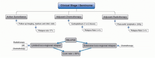
FIGURE 33A.1. Treatment algorithm after orchiectomy according to individual risk factors in patients with seminoma CSI. (Adapted from Krege S, Beyer J, Souchon R, et al. European consensus conference on diagnosis and treatment of germ cell cancer: a report of the second meeting of the European Germ Cell Cancer Consensus group (EGCCCG): part I. Eur Urol 2008;53:478.)

Other guidelines, although written prior to the EGCCG, all now recognize that the options of surveillance, chemotherapy, and radiotherapy are appropriate in the treatment of patients with clinical stage 1 seminoma (2,19,20). Nevertheless, there are some caveats. For example, in the 2009 National Comprehensive Cancer Network (NCCN) Testicular Cancer Clinical Practice Guidelines all adjuvant options are rated “category 1” according to the meaning that there is a uniform consensus among experts on their recommendation as approved treatment. However, when examining the finer detail, it emerges that they recommend surveillance as category 1 only in specific instances: for those having undergone previous radiotherapy, those with inflammatory bowel disease or a horseshoe kidney. Surveillance is recommended as category 2B for others motivated to undergo follow-up and more intensive imaging. In contrast, chemotherapy has been raised to equal status with the more traditionally used radiotherapy as category 1 for all comers. The most recent European Association of Urology guidelines have a similar approach with surveillance as a grade B recommendation behind chemotherapy and radiotherapy (Category A, Oxford Evidence Based Guidelines) (20).


Surveillance

A large body of evidence has now been published (Table 33A.1) considering surveillance of stage I seminoma. Of 2,496 patients studied, a relapse rate of 17% is expected with a survival of practically 100% for those undergoing salvage treatments for relapse. This reinforces the salvageable nature of any relapse that may be treated with chemotherapy or radiation (3). Such data are compelling when considering surveillance as an option. The benefits are also clear: avoidance of overtreatment in over 80% of patients, knowledge that relapses are salvageable, no risk to fertility, no risk of acute toxicity, and no iatrogenic risk of cardiotoxicity or second malignancy. Against this, surveillance does oblige the patient to close follow-up and imaging that is vigorous and even in a study situation, is not always achievable (21). Anxiety is also a potential effect of surveillance (22) and this is where selection of patients who accept the risk of relapse is essential. This type of anxiety falls within “Damocles syndrome” (23) where patients carry the stressful feelings of potential relapse with little control over events and in its most severe form leads to avoidant behaviors, intrusive thoughts, and heightened arousal in survivors of cancer. However, data on anxiety in surveillance patients are weak, and further study needs to be done in this area.

However, the need for more extensive treatment at relapse rather than upfront adjuvant treatment is considered a disadvantage by some clinicians. Technically, this is true for chemotherapy and to a lesser extent radiation, but the reality is that this will happen in one in five or fewer patients. At relapse, treatment will be based on the stage at that time which is normally low-burden retroperitoneal nodal disease, usually clinical stage II (see section on “Stage II Seminoma”). The need for more extensive treatment has not been realized at our center.

A surveillance policy does, undoubtedly, not only require a high knowledge of physician but also patients’ social, economic, and cultural status. Furthermore, patients’ motivation is an important factor. Awareness of having a cancer, the necessity of more frequent hospital and clinic visits, and the potential psychological stress that there is a risk of relapse of up to 20% are important factors for patient (24). Late relapses more than 5 years from original diagnosis are well known to seminoma surveillance and protocols generally follow patients for at least 10 years from the point of diagnosis. The Princess Margaret Hospital seminoma surveillance protocol is appended (Table 33A.2). Loss to follow-up and compliance with imaging in young population is always a concern, as it is in NCGCT (25). That is why patient selection is paramount, as with any treatment option, but in almost all instances, surveillance is possible once patients understand the rationale and protocols. Despite such concerns the quality of life on surveillance is excellent (26). Another point is that fertility is not affected by surveillance as it may be with radiotherapy (27,28,29).

Certainly, there will remain some patients for whom surveillance should automatically be undertaken and these are patients with a horseshoe kidney, inflammatory bowel disease, and those who have had prior radiotherapy (19). Evidence is emerging that it should be offered to all patients.


TABLE 33A.1 SUMMARY OF SURVEILLANCE OUTCOMES FOR CSI SEMINOMA


Author


Year


n


Median Follow-up (m = months)


Number of Patients that Relapsed (%)


Deaths Survival (%)


Berkmen


2006


34


24 m


30 (20)


0 (100)


Choo


2005


88


145 m


17 (19)


0 (100)


Aparicio


2005


143


52 m


23 (16)


0 (100)


Bauduceau


2003


65


37 m


3 (05)


1 (100)


Daugaard


2003


394


60 m


69 (18)


0 (100)


Parker


2002


150


113 m


30 (20)


NR


Porcaro


2002


42


85 m


8 (19)


3 (97)


Chung


2002


203


110 m


35 (17)


1 (100)


Francis


2000


120


55 m


18 (15)


0 (100)


Warde


2000


226


92 m


37 (16)


1 (100)


Jones


1999


60


52 m


18 (30)


0 (100)


Coleman


1998


94


>120 m


14 (15)


0 (100)


Oliver


1994


26


18 m


4 (0.15)


0 (100)


Germa-Lluch


1993


45


34 m


5 (11)


0 (100)


von der Masse


1993


261


48 m


49 (19)


(3) 99.9


Thomas


1993


209


48 m


13 (15)


1 (100)


Ramakrishnan


1992


72


44 m


13 (18)


0 (100)


Horwich


1992


103


62 m


17 (17)


0 (100)


Alhoff


1991


33


48 m


3 (09)


0 (100)


Duchesne


1990


113


30 m


13 (12)


0 (100)


Charig


1990


15


31 m


5 (33)


0 (100)


Totals


2496


52 m (median)


424 (17)


10 (99.6)


NR, not reported.


Source: Adapted from Lawrentschuk N, Fleshner N. Clinical stage I seminoma: the case for surveillance. World J Urol 2009;27:433.


Radiotherapy

In the past, adjuvant radiotherapy has been the most frequently used treatment for stage I seminoma which significantly reduces the relapse rate (3,7,8,9,10). As alluded to, this circumstance is changing largely due to concerns of overtreatment, cardiac toxicity (30,32,39), and second malignancy (11,12,13,14,15,16,33). In fact, the risk of developing a second malignancy compared with the general population is doubled (11,12,13,14,15). This risk becomes apparent only after 5 years and peaks between 10 and 20 years after treatment (13). Overall, patients treated with radiotherapy have a decreased survival compared with matched individuals from the general population (31). Clearly, the concern with such long-term effects is that they will ultimately result in increased mortality and this has been demonstrated when comparing with matched individuals from the general population (31).


TABLE 33A.2 PRINCESS MARGARET HOSPITAL STAGE I SEMINOMA SURVEILLANCE PROTOCOL


Year


Month 2


Month 4


Month 6


Month 8


Month 10


Month 12


1


TM, CT


TM, CXR CT


TM, CT


2


TM, CXR CT


TM, CT


TM, CXR CT


3


TM, CT


TM, CT


TM, CXR CT


4


CT


CT, CXR


5


CT


CT, CXR


6


CT


CT, CXR


7


CT


CT, CXR


8


CT, CXR


9


CT, CXR


10


CT, CXR


CT, Computed tomography of the abdomen and pelvis; CXR, chest X-ray; TM, tumor markers.


Due to the long-term toxicities, alternative modalities and modifications in technique have occurred. Testicular shielding is routinely used to minimize the risk of radiation scatter. Reductions in radiation have also been pursued to reduce morbidity yet to retain the low relapse rates. In a randomized study, patients receiving 20 Gy in 10 fractions compared to those administered 30 Gy in 15 fractions with a median follow-up of 61 months had equivalent relapse rates (34). In recognition of these effects, radiotherapy has had its fields modified from the pre-1990 model of anteriorly and posteriorly delivered so-called “dog-leg” (DL) fields that covered
lymphatic drainage of the testis to include the ipsilateral pelvis and the paraaortic (PA) region. The decision to reduce fields from DL to PA was largely based on the Medical Research Council trial reported on by Fosså et al. in 1999. This trial randomized 478 men with stage I testicular seminoma to DL or PA radiotherapy of 30 Gy. The median follow-up was 4.5 years with nine relapses in each group, yet no pelvic recurrences in the DL versus four in the PA group. However, acute toxicity was less frequent and less pronounced in PA arm with sperm counts significantly higher in PA arm. They concluded that there was no difference in overall outcome with decreased morbidity in PA-only arm and thus recommended the PA field as standard. Overall, since the general adoption of the reduced PA field, this has not resulted in a reduction in survival rates, but interestingly, cases of recurrence at the edge of the traditional DL fields have been surfacing in the literature (35,36,37,38,39,40).

Decreasing overall treatment doses is also being explored but is yet to be published beyond the reductions from 30 and 20 Gy that have occurred (6). Risks to fertility remain despite shielding of the contralateral testis, yet they may be related to the disease process itself, and not just the treatment (41).

Acute toxicities associated with radiotherapy are generally mild and usually gastrointestinal (42). Treatment relapse rates range from 3% to 4%, with recurrences typically occurring outside the irradiated area (43). The commonest sites of relapse following radiotherapy are the mediastinum, lungs, and left supraclavicular fossa (44). Finally, another issue is the potential for late gonadal toxicity and resultant impairment of spermatogenesis and fertility from radiation scatter affecting the contralateral testicle (27,28,29).


Chemotherapy

The rationale for chemotherapy was born out of the concern with radiotherapy secondary effects but also with the recognition that where tumors are sensitive to chemotherapy, reduced doses have been successful in many fields of oncology. Carboplatin has emerged as a primary adjuvant treatment alternative for stage I seminoma based on the use of combination chemotherapy for higher stage germ cell tumors.

Although the most recent publication concerning a single cycle of carboplatin has only modest follow-up of 4 years, it proved to be noninferior to radiotherapy in terms of relapse rates and survival (16). Despite this evidence, and guideline recommendations as outlined previously, many authors believe that carboplatin should not be considered the standard of care (4,45). Others have argued that two courses of treatment are necessary should carboplatin be used (46,47). Even with two cycles, a small but significant percentage of patients (3%-4%) recur in the retroperitoneum.

In 2005, Loehrer and Bosl (4) outlined issues relating to carboplatin for low-stage seminoma that remain largely unaddressed: (a) the rate and sites of relapse after single-agent carboplatin are not satisfactory, (b) carboplatin is inferior to cisplatin in disseminated germ cell tumors but has been utilized due to less acute toxicity than with cisplatin, (c) the late consequences of carboplatin therapy are unknown, and (d) the risk-adaptive model to predict relapse for seminoma has not been validated. On point of toxicity, although acute toxicity following adjuvant chemotherapy are well understood with myelosuppression and gastrointestinal side-effects predominating (47), late toxicities are poorly understood. Concerns have been based on evidence that combination chemotherapy for testis cancer has been associated with the development of hematological cancers and solid tumors (13,14,15). However, recent evidence suggests that these concerns may be unfounded. Powles et al. examined 199 patients with a median followup for the cohort being 9 years. Overall mortality had not increased, cardiotoxicity was not an issue, and there was not a greater incidence of second nontestis cancers. There was an increase in the long-term development of contralateral testis cancers. Clearly, more data are required and justified concerns about the long-term side effects associated with single-agent carboplatin (13,14,15,17,48,49,50,51) remain, but the first published data are encouraging. As with radiotherapy, large databases accumulating data over decades are required to completely answer such questions regarding second malignancies and until that occurs, speculation is the best evidence we have.

Unlike following radiotherapy, adjuvant chemotherapy requires continued surveillance of the retroperitoneum (18,19). So in summary, chemotherapy will continue to be a valid option but with some clinicians not convinced until further longer-term data on efficacy and safety emerge. We agree that the use of routine single-agent carboplatin cannot be widely recommended (17), and we would proceed to radiation in cases where surveillance was not appropriate due to the aforementioned reasons.


Points for Consideration in Stage I Seminoma

Stage I seminoma is a highly curable disease but does require some degree of follow-up. The aim of follow-up of testicular cancer patients is twofold: to detect relapses early when minimal disease is present so that they may be salvaged and to detect and manage the effects of the original definitive oncological treatment. A major criticism of surveillance is that the required number of follow-up investigations reduces quality of life from anxiety and increases costs to a healthcare system. It should be pointed out that the 2009 NCCN guidelines (19) recommend the same imaging and follow-up whether a man is treated with surveillance or chemotherapy. Among radiated men imaging of the retroperitoneum is not required due to the reduced risk of recurrence. Similar to the treatment paradigm, a nonevidence-based risk-adapted follow-up strategy has been recommended (49,52). Studies are currently underway to reduce the use of computed tomography in surveillance of testicular tumors such that more limited protocols may be developed, thus reducing potential risk of radiation (53).

In any discussion on health care today concerns regarding costs are worth considering with several studies demonstrating the higher cost of surveillance compared to other treatments (24,54). However, many studies lack the ability to factor in costs of second treatments or cardiotoxicity. Also, reducing the burden of imaging and the radiation that accompanies it is currently under study for NSGCT (53). Such approaches in the future will assist in stratifying patients who really do require retroperitoneal axial imaging and at what frequency will further diminish cost inequalities.

In conclusion, for stage I seminoma, we believe that if a patient is willing and able to comply with a surveillance policy then long-term close follow-up should be recommended after orchiectomy for CSI seminoma. The rationale, advantages and disadvantages of the treatment modalities available for CSI seminoma are summarized in Table 33A.3. The clear benefits of surveillance relate to overtreatment of over 80% of men and the reduced risk of acute and late toxicities. With chemotherapy, long-term data on survival and toxicities are not yet available for a large numbers of patients. Further, recurrences do occur in the chemotherapy and radiotherapy adjuvant arms meaning no guarantee of complete success (i.e., no relapse) may be given by having more extensive treatment upfront. Ultimately, each center must factor in their experience costs, compliance, psychosocial issues, and the impact on fertility of any individual patient prior to selecting a treatment. This is because all modalities achieve similar outcomes and are considered acceptable to patients. The future may yield risk-adapted strategies not only for providing details on prognosis and giving information for selecting the appropriate adjuvant treatment, but to help minimize follow-up in surveillance patients.


TABLE 33A.3 RATIONALE, ADVANTAGES AND DISADVANTAGES OF THE TREATMENT MODALITIES CURRENTLY AVAILABLE FOR CSI SEMINOMA


Modality of Treatment


Rationale


Acute Side Effects


Late Side Effects


Quality of Life


Acceptability


Advantages


Disadvantages


Future


Active Surveillance


˜17% will relapse on surveillance, but cure rates remain excellent even if relapse occurs


No


Possible increased risk of malignancy from radiation burden for surveillance, but no data and low radiation protocols currently being investigated


Good


Good


  • Avoid overtreatment in >80% of men


  • Cure rates maintained even if relapse


  • No second malignancy


  • No late toxicity


  • Potential anxiety from follow-up and recurrent investigations


  • Need for more extensive treatment at relapse


  • Loss to follow-up in young population


  • Cost and compliance with imaging


As late risks of adjuvant treatment effects reported, surveillance likely to become more widely accepted


Radiotherapy


Seminoma is exquisitely radiosensitive with excellent 20-yr follow-up data


Yes


Yes


Good


Good


  • Less followup than with active surveillance


  • CT of retroperitoneum not required


  • Acute and late toxicity


  • Second malignancy


  • Possible effects on fertility


  • Relapse still possible


Due to chemotherapy data and increasing surveillance practice, in decline as the primary modality


Chemotherapy


Reduced dose regimens have been extrapolated from success with higher-stage tumors, but potential morbidity not fully characterized


Yes


Likely but no data


Good


Good


  • As efficacious as radiotherapy


  • Potential for less follow-up, but currently this is not always practised


  • Acute toxicity


  • Potential late toxicity


  • Potential second malignancy


  • Relapse still possible


Increasing in practice despite a lack of longterm data with toxicity concerns


STAGE II SEMINOMA

Approximately, 15% to 20% of patients with seminoma are classified as having clinical stage II disease, or metastatic spread limited to the retroperitoneum (56). For stage II seminoma, it is divided into clinical stage IIa (nodes ≤ 2 cm); stage IIb (nodes 2-5 cm), and IIc (nodes > 5 cm) (1). There is much less treatment controversy as radiation has long-term followup with excellent results in both substage IIa and IIb while chemotherapy is an accepted alternative in stages IIb and IIc (Fig. 33A.2). However, it should also be noted that despite the good prognosis, the International Consultation on Urological Diseases (ICUD) only classifies evidence as Oxford Level III for all recommendations just described due to data limitation (57).


Radiotherapy

In stage II seminoma, there have been no randomized controlled trials to confirm the optimal radiation dose. By consensus most centers manage stages IIA and sometimes B with 25 to 30 Gy radiation alone, administering to the retroperitoneum and pelvis with a further 5 to 10 Gy boost to areas of disease. Although a randomized controlled trial demonstrates similar efficacy between 20 and 30 Gy in stage I disease, this may not necessarily be extrapolated to stage IIA or greater (34). Some centers have added prophylactic radiation to the left supraclavicular fossa in an attempt to reduce relapse rates in that region (58). This practice has not been widely adopted as <3% of patients within this group are likely to receive any material benefit. The earlier practice of prophylactic radiotherapy to the mediastinum has been discarded due to first, minimal patient benefits but more importantly an associated increased risk of treatment-related death if salvage chemotherapy is required later.

The most important prognostic variable for stage II outcome overall is the volume of lymph node involvement. For men with stage IIA disease, relapse after therapy occurs in 0% to 15% (59,60,61). Specifically, long-term relapse rates between 0% and 8% may be expected in modern practice, with in field recurrences in the order of 0% to 2% (62). Once again, cure is achieved in virtually 100% with salvage chemotherapeutic regimens if relapse occurs following radiotherapy treatment in stage IIA seminoma. In stage IIb, relapse rates approach 25% but most are amenable to salvage therapy (63).

As tumor burden in the retroperitoneum increases, the likelihood of cure with radiation alone diminishes. In the Princess Margaret Hospital experience, 5-year relapse-free survival was 91% if nodal disease with maximal diameter <5 cm was present compared with only 44% if >5 cm (64). Further to this, patients with nodes smaller than 2 cm experienced disease recurrence 11% of the time, for patients with nodes 2 to 5 cm in diameter this increased to 9% to 18%.


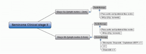
FIGURE 33A.2. Treatment algorithm after orchiectomy according to individual risk factors in patients with seminoma clinical stage II. (Adapted from Krege S, Beyer J, Souchon R, et al. European consensus conference on diagnosis and treatment of germ cell cancer: a report of the second meeting of the European Germ Cell Cancer Consensus Group (EGCCCG): part II. Eur Urol 2008;53:497.)


Chemotherapy

Chemotherapy alone is generally not employed for stage IIA disease unless there are factors complicating the delivery of radiotherapy such as previous radiation or higher than usual risk of visceral organ injury as may be seen with disease beyond the primary echelon nodes. The current standard of care for stage IIA seminoma is radiotherapy. This remains the case due to the effectiveness of radiotherapy in achieving a cure, the toxicity profile of chemotherapy, and ability of patients with disease relapse following radiotherapy to be effectively salvaged with chemotherapy.

In 2001, the Royal Marsden Hospital published their results using a combination of a single cycle of carboplatin and radiotherapy for the treatment of stage II seminoma (65). This addition prevented recurrence in 31 of 33 patients with stage IIA and B disease. The overall 6% recurrence rate at a median of 2 years follow-up compares favorably with results from this center prior to the initiation of the protocol change. Previously, stage IIA and B recurrence rates were reported as 15% and 30%, respectively. This study stands alone to date, and the regimen requires further verification but may herald a change in the paradigm of stage II seminoma in the longer term.

In stage IIb disease, chemotherapy is administered with either in combination with bleomycin, etoposide, and cisplatin (BEP) or etoposide (EP) alone (65,66). Chemotherapy remains an option used mainly in patients where a treatment alternative to radiotherapy is mandatory due to prior radiation and also is considered in patients with larger multinodal retroperitoneal disease (66).

As stated, due to excellent efficacy of both treatments the one chosen is often done so due to toxicity profiles whereby chemotherapy fares much worse. Furthermore, salvage following radiation for relapse with chemotherapy is often considered a more acceptable pathway (63).


Patients Having Relapse Undergoing Active Surveillance for Clinical Stage 1 Seminoma

For patients having relapse undergoing active surveillance, treatment will be based on the stage at that time which is
normally low-burden retroperitoneal nodal disease, usually clinical stage IIA (nodes ≤ 2 cm) or stage IIB (nodes 2-5 cm). In stage IIA radiotherapy (infradiaphragmatic 35-40 Gy to include PA and ipsilateral iliac nodes) remains the preferred treatment option over chemotherapy. In stage IIB chemotherapy with three cycles of standard-dose BEP, or four cycles of EP, represents a treatment alternative to radiotherapy, particularly in patients with larger multinodal retroperitoneal disease (a small subset of the seminoma relapse patients) but may be associated with a higher risk of acute toxicity as compared to radiotherapy (19,66). However, one must also consider that recurrences do occur in the chemotherapy and radiotherapy arms meaning no guarantee of no relapse may be given if such treatment options are chosen.


Postchemotherapy Residual Masses

For patients with a residual mass after chemotherapy originally there is a paucity of data. The ICUD recommends (Level IV evidence) that those patients with residual masses of <3 cm may be safely observed and that those with persistent masses over time may be considered for immediate retroperitoneal lymphadenectomy or close observation. However, surgery be undertaken only in experienced centers as the surgery is technically challenging and associated with a higher morbidity (57).


CONCLUSIONS

In stage I seminoma, we believe that active surveillance is the optimal choice for compliant men who are able to handle the mental burden of not receiving adjuvant treatment. This takes into consideration the fact that a small number of men with CSI seminoma on surveillance will recur, but are salvageable with equivalent outcome compared to those having adjuvant treatment which exposes eight in ten men to unnecessary short- and long-term risks with still the possibility of recurrence requiring salvation. Our second choice considering the current efficacy and safety data is radiotherapy, with singleagent chemotherapy emerging as a possible alternative.

Stage IIa seminoma has radiotherapy as its mainstay and this has not been challenged in recent years. Stage IIb has the alternatives of radiation or chemotherapy while stage IIc has chemotherapy as the first option. Concerns about long-term toxicity remain but are being better understood and catered for in current treatment and follow-up regimens.


References

1. UICC. In: Sobin LH, Wittekind CH, eds. TNM classification of malignant tumours, 6 ed. New York, NY: Wiley-Liss, 2002.

2. Krege S, Beyer J, Souchon R, et al. European consensus conference on diagnosis and treatment of germ cell cancer: a report of the second meeting of the European Germ Cell Cancer Consensus group (EGCCCG): part I. Eur Urol 2008;53:478.

3. Warde P, Specht L, Horwich A, et al. Prognostic factors for relapse in stage I seminoma managed by surveillance: a pooled analysis. J Clin Oncol 2002;20:4448.

4. Loehrer PJ, Sr, Bosl GJ. Carboplatin for stage I seminoma and the sword of Damocles. J Clin Oncol 2005;23:8566.

5. Warde P, Gospodarowicz MK, Banerjee D, et al. Prognostic factors for relapse in stage I testicular seminoma treated with surveillance. J Urol 1997;157:1705.

6. Sava T, Consoli F, Santo A, et al. Adjuvant treatment in the management of testis-confined germ cell tumours after orchidectomy. BJU Int 2008;101:155.

7. Bosl GJ, Bajorin DF, Sheinfeld J, et al.: Cancer of the testis. In: Devita VT Jr, Hellman Rosenberg SA, eds.: Cancer: principles and practice of oncology. 7th ed. Philadelphia, Pa: Lippincott Williams & Wilkins, 2005, pp 1269-1290.

8. Chung P, Parker C, Panzarella T, et al. Surveillance in stage I testicular seminoma – risk of late relapse. Can J Urol 2002;9:1637.

9. Gurkaynak M, Akyol F, Zorlu F, et al. Stage I testicular seminoma: para-aortic and iliac irradiation with reduced dose after orchiectomy. Urologia Internationalis 2003;71:385.

10. Garcia-Serra AM, Zlotecki RA, Morris CG, et al. Long-term results of radiotherapy for early-stage testicular seminoma. Cancer clinical trials. Am J Clin Oncol 2005;28:119.

11. Bokemeyer C, Schmoll HJ. Secondary neoplasms following treatment of malignant germ cell tumors. J Clin Oncol 1993;11:1703.

12. Travis LB, Curtis RE, Storm H, et al. Risk of second malignant neoplasms among long-term survivors of testicular cancer. J Natl Cancer Inst 1997;89:1429.

13. Travis LB, Fossã SD, Schonfeld SJ, et al. Second cancers among 40, 576 testicular cancer patients: Focus on long-term survivors. J Natl Cancer Inst 2005;97:1354.

14. Van Leeuwen FE, Stiggelbout AM, Van den Belt-Dusebout AW, et al. Second cancer risk following testicular cancer: a follow-up study of 1,909 patients. J Clin Oncol 1993;11:415.

15. Wanderas EH, Fossa SD, Tretli S. Risk of subsequent non-germ cell cancer after treatment of germ cell cancer in 2006 Norwegian male patients. Eur J Cancer Part A 1997;33:253.

16. Oliver RTD, Mason MD, Mead GM, et al. Radiotherapy versus single-dose carboplatin in adjuvant treatment of stage I seminoma: a randomised trial. Lancet 2005;366:293.

17. Loehrer PJ Sr, Bosl GJ. Carboplatin for stage I seminoma and the sword of damocles. J Clin Oncol 2005;23:8566.

18. Groll RJ, Warde P, Jewett MA. A comprehensive systematic review of testicular germ cell tumor surveillance. Crit Rev Oncol Hematol 2007;64:182.

19. National Comprehensive Cancer Network: The NCCN Testicular Cancer Clinical Practice Guidelines in Oncology http://www.nccn.org, vol. 1, Version 2, 2009.

20. Albers P, Albrecht W, Algaba F, et al. Guidelines on testicular cancer. Arnhem, The Netherlands: European Association of Urology (EAU), 2008:54.

21. Ernst DS, Brasher P, Venner PM, et al. Compliance and outcome of patients with stage 1 non-seminomatous germ cell tumors (NSGCT) managed with surveillance programs in seven Canadian centres. Can J Urol 2005;12:2575.

22. Moynihan C. Testicular cancer: the psychosocial problems of patients and their relatives. Cancer Surv 1987;6:477.

23. Koocher GP, O’Malley JE, Gogan JL, et al. Psychological adjustment among pediatric cancer survivors. J Child Psychol Psychiatry 1980;21:163.

24. Berkmen F, Hizli F. Stage I testicular seminoma: surveillance versus adjuvant radiotherapy. Turk J Cancer 2006;36:169.

25. Fleshner N, Warde P. Controversies in the management of testicular seminoma. Semin Urol Oncol 2002;20:227.

26. Rudberg L, Carlsson M, Nilsson S, et al. Selfperceived physical, psychologic, and general symptoms in survivors of testicular cancer 3 to 13 years after treatment. Cancer Nurs 2002;25:187.

27. Fossa SD, Abyholm T, Normann N, et al. Posttreatment fertility in patients with testicular cancer. III. Influence of radiotherapy in seminoma patients. Br J Urol 1986;58:315.

28. Fosså SD, Horwich A, Russell JM, et al. Optimal planning target volume for stage I testicular seminoma: a medical research council randomized trial. J Clin Oncol 1999;17:1146.

29. Joos H, Sedlmayer F, Gomahr A, et al. Endocrine profiles after radiotherapy in stage I seminoma: impact of two different radiation treatment modalities. Radiother Oncol 1997;43:159.

30. Huddart RA, Norman A, Shahidi M, et al. Cardiovascular disease as a long-term complication of treatment for testicular cancer. J Clin Oncol 2003;21:1513.

31. Zagars GK, Ballo MT, Lee AK, et al. Mortality after cure of testicular seminoma. J Clin Oncol 2004;22:640.

32. Van Den Belt-Dusebout AW, Nuver J, De Wit R, et al. Long-term risk of cardiovascular disease in 5-year survivors of testicular cancer. J Clin Oncol 2006;24:467.

33. Horwich A. Mortality and cancer incidence following radiotherapy for seminoma of the testis. Radiother Oncol 1994;30:193.

34. Jones WG, Fossa SD, Mead GM, et al. Randomized trial of 30 versus 20 Gy in the adjuvant treatment of stage I testicular seminoma: a report on Medical Research Council Trial TE18, European Organisation for the Research and Treatment of Cancer Trial 30942 (ISRCTN18525328). J Clin Oncol 2005;23:1200.

35. Jones RH, Vasey PA. Part I: testicular cancer-management of early disease. Lancet Oncol 2003;4:730.

36. Horwich A. Radiotherapy in stage I seminoma of the testis. J Clin Oncol 2004;22:585.

37. Fossa SD, Aass N, Kaalhus O. Radiotherapy for testicular seminoma stage I: treatment results and long-term post-irradiation morbidity in 365 patients. Int J Radiat Oncol Biol Phys 1989;16:383.

38. Niewald M, Freyd J, Fleckenstein J, et al. Low-dose radiotherapy for Stage I seminoma-long-term results. Int J Radiat Oncol Biol Phys 2006;66:1112.

39. Fatigante L, Ducci F, Campoccia S, et al. Longterm results in patients affected by testicular seminoma treated with radiotherapy: risk of second malignancies. Tumori 2005;91:144.


40. Classen J, Schmidberger H, Meisner C, et al. Para-aortic irradiation for stage I testicular seminoma: results of a prospective study in 675 patients. A trial of the German testicular cancer study group (GTCSG). Br J Cancer 2004;90:2305.

41. Raman JD, Nobert CF, Goldstein M. Increased incidence of testicular cancer in men presenting with infertility and abnormal semen analysis. J Urol 2005;174:1819.

42. Yeoh E, Razali M, O’Brien PC. Radiation therapy for early stage seminoma of the testis. Analysis of survival and gastrointestinal toxicity in patients treated with modern megavoltage techniques over 10 years. Aust Radiol 1993;37:367.

43. Schmoll HJ, Souchon R, Krege S, et al. European consensus on diagnosis and treatment of germ cell cancer: a report of the European Germ Cell Cancer Consensus Group (EGCCCG). Ann Oncol 2004;15:1377.

44. Schmoll HJ, Souchon R, Krege S, et al. European consensus on diagnosis and treatment of germ cell cancer: a report of the European Germ Cell Cancer Consensus Group (EGCCCG). Ann Oncol 2004;15:1377.

45. Feldman DR, Bosl GJ. Treatment of stage I seminoma: is it time to change your practice? J Hematol Oncol 2008;1:22.

46. Dieckmann KP, Bruggeboes B, Pichlmeier U, et al. Adjuvant treatment of clinical Stage I seminoma: is a single course of carboplatin sufficient? Urology 2000;55:102.

47. Oliver RTD, Edmonds PM, Ong JYH, et al. Pilot studies of 2 and 1 course carboplatin as adjuvant for stage I seminoma: should it be tested in a randomized trial against radiotherapy? Int J Radiat Oncol Biol Phys 1994;29:3.

48. Fosså SD, Gilbert E, Dores GM, et al. Noncancer causes of death in survivors of testicular cancer. J Natl Cancer Inst 2007;99:533.

49. Martin JM, Panzarella T, Zwahlen DB, et al. Evidence-based guidelines for following stage 1 seminoma. Cancer 2007;109:2248.

50. Meinardi MT, Gietema JA, Van Der Graaf WTA, et al. Cardiovascular morbidity in longterm survivors of metastatic testicular cancer. J Clin Oncol 2000;18:1725.

51. Travis LB, Andersson M, Gospodarowicz M, et al. Treatment-associated leukemia following testicular cancer. J Natl Cancer Inst 2000;92:1165.

52. Tarin TV, Sonn G, Shinghal R. Estimating the risk of cancer associated with imaging related radiation during surveillance for stage I testicular cancer using computerized tomography. J Urol 2009;181:627.

53. Rustin GJ, Mead GM, Stenning SP, et al. Randomized trial of two or five computed tomography scans in the surveillance of patients with stage I nonseminomatous germ cell tumors of the testis: medical research council trial TE08, ISRCTN56475197—The National Cancer Research Institute Testis Cancer Clinical Studies Group. J Clin Oncol 2007;25:1310.

54. Warde P, Gospodarowicz MK, Panzarella T, et al. Long term outcome and cost in the management of stage I testicular seminoma. Can J Urol 2000;7:967.

55. Lawrentschuk N, Fleshner N. Clinical stage I seminoma: the case for surveillance. World J Urol 2009;27:433.

56. Cheville JC. Classification and pathology of testicular germ cell and sex cord-stromal tumors. Urol Clin North Am 1999;26:595.

57. International Consultation on Urological Diseases (ICUD): Report of Consensus: Management of Stage I & Stage II Testicular Seminoma. Shanghai, Société Internationale d’Urologie 30th World Congress, Presented by Warde P, 2009.

58. Zagars GK, Pollack A. Radiotherapy for stage II testicular seminoma. Int J Radiat Oncol Biol Phys 2001;51:643.

59. Chung PW, Warde PR, Panzarella T, et al. Appropriate radiation volume for stage IIA/B testicular seminoma. Int J Radiat Oncol Biol Phys 2003;56:746.

60. Classen J, Schmidberger H, Meisner C, et al. Radiotherapy for stages IIA/B testicular seminoma: final report of a prospective multicenter clinical trial. J Clin Oncol 2003;21:1101.

61. Zagars GK, Pollack A. Radiotherapy for stage II testicular seminoma. Int J Radiat Oncol Biol Phys 2001;51:643.

62. Classen J, Schmidberger H, Meisner C, et al. Radiotherapy for stages IIA/B testicular seminoma: final report of a prospective multicenter clinical trial. J Clin Oncol 2003;21:1101.

63. Detti B, Livi L, Scoccianti S, et al. Management of Stage II testicular seminoma over a period of 40 years. Urol Oncol 2009;27:534.

64. Chung PWM, Gospodarowicz MK, Panzarella T, et al. Stage II testicular seminoma: patterns of recurrence and outcome of treatment. Eur Urol 2004;45:754.

65. Patterson H, Norman AR, Mitra SS, et al. Combination carboplatin and radiotherapy in the management of stage II testicular seminoma: comparison with radiotherapy treatment alone. Radiother Oncol 2001;59:5.

66. Krege S, Beyer J, Souchon R, et al. European consensus conference on diagnosis and treatment of germ cell cancer: a report of the second meeting of the European Germ Cell Cancer Consensus Group (EGCCCG): part II. Eur Urol 2008;53:497.


▪ 33B Management of Low-Stage Nonseminomatous Germ Cell Tumors

Andrew J. Stephenson

Timothy D. Gilligan


INTRODUCTION

Clinical stage (CS) I testis cancer consists of cancers that originate in the testicles and appear not to have spread beyond the spermatic cord and scrotum. Radiographic imaging of the chest, abdomen, and pelvis must show no evidence of metastatic disease. CS II disease has radiographic evidence of regional metastases to retroperitoneal lymph nodes but not to any other site. The pelvic lymph nodes distal to the common iliac bifurcation are not considered a primary drainage site for the testis. Thus, men with pelvic lymphadenopathy in this region are classified as having CS III disease (distant metastases). For right-sided tumors, the primary lymphatic drainage is to the interaortocaval and precaval lymph nodes. For left-sided tumors, the primary lymphatic drainage site is the paraaortic region. CS II is subdivided into IIA (no lymph node larger than 2 cm), IIB (largest lymph node 2-5 cm), and IIC (any lymph node larger than 5 cm). Men with CS I nonseminomatous germ cell tumors (NSGCT) who have persistently elevated and rising serum levels of alpha fetoprotein (AFP) and/or human choriogonadotropin (HCG) are classified as having CS IS disease. Men with CS IS or IIC NSGCT are presumed to have systemic disease, and the standard primary treatment is induction chemotherapy, similar to those with CS III NSGCT. The specific chemotherapy regimen and number of cycles are dictated by the postorchiectomy serum levels of AFP, HCG, and lactate dehydrogenase (as discussed in Chapter XX). This chapter discusses the management of men with low-stage NSGCT, defined as CS I, IIA, and IIB.

Men with low-stage NSGCT have an excellent prognosis. The 5-year overall survival is about 99% for CS I disease and 95% for CS IIA-B disease. Despite the extremely favorable outcomes seen with low-stage NSGCT, the optimal treatment algorithm remains highly controversial, particularly for CS I tumors.


CLINICAL STAGE I NSGCT

Men with CS I NSGCT and normal postorchiectomy AFP and HCG have three standard treatment options: surveillance, primary chemotherapy, and retroperitoneal lymph node dissection (RPLND). Primary chemotherapy consists of one or two cycles of bleomycin, etoposide, and cisplatin (BEP). Each treatment approach has been associated with an overall 5-year survival of about 99%, and each has its own advantages and disadvantages. Among men diagnosed with CS I NSGCT, roughly 25% to 30% has occult metastases that will manifest in the future if no additional treatment is administered. Therefore, if all patients are actively treated, 70% to 75% receives unnecessary and potentially harmful treatment. Malignant GCT elements accumulate fluorodeoxyglucose (FDG) and several studies have investigated positron emission tomography (FDG-PET) in the staging of NSGCT at
diagnosis. In a prospective trial of centrally reviewed FDG-PET studies in 111 contemporary patients with CS I NSGCT on surveillance, relapse was observed in 33 of 87 patients who were PET negative with an estimated relapse-free rate of 63% (1). The investigators concluded that the FDG-PET is not sufficiently sensitive to accurately stage CS I NSGCT. De Wit et al. (2) also reported that FDG-PET yielded only slightly better results than computed tomography (CT) as a primary staging tool for low-stage NSGCT. Thus, there is currently no role for FDG-PET in the routine evaluation of NSGCT at the time of diagnosis. Abdominal-pelvic CT imaging remains the standard for staging NSGCT.


Prognostic Factors

A variety of risk factors have been studied in attempts to identify CS I NSGCT patients with a high and low risk of relapse. The published studies have looked at men undergoing either surveillance or RPLND. Surveillance studies use relapse rate as the endpoint, while RPLND studies use the number of patients with pathologically involved (positive) lymph nodes ± the number of postoperative relapses. In an early large prospective study of 259 men with CS I NSGCT on surveillance, the Medical Research Council identified four histopathological features in the primary tumor that were associated with an increased risk of relapse: the presence of embryonal carcinoma (EC), lymphatic invasion, vascular invasion, and the absence of yolk sac tumor elements (3). These risk factors were then prospectively confirmed in a follow-up study of 373 men, but no very high-risk group was identified. Among the 23% of patients who had either three or four risk factors, the relapse rate was 54%, meaning that treatment of even this select group would result in overtreatment of roughly half the patients (4). The relapse risk at 2 years for men with one or two risk factors was 16% and 21%, respectively. EC appears to be a more powerful prognostic marker when reported as a percentage of the primary tumor. An Italian study reported that relapse during surveillance for men with CS I NSGCT occurred in 60% of men with pure EC, 37% of men with 50% to 99% EC, and 15% of men with <50% EC (5).

Subsequent studies focused on lymphovascular invasion and EC predominance, rather than simply its presence or absence. In a pooled analysis of 23 studies assessing predictors of occult metastasis in CS I NSGCT, Vergouwe et al. identified lymphovascular invasion (odds ratio [OR] 5.2), MIB-1 staining >70% (OR 4.7), and EC predominance (OR 2.8) as the strongest predictors, and these factors were present in 36%, 55%, and 51% of patients (6).

Analyses of data from the Armed Forces Institute of Pathology reported that lymphovascular invasion and the proportion of EC identified men with a very high (88%) and very low (8%) risk of occult retroperitoneal metastases, but these results have not been independently confirmed (7). Indiana University also reported that the presence of lymphovascular invasion and EC predominance in the primary tumor predicted metastatic disease. A series of 292 men with CS I disease and at least 2 years follow-up after RPLND reported that the rate of retroperitoneal or distant metastatic disease was 62% in men with both EC predominance and lymphovascular invasion, compared to about 30% and 16% in those with one or neither of these risk factors, respectively (8). Among men with EC predominance undergoing RPLND, 38% received chemotherapy either as adjuvant therapy for pathological stage II disease or as definitive treatment of relapsed cancer. EC predominance and lymphovascular invasion each predicted a higher risk of systemic relapse in men who were pathological stage I (pathologically negative retroperitoneal lymph nodes) and also in men who were pathological stage II (9). Similar results confirming that EC predominance and lymphovascular invasion are risk factors for relapse in CS I NSGCT were reported by Memorial Sloan-Kettering Cancer Center. Of 26 patients with CS I pure EC undergoing RPLND, 73% were found to have lymph node metastases at RPLND (10). A subsequent study at the same center reported that patients with both lymphovascular invasion and EC predominance (n = 84) had a 54% risk of having pathological stage II disease compared to a 37% risk if only one of the two risk factors was present (n = 183) (11). The Memorial Sloan-Kettering data did not confirm the early findings that EC predominance was associated with a higher risk of relapse after RPLND in men with pathological stage I disease.

The results of abdominal-pelvic CT imaging should also be considered when assessing the risk of occult retroperitoneal metastases as there is no consensus regarding size criteria for lymph nodes that constitutes a “normal” CT scan and a size cutoff of 10 mm is associated with false-negative rates up to 63%. An understanding of the primary drainage sites for left- and right-sided tumors has led to efforts to increase the sensitivity of abdominal-pelvic CT imaging by decreasing the size criteria for clinically positive lymph nodes in the primary landing zone and size criterion as small as 4 mm have been proposed. Leibovitch et al. showed that using a size cutoff of 4 mm in the primary landing zone and 10 mm outside this region was associated with a sensitivity and specificity for pathologic stage II disease of 91% and 50%, respectively (12). In a similar study, Hilton et al. reported a sensitivity and specificity of 93% and 58%, respectively, using a cut-off of 4 mm for lymph nodes in the primary landing zone that were anterior to a horizontal line bisecting the aorta (13). Based on this evidence, retroperitoneal lymph nodes >5 to 9 mm in the primary landing zone, particularly if they are anterior to the great vessels on transaxial CT images, should be viewed with suspicion for regional lymph node metastasis.


Treatment Options


Surveillance

With the development of curative systemic therapy for metastatic GCT, surveillance is an attractive option for men with CS I NSGCT, because it spares over 70% of patients any postorchiectomy treatment and nearly all of those who relapse on surveillance can be successfully salvaged. Nineteen published surveillance studies have reported outcomes on a total of 3,921 men, of whom 957 (24%) relapsed and 36 (0.9%) died of progressive GCT (Table 33B.1) (3,4,5,14,15,16,17,18,19,20,21,22,23,24,25,26,27,28,29). The median time to relapse ranges from 4 to 8 months. About 80% of relapses occurred in the first 12 months of follow-up and about 93% occurred within the first 24 months. Relapses most commonly occur in the retroperitoneal lymph nodes. Whether relapses are first detected by radiographic imaging or by serum tumor markers (STM) depends to a large extent on the frequency with which imaging and serologic testing are performed. Only 15% to 40% of relapsing patients will have normal STM at the time of relapse, and most patients who have radiographic abnormalities at the time of relapse will have no detectable disease outside the retroperitoneum. Metastases to the lungs or mediastinal nodes are seen in 10% to 30% of relapsing patients although the contribution of chest imaging in the detection of relapses remains unclear (16,17,29). Isolated pelvic relapses are rare, and there is little evidence that routine pelvic imaging has a significant impact on the detection of relapses (30). Surveillance of the pelvis should be considered for patients with a history of scrotal or inguinal surgery and physical examination of all patients should include palpation of inguinal lymph nodes.

The optimal surveillance schedule has not been well defined. Frequent testing of STM is universal because elevated markers
are frequently the earliest sign of relapse and these tests are low risk, relatively inexpensive, and widely available. Measurement of STM is typically performed every 1 to 2 months during year 1, every 2 to 3 months in year 2, every 3 to 6 months in years 3 to 5, and annually thereafter. The schedule of imaging tests is much more variable and surveillance schedules using less frequent cross-sectional imaging have reported similar outcomes compared to centers using more frequent imaging. The issue of greatest concern has been the hypothetical risk of the radiation from CT scans inducing secondary malignant neoplasms (31). A modeling study at Stanford estimated that very aggressive surveillance schedules such as the one recommended by the National Comprehensive Cancer Center Network would result in a 1.9% and 1.2% lifetime cancer risk for an 18- and 40-year-old, respectively (32,33). These numbers are significant relative to the <1% risk of dying of CS I NSGCT and this concern has led some centers to reduce the frequency and duration of CT imaging and/or to switch from CT to magnetic resonance imaging (14,17). Another approach has been the use of low-radiation-dose CT scans (30,34). Investigators at Princess Margaret Hospital have reported that radiation dose could be cut by 55% while maintaining adequate image quality in 99% of patients (34). A second concern is the cost of imaging studies. Given the very small number of relapses after 2 years of follow-up, demonstrating the cost-effectiveness or even any medical benefit from prolonged imaging schedules is difficult. The point at which additional imaging does more harm than good remains undefined. Long-term surveillance CT imaging entails exposing the whole population of patients to the radiation and costs of imaging in the hope of benefiting the few percent who will have late relapses with normal STM.


TABLE 33B.1 SURVEILLANCE OF CS I NSGCT OF THE TESTIS


Author


No. of Patients


Relapse Rate (%)


Median Time to Relapse (months)


Median Follow-up (months)


Disease-specific Survival (%)


Tandstad (14)a


129


15a


8


123


100


Kollmannsberger (17)


223


26


4


52


100


Tandstad (16)a


338


12a


8


60


99.7


Rustin (18)b


414


17b


N/A


40


100


Duran (15,19)


305


25


7


76


99


Oliver (60)


234


30


N/A


84


97


Daugaard (20)


301


29


5


60


100


Atsu (21)


132


24


5


38


99


Spermon (22)


90


26


7


92


99


Alexandre (24)


88


27


52


99


Roeleveld (23)


90


26


7


97


99


Francis (25)


183


28


6


70


99


Sharir (26)


170


28


7


76


99


Colls (27)


248


28


6


53


98


Sogani (28)


105


26


5


136


97


Gels (29)


154


27


4


84


99


Nicolai (5)


85


29


7


96


Read (4)


373


27


6


60


99


Freedman (3)


259


32


6


30


98


TOTAL


3,921


25


N/A


N/A


99


a These series consisted of low-risk patients only and reported lower relapse rates.

b Most of the patients in this study were low risk: only 10% had lymphovascular invasion.


N/A: Not available.


The clearest evidence that many of the published surveillance schedules use an excessive number of CT scans comes from an international randomized controlled trial conducted by the Medical Research Council (18). Two hundred fortyseven patients were randomized to CT scans of the chest and abdomen at 3 and 12 months, while 167 were randomized to scans at 3, 6, 9, 12, and 24 months. With a median follow-up of 40 months, there were 37 relapses (15%) in the two-scan arm and 33 relapses (20%) in the five-scan arm. There was no difference in the proportion of patients with intermediaterisk disease at relapse (0.8% vs. 0.6%, respectively) and no deaths were reported. All three patients who were intermediate risk were classified as such based on a serum LDH level >1.5 times the upper limit of normal. Of note, in the two-scan arm, 14% of relapses were detected by chest x-ray, suggesting that as CT scan frequency declines, chest imaging may become more important in detecting marker-negative relapses. In the 5-scan arm, chest CT detected 9% of the relapses. A total of five patients in the study (7% of relapsing subjects) had chestonly marker-negative relapses.

Only gold members can continue reading. Log In or Register to continue

Stay updated, free articles. Join our Telegram channel

Jul 15, 2016 | Posted by in ONCOLOGY | Comments Off on Management of Low Stage Germ Cell Tumors of the Testis

Full access? Get Clinical Tree

Get Clinical Tree app for offline access